ISSN 0378-6471 (Print)⋅ISSN 2092-9374 (Online)
http://dx.doi.org/10.3341/jkos.2015.56.5.694
Original Article
서로 다른 세 가지 기기로 측정한 중심각막두께 및 전방깊이의 비교
Comparison of Central Corneal Thickness and Anterior Chamber Depth Measured Using Three Different Devices
한세훈⋅황호식⋅신민철⋅한경은
Se Hun Han, MD, Ho Sik Hwang, MD, Min Chul Shin, MD, PhD, Kyung Eun Han, MD
한림대학교 의과대학 춘천성심병원 안과학교실
Department of Ophthalmology, Chuncheon Sacred Heart Hospital, Hallym University College of Medicine, Chuncheon, Korea
Purpose: To compare measurements of central corneal thickness (CCT) and anterior chamber depth (ACD) obtained using Galilei™, Pentacam® (Oculus, Wetzlar, Germany) and Lenstar® (Haag-Streit, Koeniz, Switzerland) and analyze the measure- ment agreements.
Methods: CCT and ACD were measured using Galilei™, Pentacam® and Lenstar® in 47 eyes of 25 healthy subjects. The meas- urements were compared among the 3 devices.
Results: The average CCT measurements using Galilei™, Pentacam® and Lenstar® were 552.6 ± 29.41 μm, 543.9 ± 30.50 μm and 537.5 ± 30.26 μm, respectively. The measurements significantly correlated with each other (r > 0.9, p < 0.001), but were stat- istically significantly different (p < 0.001). The average ACD measurements using Galilei™, Pentacam® and Lenstar® were 3.23
± 0.360 mm, 3.22 ± 0.403 mm and 3.19 ± 0.367 mm, respectively. The measurements significantly correlated with each other (r
> 0.9, p < 0.001), but were statistically significantly different (p = 0.034). The CCT 95% limits of agreement (LoA) between Galilei™
and Pentacam®, Pentacam® and Lenstar® and Lenstar® and Galilei™ were 31.95 μm, 44.76 μm and 46.57 μm, respectively and 95% ACD LoA were 0.46 mm, 0.32 mm and 0.28 mm, respectively.
Conclusions: CCT and ACD measured using the 3 devices were highly correlated with each other but the measurements were statistically different. Therefore, the measurements were not interchangeable and these differences should be considered in clin- ical use.
J Korean Ophthalmol Soc 2015;56(5):694-701
Key Words: Anterior chamber depth, Central corneal thickness, Galilei™, Lenstar®, Pentacam®
■Received: 2014. 11. 14. ■ Revised: 2015. 2. 24.
■Accepted: 2015. 4. 9.
■Address reprint requests to Kyung Eun Han, MD
Department of Ophthalmology, Hallym University Chuncheon Sacred Heart Hospital, #77 Sakju-ro, Chuncheon 200-704, Korea
Tel: 82-33-240-5176, Fax: 82-33-255-6244 E-mail: [email protected]
ⓒ2015 The Korean Ophthalmological Society
This is an Open Access article distributed under the terms of the Creative Commons Attribution Non-Commercial License (http://creativecommons.org/licenses/by-nc/3.0/) which permits unrestricted non-commercial use, distribution, and reproduction in any medium, provided the original work is properly cited.
각막두께 및 전방깊이를 포함한 안구계측을 정확하게 하 는 것은 각막굴절교정 수술, 백내장 수술 및 유수정체용 인 공수정체 삽입을 위한 수술 전 검사뿐 아니라 수술 후 상태
평가에 매우 중요한 정보를 제공한다. 뿐만 아니라 각막 질 환 및 녹내장 등 안과적으로 다양한 질환의 평가에도 사용 된다.
전통적으로 생체 계측에 검사 방법이 간단하고 비용이 저렴한 초음파각막두께측정계, A-scan 등의 초음파를 이용 하였지만 초음파를 이용한 방식은 안구에 기계가 접촉하기 때문에 환자에게 불편감을 초래할 수 있고 안구를 함입함 으로써 측정값의 오차에 대한 우려가 제기되어 왔다.1,2 또 한 굴절교정수술 및 백내장 수술의 발전과 함께 더욱 정확 하고 다양한 전안부 계측치들이 필요하게 되었으며,3 이에
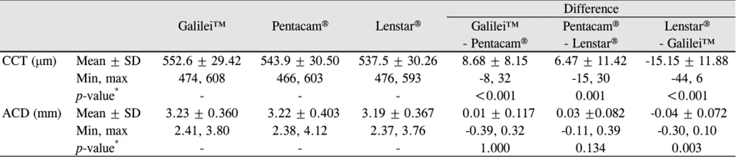
Table 1. The central corneal thickness (μm) and anterior chamber depth (mm) measured by Galilei™, Pentacam® and Lenstar®
Galilei™ Pentacam® Lenstar®
Difference Galilei™
- Pentacam®
Pentacam®
- Lenstar®
Lenstar®
- Galilei™
CCT (μm) Mean ± SD 552.6 ± 29.42 543.9 ± 30.50 537.5 ± 30.26 8.68 ± 8.15 6.47 ± 11.42 -15.15 ± 11.88
Min, max 474, 608 466, 603 476, 593 -8, 32 -15, 30 -44, 6
p-value* - - - <0.001 0.001 <0.001
ACD (mm) Mean ± SD 3.23 ± 0.360 3.22 ± 0.403 3.19 ± 0.367 0.01 ± 0.117 0.03 ±0.082 -0.04 ± 0.072
Min, max 2.41, 3.80 2.38, 4.12 2.37, 3.76 -0.39, 0.32 -0.11, 0.39 -0.30, 0.10
p-value* - - - 1.000 0.134 0.003
CCT = central corneal thickness; ACD = anterior chamber depth.
*Repeated measures analysis of variance with Bonferroni post hoc analysis.
따라 측정 오차를 줄이기 위한 비접촉 광학 기기들이 개발 되었다. 현재 slit-scanning 방식을 이용한 ORBscan II® (Bausch & Lomb, Rochester, NY, USA), 부분결합간섭계 원리를 이용한 기기인 IOL Master® (Carl Zeiss Meditec, Dublin, CA, USA) 및 저간섭반사계를 이용한 Lenstar® (Haag-Streit, Koeniz, Switzerland), Scheimpflug camera를 이용 한 Pentacam® (Oculus, Wetzlar, Germany), Galilei™ (Ziemer, Port, Switzerland), Sirius® (Costruzione Strumenti Oftalmici, Florence, Italy) 등을 포함한 다양한 기기들이 개발되어 임 상에서 사용되고 있다.
이 중 Pentacam®은 하나의 Scheimpflug camera가 475 nm 의 청색 LED 빛을 이용하여 360도 회전하며 얻는 25-30개의 각막 전면 및 후면의 정보를 포함한 전안부 이미지를 3차원 적 구조로 재구성하는 기기이며, 기기의 중심부에 존재하는 제2의 카메라(pupil camera)로 안구의 움직임을 보정한다.
Galilei G2는 dual rotating Scheimpflug camera와 placid disc topography가 결합된 방식으로 두 개의 Scheimpflug camera가 470 nm의 청색 LED 빛을 이용하여 180도 회전 하며 얻는 15-60개의 전안부 이미지를 분석하여 생체계측 치를 얻는다. 이 기기는 Placido disc image를 보정하여 각 막 전면의 곡률을 얻으며, Scheimpflug 이미지를 통해 높이 (elevation data)에 대한 정확한 정보를 제공한다고 알려졌 다.4 Lenstar는 인공수정체 도수 측정을 위해 기존에 개발되 어 널리 사용되고 있는 IOL Master의 전안부 계측치 및 인 공수정체 도수의 일치도가 매우 높을 뿐 아니라, IOL Master의 파장보다 긴 820 nm의 파장을 이용하기 때문에 투과력이 좋다고 알려졌다.5
상기 세 가지 기기 모두 비접촉 광학 방식의 기기로 점안 마취가 필요하지 않고 안구모양 변형을 일으키지 않으며, 수 초 이내의 한 번의 검사로 각막두께, 전방깊이 이외에도 동공크기, 각막윤부직경, 수정체두께 등의 생체 계측치를 제공하는 이점이 있어 임상에서 다양한 목적으로 사용되고 있다. 기존의 여러 연구를 통해 각 기기들의 재현성 및 반 복성이 매우 높은 것이 밝혀져 있지만, 이러한 기기들의 측
정치를 비교하였을 때, 대치하여 사용할 수 있는지에 대해 서는 이견이 있는 상태이다.6-20
이에 본 연구에서는 Galilei™와 Pentacam®, Lenstar®로 중 심각막두께와 전방깊이를 비교하여, 각 기기들 간의 측정 치의 차이가 있는지 알아보고 세 가지 기기들을 임상적으 로 대치하여 쓸 수 있는지에 대하여 알아보고자 하였다.
대상과 방법
2013년 6월 1일부터 31일까지 25명의 건강한 성인 지원 자를 대상으로 Galilei™와 Pentacam®, Lenstar®를 이용하여 중심각막두께와 전방깊이를 측정하였다. 안과적 수술력이나 외상력, 각막 질환의 과거력이 있는 안은 대상에서 제외하였 으며, 총 47안의 중심각막두께와 전방깊이를 분석하였다.
중심각막두께(central corneal thickness, CCT)는 각막의 중앙부전면(각막상피)에서 각막의 중앙부후면(각막내피)까 지의 거리로 정의하였고, 전방깊이(anterior chamber depth, ACD)는 각막의 중앙부전면(각막상피)에서 수정체중앙부전 면(수정체 전낭)까지의 거리로 정의하였다.
대상자에 대한 검사는 암실에서 산동하지 않은 대상안에 대해 한 명의 검사자가 각 1회씩 검사를 시행하였으며, 검 사 순서는 Galilei™, Pentacam®, Lenstar®의 순으로 시행하 였다. Galilei™와 Pentacam®은 각막지형도 검사를 시행하여 컴퓨터 화면에 각막중심과 동공중심이 중앙에 위치하고, 내외측과 상하측이 대칭이 되는 시점에서 스캔을 하였다. Lenstar®는 광원이 동공 중심에 오게 하여 4회 측정 후 평 균값을 측정치로 하였다. 검사 시 대상자의 머리와 턱을 각 각의 고정대에 고정하고 눈높이를 적절하게 위치시킨 후 검사하는 동안 시스템 중앙의 주시 광선을 보게 하고 눈을 움직이지 않도록 하였다. 검사 전에 눈을 깜박거리게 하여 눈물층을 균일하게 하였으며, 속눈썹이 각막을 가리지 않 도록 눈을 크게 뜨도록 지시한 후 안구에 압박이 가해지지 않도록 조심스럽게 눈꺼풀을 벌리면서 측정하였다.
통계학적인 분석은 SPSS 21.0 (SPSS Inc., Chicago, IL,
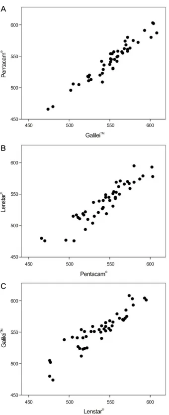
Figure 1. Scatter plots of central corneal thickness (μm) be-
tween (A) Galilei™ and Pentacam®, (B) Pentacam® and Lenstar®, (C) Lenstar® and Galilei™.USA)을 사용하였다. 세 가지 방법을 이용해 측정한 중심각 막두께 및 전방깊이는 repeated-measures analysis of variance (ANOVA)를 이용하여 비교하였고 사후 분석은 Bonferroni post hoc analysis를 이용하였다. 세 가지 검사 방법 간의 일
치도는 Bland-Altman plots를 이용하여 분석하였고, Pearson correlation을 통해 상관관계를 알아보았으며, p<0.05일 경 우 통계적으로 유의한 것으로 판정하였다.21,22
결 과
연구대상은 정상 성인 25명(남자 13명, 여자 12명) 47안이 었고, 평균 연령은 68.4 ± 11.7세였다. Galilei™, Pentacam®, Lenstar®를 이용하여 측정한 중심각막두께(평균 ± 표준편차) 는 각각 552.6 ± 29.41 μm, 543.9 ± 30.50 μm, 537.5 ± 30.26 μm 였다. Galilei™로 측정한 값이 가장 두꺼웠고, Lenstar®로 측 정한 값이 가장 얇게 나타났으며, 각 측정치 사이에 통계적 으로 유의한 차이를 보였다(repeated-measures ANOVA, p<0.05). 측정치 간의 차이를 Bonferroni 사후분석으로 비교 하였을 때, p-value는 Galilei™와 Pentacam®이 0.001 미만, Pentacam®과 Lenstar®가 0.001, Lenstar®와 Galilei™가 0.001 미만으로 모두 통계적인 차이를 보였다(Table 1). 모든 측정 치는 통계적으로 유의하게 높은 상관관계를 보였으며(Pearson correlation, Galilei™ vs. Pentacam®: r=0.964, Pentacam® vs.
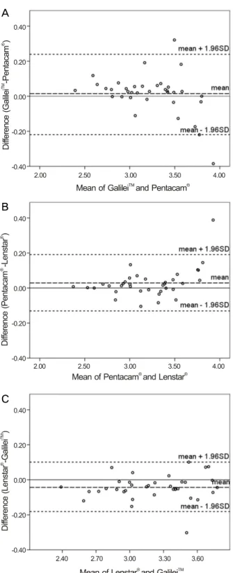
Lenstar®: r=0.929, Lenstar® vs. Galilei™: r=0.921, all p-val- ue<0.001) (Fig. 1), Bland-Altman 일치도 분석 결과 Galilei™ 와 Pentacam® 간의 95% 일치도 범위는 31.95 μm (-7.30~
24.65), Pentacam®과 Lenstar®는 44.76 μm (-15.91~28.85), Lenstar®와 Galilei™는 46.57 μm (-38.43 ~8.14)였다(Fig. 2).
전방깊이는 Galilei™, Pentacam®, Lenstar®에서 각각 3.23
± 0.360 mm, 3.22 ± 0.403 mm, 3.19 ± 0.367 mm로 Galilei™ 에서 가장 깊게, Lenstar®에서 가장 얕게 측정되었고, 통계 적인 차이를 보였다(repeated-measures ANOVA, p=0.034).
측정치 간의 차이를 Bonferroni 사후분석으로 비교하였을 때, p-value는 Galilei™와 Pentacam®이 1.000, Pentacam®과 Lenstar®가 0.134, Lenstar®와 Galilei™가 0.003으로, 세 기기 중 Lenstar®와 Galilei™만이 차이가 있는 것으로 나타났다 (Table 1). 모든 측정치는 통계적으로 유의하게 높은 상관 관계를 보였으며(Pearson correlation test, Galilei™ vs. Pentacam®: r=0.959, Pentacam® vs. Lenstar®: r=0.981, Lenstar® vs. Galilei™: r=0.981, all p<0.001) (Fig. 3), Bland-Altman 일치도 분석 결과 Galilei™와 Pentacam® 간의 95% 일치도 범위는 0.46 mm (-0.22~0.24), Pentacam®과 Lenstar®는 0.32 mm (-0.13~
0.19), Lenstar®와 Galilei™는 0.28 mm (-0.18~0.10)였다(Fig. 4).
고 찰
정확한 전안부 측정은 굴절이상을 교정하기 위한 각막굴 절교정수술이나 유수정체용 인공수정체 삽입술뿐 아니라,
A
B
C
Figure 2. Bland-Altman plots of CCT (μm) between (A)
Galilei™ and Pentacam®, (B) Pentacam® and Lenstar®, (C) Lenstar® and Galilei™. CCT = central corneal thickness.Figure 3. Scatter plots of anterior chamber depth (mm) be-
tween (A) Galilei™ and Pentacam®, (B) Pentacam® and Lenstar®, (C) Lenstar® and Galilei™.최근 빈도가 증가하고 있는 프리미엄 인공수정체 사용 등 과 더불어 그 중요성이 더욱 부각되고 있다. 비교적 최근까 지 안구의 생체 계측치는 초음파를 이용한 방식으로 측정 하여 왔으나, 최근 다양한 원리와 측정 방법을 이용한 전안
부 측정 기기들이 개발되고 임상에서 사용되고 있다.1,2,23-25 또한 이러한 기기들은 한 번의 검사로 여러 가지 생체 계측 정보를 얻을 수 있어 검사 시간을 단축하며 비접촉방식을 이용하여 환자 편의성도 증대되었다. 하지만 이러한 기기
A
B
C A
B
C
Figure 4. Bland-Altman plots of ACD (mm) between (A)
Galilei™ and Pentacam®, (B) Pentacam® and Lenstar®, (C) Lenstar® and Galilei™. ACD = anterior chamber depth.중 널리 사용되고 있는 두 가지 샤임플러그 기기와 저간섭 반사계의 측정치를 비교한 연구는 없었다. 각각의 병원이 모든 기기를 구비하기에는 현실적으로 어려움이 있으며 병 원 간 환자 정보 교류가 필요한 상황에서 각 병원이 서로
다른 기기를 사용하였다면, 기기별 측정 결과를 분석하는 데 혼란이 발생할 수 있다. 이에 본 연구에서는 상기 세 가 지 비접촉성 기기로 측정한 중심각막두께 및 전방깊이의 측정치를 비교하고 서로 대치하여 사용할 수 있는지에 대 해 알아보았다.
본 연구에서 중심각막두께는 Galilei™, Pentacam®, Lenstar® 순서로 두껍게 측정되었고, Galilei™는 Pentacam®보다 8.68 μm, Pentacam®은 Lenstar®보다 6.47 μm 컸으며, Galilei™와 Lenstar®의 차이가 15.15 μm로 가장 큰 차이를 보였다. 각 기기 간의 측정치를 비교했던 기존의 연구들에서는 대부분 본 연구와 비슷한 결과를 보였는데, 정상 성인 66명 66안에 서 Pentacam®, Galilei™, Sirius, RT-vue OCT (Optovue Inc., Freemont, CA, USA)를 비교한 Huang et al8,9의 연구에서는 Pentacam®과 Sirius의 중심각막두께만 상호 호환이 가능하 며, Galilei™가 Pentacam®보다 9.7 μm 두껍게 측정되고 95% 일치도 범위가 24.7 μm로 넓어 대치하여 사용할 수 없다고 하였고, Anayol et al6이 Galilei™와 Pentacam®, Sirius를 비교한 연구에서도 중심각막두께와 최소각막두께, 전방깊이를 비교할 때 Pentacam®과 Sirius의 측정치만이 거의 동일하며, 이 중 중심각막두께의 경우 Galilei™는 Pentacam® 이나 Sirius보다 13.9 μm 정도 더 두껍게 측정되고, 일치도 또한 높지 않다고 보고하였다. Crawford et al26의 연구에서 는 Pentacam®, Galilei™의 중심각막두께가 차이는 없으나, 95% 일치도 범위가 58 μm (-11~47)로 넓어 임상에서 대치 하여 사용하기에는 어려울 것이라고 보고하였다. 오직 Aramberri et al27의 연구에서만 Pentacam®과 Galieli™의 측 정치가 차이를 보이지 않으며, 일치도가 높다고 보고하였 다. Pentacam®과 Lenstar®를 비교한 경우 Borrego-Sanz et al7와 Tai et al14의 연구에서는 각각 23.3 μm, 6.6 μm 정도 Pentacam®이 더 두껍게 측정되었고, 95% 일치도 범위가 각 연구에서 50.79 μm (-3.09~47.7),7 36.84 μm (-11.88~24.96)14 넓어 대치하여 사용할 수 없다고 보고한 반면, Huang et al10의 연구에서는 두 기기 간에 차이가 약 3.7 μm이나, 일 치도가 23.9 μm (-8.2~15.7)10로 좋아 상호 호환이 가능하다 고 보고하였다. 본 연구에서는 Pentacam®의 측정치가 Lenstar®보다 6.47 μm 두꺼우며, 95% 일치도 범위가 44.76 μm (-15.91~28.85)로 넓어 측정치를 대치하여 사용할 수 없을 것으로 생각한다. OʼDonnell et al12은 Pentacam®과 Lenstar®, Visante OCT (Carl Zeiss, Dublin, CA, USA)를 비교하여 Lenstar®가 Pentacam®보다 8 μm 더 두껍게 측정되었다고 보고하여 본 연구와 상반되는 결과를 보였지만, 이 연구에 서도 역시 일치도가 낮아 대치하여 사용할 수는 없다고 하 였다. Galilei™와 Lenstar®의 전안부 측정치를 비교한 연구 는 현재까지 한 가지 연구가 존재하는데, Huerva et al11이
A
B
C
정상 성인 100명 100안에서 비교하였을 때 Galilei™의 측정 치가 약 17 μm 더 두껍게 측정되었다고 보고하여 본 연구 의 결과와 비슷한 정도의 차이를 보였고, 95% 일치도 범위 역시 34.2 μm (-18.8~-15.4)로 일치도가 높지는 않은 것으 로 나타났다. 본 연구에서 세 가지 기기 중에서는 중심각막 두께는 Galilei™와 Pentacam®의 일치도가 가장 높기는 하였 으나, 95% 일치도 범위가 넓어 임상에서 사용할 때 서로 대치하여 사용할 수는 없을 것으로 생각한다.
본 연구에서 전방 깊이는 Galilei™와 Pentacam®은 차이가 없었고, Galilei™와 Lenstar®만이 통계적인 차이를 보였다.
Anayol et al6과 Aramberri et al27의 연구에서는 본 연구 결 과와는 달리 Galilei™의 전방 깊이가 Pentacam®보다 약 0.1 mm 정도 더 깊게 측정되어 차이가 있었으며, Salouti et al13이 37명 74안을 대상으로 Pentacam®과 Galilei™, Orbscan®의 전방 깊이를 비교한 연구에서는 Orbscan의 측정치가 Pentacam®이나 Galilei™보다 0.3 mm 정도 깊게 측정되었 고, Pentacam과 Galilei™의 차이는 0.02 mm 정도의 통계적 인 차이가 있었으나, 이는 임상적으로는 큰 차이로 생각되 지 않는다. Salouti et al13의 연구에서는 전방깊이 3.0 mm 이내, 3.0-3.5 mm, 3.5 mm 이상의 경우 각 구간별로 전방 깊이가 깊을수록 일치도 범위가 늘어난다고 하였으나, 본 연구에서는 Fig. 4의 Bland-Altman plot 결과에서 보다시피, 전방 깊이에 따른 일치도의 비례오차(proportional error)를 보이지는 않았다. Pentacam®과 Lenstar®의 측정치를 비교한 연구들 중 Huang et al10, OʼDonnell et al12, Uçakhan et al15 은 각각 0.01 mm, 0.03 mm, 0.09 mm 정도 Pentacam®의 전 방깊이가 Lenstar®보다 더 깊게 측정되었고 통계적인 차이가 있었다. 본 연구에서도 Pentacam®의 전방깊이가 Lenstar®보 다 0.03 mm 깊게 측정되어 기존의 연구 결과와 비슷하였지 만, 이 역시 차이가 적어 임상적으로는 큰 의미는 없을 것으 로 생각한다. 일치도에 관하여서는 기존 연구에서 95% 일 치도 범위가 Pentacam®과 Lenstar® 간에 0.16-0.28 mm10,12,15 로 중등도 이상이었으나, 본 연구에서는 0.32 mm 정도로 일 치도가 높지 않았다. Galilei™와 Lenstar®의 측정치를 비교한 Huerva et al11은 95% 일치도 범위 자체는 0.04 mm로 좁으 나 Galilei™가 3.04 mm, Lenstar®가 2.92 mm로 Galilei™가 Lenstar®보다 0.12 mm 깊게 측정되어 대치하여 사용할 수 없다고 보고하였으며, 본 연구에서는 두 기기 간 차이가 0.04 mm로 기존 연구보다 차이가 적으나, 95% 일치도 범 위가 0.28 mm (-0.18~0.10)로 역시 측정치를 대치하여 사용 하기에는 힘들 것으로 생각한다. 전방깊이의 경우 Galilei™ 와 Pentacam®의 측정치는 비슷하고, Lenstar®는 차이를 보 이며, 세 기기 간의 일치도는 범위가 넓어 측정치를 대치하여 사용할 수는 없을 것으로 생각한다.
본 연구에는 몇 가지 제한점이 있는데 첫째로 각 기기들 의 반복성(Repeatability) 및 재현성(Reproducibility)에 대한 재검증을 시행하지는 않았다는 점이다. 하지만 기존 연구 들에서 Galilei™16-18와 Pentacam®,16,18 Lenstar®19,20 모두 정 상인에서 반복성 및 재현성이 뛰어남이 보고되어 있다. 둘 째, 본 연구는 병원 내원객 중 일부 지원자를 대상으로 하 였기 때문에 대상군의 평균 나이가 68세로, 주로 20-30대를 대상으로 한 기존의 연구들에 비해 연령대가 높아 본 연구 의 대상자로 지정된 표본 집단이 대한민국 국민이라는 모 집단을 대변하지는 못한다는 점이다. 검사 대상자의 연령 이 증가할수록 주시 저하, 눈물층의 불안정성으로 인한 빠 른 증발, 검사시의 눈깜박임 등에 의해 영향을 받을 가능성 이 커질 수 있다. 그러나 Pentacam®은 quality “OK”, Galieli™ 는 quality factor 90% 이상의 검사 결과만을 채택하였고, Lenstar®의 경우 눈깜박임이나 주시가 되지 않는 경우는 자 동으로 측정치에서 제외되기 때문에 이에 의한 영향은 많 지 않으리라고 생각한다. 추후 더 많은 수의 참여자를 대상 으로 한 연령별, 성별 분석 및 백내장이 있는 경우, 각막 질 환이 있는 경우, 위수정체안에서 기기 간 측정치를 비교한 연구가 추가적으로 필요할 것으로 생각한다. 셋째, 일반적 으로 검사를 위한 시간이 길수록 주시점 이탈의 기회가 많 아지고 눈물층 증발이 많아지게 되어 오차가 발생할 가능 성이 커지게 되는데, 본 연구에서는 검사에 소요된 시간을 검사자마다 따로 측정하지 못하여 이를 비교할 수는 없었 다. 이론적으로 단일샤임플러그 방식인 Pentacam®에 비해 Galilei™는 이중샤임플러그 방식으로 인해 검사 시간이 짧 고 안구 중심축 감지에 민감하여 생체계측에 있어 유리하 였을 것으로 생각되지만, 각 기기의 user’s manual에 따르 면 Galieli™와 Pentacam®의 측정 소요시간은 모두 2초 이 내, Lenstar® 또한 수 초 이내로 보고되어 있어 검사 시간에 따른 영향은 적었을 것으로 생각한다.
본 연구를 통해 세 가지 기기로 측정한 중심각막두께와 전방깊이 중 어떠한 것을 임상에서 택해서 사용해야 할지 에 대해서는 결론을 내릴 수는 없을 것이다. 하지만, 임상 에서 제한된 정보를 가지고 측정치를 비교함에 있어, 각 기 기 간의 측정치 간의 상관관계, 측정치 간의 차이, 일치도 의 정도를 보여주는 데 본 연구의 의의가 있을 것으로 생각 한다.
REFERENCES
1) Reddy AR, Pande MV, Finn P, El-Gogary H. Comparative estima- tion of anterior chamber depth by ultrasonography, Orbscan II, and IOLMaster. J Cataract Refract Surg 2004;30:1268-71.
2) Vetrugno M, Cardascia N, Cardia L. Anterior chamber depth meas-
ured by two methods in myopic and hyperopic phakic IOL implant.
Br J Ophthalmol 2000;84:1113-6.
3) Olsen T. Sources of error in intraocular lens power calculation. J Cataract Refract Surg 1992;18:125-9.
4) Savini G, Carbonelli M, Barboni P, Hoffer KJ. Repeatability of au- tomatic measurements performed by a dual Scheimpflug analyzer in unoperated and post-refractive surgery eyes. J Cataract Refract Surg 2011;37:302-9.
5) Jasvinder S, Khang TF, Sarinder KK, et al. Agreement analysis of LENSTAR with other techniques of biometry. Eye (Lond) 2011;
25:717-24.
6) Anayol MA, Güler E, Yağc R, et al. Comparison of central corneal thickness, thinnest corneal thickness, anterior chamber depth, and simulated keratometry using galilei, Pentacam, and Sirius devices.
Cornea 2014;33:582-6.
7) Borrego-Sanz L, Sáenz-Francés F, Bermudez-Vallecilla M, et al.
Agreement between central corneal thickness measured using Pentacam, ultrasound pachymetry, specular microscopy and optic biometer Lenstar LS 900 and the influence of intraocular pressure.
Ophthalmologica 2014;231:226-35.
8) Huang J, Ding X, Savini G, et al. Central and midperipheral corneal thickness measured with Scheimpflug imaging and optical coher- ence tomography. PLoS One 2014;9:e98316.
9) Huang J, Ding X, Savini G, et al. A Comparison between Scheimpflug imaging and optical coherence tomography in measuring corneal thickness. Ophthalmology 2013;120:1951-8.
10) Huang J, Pesudovs K, Wen D, et al. Comparison of anterior seg- ment measurements with rotating Scheimpflug photography and partial coherence reflectometry. J Cataract Refract Surg 2011;
37:341-8.
11) Huerva V, Ascaso FJ, Soldevila J, Lavilla L. Comparison of ante- rior segment measurements with optical low-coherence reflec- tometry and rotating dual Scheimpflug analysis. J Cataract Refract Surg 2014;40:1170-6.
12) OʼDonnell C, Hartwig A, Radhakrishnan H. Comparison of central corneal thickness and anterior chamber depth measured using LenStar LS900, Pentacam, and Visante AS-OCT. Cornea 2012;
31:983-8.
13) Salouti R, Nowroozzadeh MH, Zamani M, et al. Comparison of an- terior chamber depth measurements using Galilei, HR Pentacam, and Orbscan II. Optometry 2010;81:35-9.
14) Tai LY, Khaw KW, Ng CM, Subrayan V. Central corneal thickness measurements with different imaging devices and ultrasound
pachymetry. Cornea 2013;32:766-71.
15) Uçakhan OÖ, Akbel V, Bıyıklı Z, Kanpolat A. Comparison of cor- neal curvature and anterior chamber depth measurements using the manual keratometer, Lenstar LS 900 and the Pentacam. Middle East Afr J Ophthalmol 2013;20:201-6.
16) Hernández-Camarena JC, Chirinos-Saldaña P, Navas A, et al.
Repeatability, reproducibility, and agreement between three differ- ent Scheimpflug systems in measuring corneal and anterior seg- ment biometry. J Refract Surg 2014;30:616-21.
17) Lopez de la Fuente C, Sanchez-Cano A, Segura F, et al. Repeatability of ocular measurements with a dual-Scheimpflug analyzer in healthy eyes. Biomed Res Int 2014;2014:808646.
18) Wang Q, Ding X, Savini G, et al. Anterior chamber depth measure- ments using Scheimpflug imaging and optical coherence tomog- raphy: repeatability, reproducibility, and agreement. J Cataract Refract Surg 2015;41:178-85.
19) Shammas HJ, Hoffer KJ. Repeatability and reproducibility of bio- metry and keratometry measurements using a noncontact optical low-coherence reflectometer and keratometer. Am J Ophthalmol 2012;153:55-61.e2.
20) Bayhan HA, Aslan Bayhan S, Can I. Comparison of central corneal thickness measurements with three new optical devices and a standard ultrasonic pachymeter. Int J Ophthalmol 2014;7:302-8.
21) Bland JM, Altman DG. Statistical methods for assessing agree- ment between two methods of clinical measurement. Lancet 1986;1:307-10.
22) Bland JM, Altman DG. Measurement error. BMJ 1996;313:744.
23) Doughty MJ, Zaman ML. Human corneal thickness and its impact on intraocular pressure measures: a review and meta-analysis approach. Surv Ophthalmol 2000;44:367-408.
24) Holladay JT. Standardizing constants for ultrasonic biometry, kera- tometry, and intraocular lens power calculations. J Cataract Refract Surg 1997;23:1356-70.
25) Kim YY, Jung HR. Clarifying the nomenclature for primary an- gle-closure glaucoma. Surv Ophthalmol 1997;42:125-36.
26) Crawford AZ, Patel DV, McGhee CN. Comparison and repeat- ability of keratometric and corneal power measurements obtained by Orbscan II, Pentacam, and Galilei corneal tomography systems.
Am J Ophthalmol 2013;156:53-60.
27) Aramberri J, Araiz L, Garcia A, et al. Dual versus single Scheimpflug camera for anterior segment analysis: precision and agreement. J Cataract Refract Surg 2012;38:1934-49.
= 국문초록 =
서로 다른 세 가지 기기로 측정한 중심각막두께 및 전방깊이의 비교
목적: Galilei™ (Ziemer, Port, Switzerland)와 Pentacam® (Oculus, Wetzlar, Germany), Lenstar® (Haag-Streit, Koeniz, Switzerland) 로 측정한 중심각막두께와 전방깊이의 측정치를 비교하고, 일치도를 분석하고자 한다.
대상과 방법: 정상 성인 25명의 47안을 대상으로 세 기기를 이용하여 측정한 중심각막두께와 전방깊이를 비교하였다.
결과: Galilei™, Pentacam®, Lenstar®를 이용하여 측정한 중심각막두께는 각각 552.6 ± 29.41 μm, 543.9 ± 30.50 μm, 537.5 ± 30.26 μm였고, 측정치 간에 높은 상관관계를 보였으나(r>0.9, p<0.001), 통계적으로 유의한 차이를 보였다(p<0.001). 전방깊이는 Galilei™가 3.23 ± 0.360 mm, Pentacam®이 3.22 ± 0.403 mm, Lenstar®가 3.19 ± 0.367 mm였고, 측정치 간에 높은 상관관계를 보였으나(r>0.9, p<0.001), 통계적으로 유의한 차이를 보였다(p=0.034). 95% 일치도 범위는 Galilei™와 Pentacam®, Pentacam®과 Lenstar®, Lenstar®와 Galilei™ 사이에서 중심각막두께는 31.95 μm, 44.76 μm, 46.57 μm, 전방깊이는 0.46 mm, 0.32 mm, 0.28 mm 였다.
결론: 세 기기로 측정한 중심각막두께 및 전방깊이는 상관성이 높으나, 측정값에 차이를 보이고 일치도가 낮아 서로 대치하여 사용할 수 없을 것으로 생각한다.
<대한안과학회지 2015;56(5):694-701>