pISSN: 0378-6471⋅eISSN: 2092-9374
http://dx.doi.org/10.3341/jkos.2014.55.7.1007
Original Article
전방깊이 및 전방각 측정에서 앉은 자세로 촬영된 초음파생체현미경의 유용성
Clinical Usefulness of UBM in the Sitting Position in Anterior Chamber Depth and Angle Measurements
김태기⋅문성운⋅양지호⋅진경현
Tae Gi Kim, MD, Sung Woon Moon, MD, Ji Ho Yang, MD, Kyung Hyun Jin, MD, PhD
경희대학교 의학전문대학원 경희대학교병원 안과학교실
Department of Ophthalmology, Kyung Hee University Medical Center, Kyung Hee University School of Medicine, Seoul, Korea
Purpose: We compared the measurements of anterior chamber depth (ACD) and anterior chamber angle (ACA) using ultra- sound biomicroscopy (UBM) in the sitting position compared with IOL Master®, Pentacam®, and Spectralis optical coherence to- mography (OCT) to evaluate the clinical usefulness of UBM in the sitting position.
Methods: We evaluated 92 eyes in 47 healthy adults. ACD was measured by IOL Master®, Pentacam®, and UBM. ACA was measured using Pentacam®, UBM, and Spectralis OCT. UBM was performed in the sitting position using bag/balloon technology. Measured values were compared statistically.
Results: ACD measured by IOL Master®, Pentacam®, and UBM was 3.57 ± 0.32 μm, 3.64 ± 0.33 μm and 3.51 ± 0.32 μm, respectively. UBM measurements of ACD were significantly shallower than with the other methods (p < 0.001). The results among the 3 methods were strongly correlated (r > 0.8, p < 0.05 in all groups). Mean ACA of 4 directions was not significantly dif- ferent among the 3 methods (p > 0.05). There was strong correlation between UBM and Spectralis OCT (r = 0.957) but moderate correlation between Pentacam® and UBM and Pentacam® and Spectralis OCT (r = 0.557, 0.571, respectively, p < 0.05).
Specifically, ACA of the superior quadrant showed a low correlation between Pentacam® and UBM and Pentacam® and Spectralis OCT (r = 0.257, 0.295, respectively).
Conclusions: ACD measured by UBM in the sitting position was shallower compared to the other methods; however, ACD meas- ured by IOL Master®, Pentacam®, and UBM showed significant correlations among the methods. The mean ACA measured by Pentacam®, UBM, and Spectralis OCT showed no significant differences. Due to the high correlation of ACA measurements between UBM and Spectralis OCT in the present study, UBM is expected to be a good tool for measuring anterior segment parameters.
J Korean Ophthalmol Soc 2014;55(7):1007-1016
Key Words: Anterior chamber angle, Anterior chamber depth, Sitting position, UBM
■Received: 2013. 11. 23. ■ Revised: 2014. 2. 19.
■Accepted: 2014. 6. 9.
■Address reprint requests to Kyung Hyun Jin, MD, PhD Department of Ophthalmology, Kyung Hee University Medical Center, #23 Kyungheedae-ro, Dongdaemun-gu, Seoul 130-872, Korea
Tel: 82-2-958-8451, Fax: 82-2-966-7340 E-mail: [email protected]
* This study was presented as a narration at the 110th Annual Meeting of the Korean Ophthalmological Society 2013.
ⓒ2014 The Korean Ophthalmological Society
This is an Open Access article distributed under the terms of the Creative Commons Attribution Non-Commercial License (http://creativecommons.org/licenses/by-nc/3.0/) which permits unrestricted non-commercial use, distribution, and reproduction in any medium, provided the original work is properly cited.
전방 깊이와 전방각은 유수정체용 인공수정체의 삽입, 인공수정체의 도수결정 및 녹내장 연구에 있어서 매우 중 요한 계측치이다.1-4 백내장 수술 시 전방 깊이의 오류가 목 표 굴절력 편향의 20-40%를 차지한다는 보고가 있으며,1,2 전방 깊이가 얕고, 전방각이 작은 경우 폐쇄각 녹내장의 발 생 빈도가 높다고 한다. 특히, 급성 폐쇄각 녹내장의 경우 서양인에 비해 동양인에서 흔하게 나타나며, 전방 깊이 및
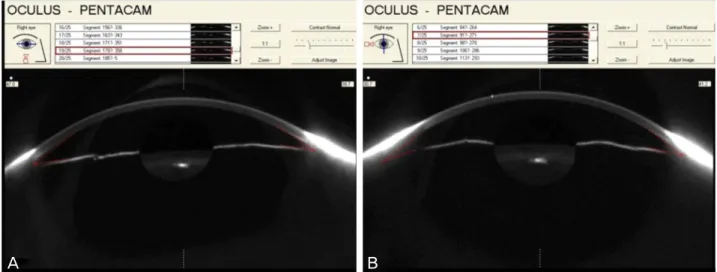
A B
Figure 1. An example of the result of anterior chamber angle (ACA) measurement by Pentacam®. (A) Nasal and temporal angle. (B)
Superior and inferior angle. ACA is calculated by integrated software of Pentacam®.전방각의 구조를 파악하는 것은 급성 폐쇄각 녹내장의 진 단 및 치료에 중요한 정보를 제공한다.5,6
이처럼 전방 깊이 및 전방각의 정확한 측정은 여러 안과 분야에서 중요한 의미가 있어 현재 다양한 검사 장비에 의 해 계측이 이루어지고 있다. 전방 깊이는 초음파를 사용한 A-scan이 주로 사용되었고,7 전방각은 과거에는 전방각경을 사용한 검사를 통해 Van Herick and Shaffer grade, 또는 Spaeth grading system에 의해 구분하는 방법이 많이 사용 되었다. 근래에는 전방의 구조를 좀 더 정확하고 정량적으로 계측하려는 시도가 이루어지고 있다. 현재 전방 깊이의 측정 은 IOL master®, 초음파생체현미경(ultrasound biomicroscope, UBM), 샤임플러그 사진기 원리를 사용하여 전방을 계측하 는 Pentacam® 등의 장비가 개발되어 사용되고 있으며, 전 방각 측정에는 초음파생체현미경, Pentacam®, Visante OCT 등과 같은 장비가 다양하게 사용되고 있다.8-10
초음파생체현미경은 고주파의 초음파를 이용하는 검사 기계로 전방각 및 전방깊이를 포함하여 기존 안과 장비로 는 관찰할 수 없는 홍채 뒷부분의 조직까지 촬영할 수 있 다. 초음파 생체현미경과 다른 검사 장비 사이에 측정치의 정확도 및 일치도에 대해서 다양한 연구가 이루어지고 있 으며, 검사기 기간에 신뢰할 만한 일치도를 보였다는 연구 가 많다.11-13 그러나 기존의 비교 연구에서 사용되었던 초음 파생체현미경의 경우 누운 자세에서 촬영하였고 중력의 영 향으로 누운 자세에서 전방 계측치가 영향을 받는다는 연 구 결과도 있어 일반적으로 앉아서 측정하게 되는 다른 기 기와의 비교연구에서 자세변화에 계측치의 변화가 변수로 고려되지 않았을 가능성도 있다.14-16
기존 초음파생체현미경은 open-shell immersion technique 을 이용하여 컵을 눈꺼풀 사이에 끼고 누운 자세에서 검사
를 받아야 하기 때문에 앉아서 촬영하는 다른 기계들에 비 해 불편함이 있었다. 최근 bag/balloon cover (ClearScan;
ESI, Inc., Plymouth, MN)를 이용하여 앉은 자세에서도 초 음파 생체현미경을 촬영하기 위한 방법이 소개되었다.17 SONOMED사의 VuMax-II UBM은 탐침에 장착하는 특수 한 캡(water-filled bag)으로 앉은 자세에서도 촬영이 가능하 여 다른 기기와의 비교 연구에서 자세변화 의한 변수를 최 소화하여 비교할 수 있다. 또한 하루의 대부분을 일어난 자 세에서 생활하는 것을 감안하면 초음파생체현미경의 경우 도 누운 자세에서 측정하는 것보다 앉은 자세에서 측정하 여 비교하는 것이 더 의미가 크다. SONOMED UBM의 임 상적 유용성에 대해서는 최근 연구가 진행되었으나 국내를 비롯하여 동양인에서는 아직 보고된 바가 없다.16,18
본 연구는 특수 캡을 이용하여 앉은 자세에도 촬영이 가 능한 SONOMED사의 UBM과 IOL master®, Pentacam®, 후 안부 OCT인 Spectralis OCT를 이용하여 전방각 및 전방 깊 이를 측정하여 결과를 비교하여 보고자 하였다.
대상과 방법
2012년 6월부터 2013년 5월까지 건강한 성인 지원자를 대 상으로 IOL master® (Carl Zeiss Jena GmbH, Jena, Germany), Pentacam® (Oculus, Wetzlar, Germany), 초음파생체현미경 (VuMax-II, SONOMED, US)을 이용하여 전방 깊이를 측정 하였고 전방각의 경우 Pentacam®, 초음파생체현미경, Spectralis HRA+OCT (Heidelberg Engineering, Heidelberg, Germany)의 Anterior Segment Module을 이용하여 측정하 였다. 대상 안은 모두 유수정체 안으로 안과적 수술력, 외 상, 녹내장과 같이 전안부 계측치에 영향을 줄 수 있는 경
Figure 2. ClearScan bag/balloon technology.
Figure 3. Ultrasound biomicroscopic image of anterior cham-
ber and lens. Anterior chamber depth 1 (ACD1) is defined as distance from the central corneal epithelium to the anterior surface of lens capsule and ACD2 is defined as distance from the central corneal endothelium to the anterior surface of lens capsule.Figure 4. Ultrasound biomicroscopic (A) and spectralis optical coherence tomography (OCT), (B) image of anterior chamber angle.
Anterior chamber angle (ACA) is measured by trabecular iris angle (TIA). TIA is defined as the angle between the iris and tra- becular meshwork, with the apex of the angle lying in the angle recess and arms lying at a point 500 μm away from the scleral spur.
우는 대상에서 제외하였다.
초음파생체현미경 검사 시 접촉에 의한 측정값 변화의 가능성을 배제하기 위해 검사는 IOL master®, Pentacam®,
Spectralis OCT, 초음파생체현미경 순서로 진행하였고, 검 사는 동일한 임상 조건하에서 동일한 장비를 사용하여 실 시되었다. 모든 검사 시 조절에 의한 영향을 최소화하기 위 해 1.5 m 거리에 있는 목표점을 응시하도록 하였다.
IOL master®를 이용한 전안부 계측은 내장된 소프트웨어 에 의해 자동 계산된 전방 깊이를 이용하였다. IOL master® 의 경우 전방 깊이를 계산할 때, 각막 표면과 수정체 표면 에서 나타나는 반사광을 인지하여 계산되도록 프로그램 되 어 있다.
Pentacam® 촬영 시 전방 깊이의 경우 mm 단위로, 전방 각의 경우 비측, 상측, 이측 및 하측에서 degree 단위로 자 동 계산된 값을 이용하였다(Fig. 1). 전방 깊이 계측치는 IOL master®와 비교하기 위해 두 가지 방법으로 측정되었 다. Anterior chamber depth 1 (ACD 1)은 각막 상피에서부 터 수정체전낭까지의 거리로 내장된 소프트웨어의 external anterior chamber depth에 의해 자동 계산하였고 Anterior chamber depth 2 (ACD 2)는 각막 내피에서부터 수정체전 낭까지의 거리로 internal anterior chamber depth를 이용하 여 자동 계산된 전방 깊이를 이용하였다. 전방각은 측정값 은 360도 방위로 스캔된 이미지 중 수평면 및 수직면에서 얻어진 화상을 바탕으로 내장된 소프트웨어에 의해 자동 계산하였다. 수직면 영상 촬영 시 눈꺼풀에 의한 영향을 최 소화하기 위해 눈꺼풀을 최대한 벌린 후 촬영하였다.
초음파생체현미경을 통한 전안부 계측은 0.5% propar- acaine (Alcaine, Alcon Inc., Cleveland, USA)으로 점안 마 취 후 시행하였다. 본 연구에 사용된 초음파생체현미경은 SONOMED사의 VuMax-II로 기존 초음파생체현미경의 경 우 누운 자세로 눈을 물에 잠기게 한 후 촬영해야 하였으 나, VuMax-II의 경우 탐침에 bag/balloon cover (ClearScan;
ESI, Inc., Plymouth, MN)라고 불리는 특수 캡(Cap)을 장착
A B
Table 1. Comparison of different techniques of anterior chamber depth (ACD)
IOL master® Pentacam® UBM p-value*
ACD1 Male 3.60 ± 0.36 3.66 ± 0.36 3.52 ± 0.36 <0.001
Female 3.51 ± 0.20 3.59 ± 0.23 3.48 ± 0.25 <0.001
Mean 3.57 ± 0.32 3.64 ± 0.33 3.51 ± 0.32 <0.001
ACD2 Male NA 3.10 ± 0.36 2.95 ± 0.25 <0.001
Female NA 3.04 ± 0.23 2.94 ± 0.35 <0.001
Mean NA 3.08 ± 0.33 2.95 ± 0.33 <0.001
Values are presented as mean ± SD; ACD1: From the central corneal epithelium to the anterior surface of lens capsule; ACD2: From the central corneal endothelium to the anterior surface of lens capsule.
ACD = anterior chamber depth; UBM = ultrasound biomicroscopy.
*Paired t-test, comparing IOL master®, Pentacam® and UBM.
하여 10 mL의 물을 채운 후 앉은 자세에서도 촬영이 가능 하다(Fig. 2). 검사 시에 전방에 있는 주시점을 바라보게 한 상태에서 탐침을 움직여 주시 방향과 동일선상의 중심부위 와 이측, 상측, 비측 및 하측의 영상을 촬영하였다. 촬영 시 탐침의 주파수는 35 Hz로 동일하였다. 전방 깊이는 마찬가 지로 두 가지 방법으로 측정하였고, 초음파생체현미경에 내장되어 있는 caliber를 이용하여 중심 각막 상피부터 수 정체전낭까지의 수직 거리를 측정하였고(ACD 1), ACD 2 는 각막 내피에서부터 수정체전낭까지의 수직 거리를 측정 하였다(Fig. 3). 전방각(anterior chamber angle, ACA)은 공 막극에서 500 μm 떨어진 섬유주의 한 점과 그 점에서 수직 으로 내려와 반대편 홍채와 만나는 지점에서 iris recess의 꼭지에 이르는 선이 만드는 각도로 측정하였다(Fig. 4).
Spectralis HRA+OCT (Heidelberg Engineering, Heidelberg, Germany)를 이용한 촬영에서는 Anterior Segment Module을 이용하여 이측, 상측, 비측 및 하측의 전방각 영상을 촬영 하여 초음파생체현미경에서 측정한 방법과 동일하게 계산 하였다. 각각의 측정값은 동일한 검사자에 의해 3회씩 반복 측정하여 평균값을 구하였다(Fig. 4).
각 측정값에 대한 평균값 및 표준편차를 계산하였고, 통 계는 SPSS 12.0 for Windows (SPSS INC)를 이용하여 분석 하였다. 각각의 방법에 따른 측정값 사이의 차이를 paired t-test를 통해 측정값의 평균의 차이를 비교하였으며, p-val- ue가 0.05 이하인 경우에 통계적으로 유의하다고 판단하였 다. 각 방법 사이의 상관관계에 대해서는 Pearson의 단순상 관계수를 이용하여 구하였으며, p-value가 0.05 이하인 경 우에 통계적으로 유의하다고 판단하였다. 기기 간 일치성 을 평가하기 위해 Bland-Altman plot을 이용하였으며 95%
신뢰구간에서 평균 차이는 ±1.96 SD였다.
결 과
대상 안은 남자 31명(61안), 여자 16명(31안), 총 92안이
었고 평균 연령은 30.35 ± 2.88세였다.
전방 깊이의 비교 및 상관관계 분석
전방 깊이는 IOL master®, Pentacam®, 초음파생체현미경 을 비교하였다. 각각의 ACD 1의 평균값은 3.57 ± 0.32 μm, 3.64 ± 0.33 μm, 3.51 ± 0.32 μm이었고 남자의 경우 각각 3.60 ± 0.36 μm, 3.66 ± 0.36 μm, 3.52 ± 0.36 μm였고 여자 의 경우 3.51 ± 0.20 μm, 3.59 ± 0.23 μm, 3.48 ± 0.25 μm로 젊은 남자가 여자보다 전방 깊이가 유의하게 깊게 측정되 었다(Table 1). 전방 깊이는 Pentacam®으로 측정한 값이 가 장 깊었고, 초음파생체현미경으로 측정한 값이 가장 얕게 나타났으며 각 측정치 사이에 통계적으로 유의한 차이를 보였다(p<0.001) (Table 1). ACD 2를 이용한 전방 깊이의 평균값은 Pentacm®, 초음파생체현미경의 경우 각각 3.08 ± 0.33 μm, 2.95 ± 0.33 μm으로 측정되었고 남자의 경우 각 각 3.10 ± 0.36 μm, 2.95 ± 0.25 μm였고 여자의 경우 3.04
± 0.23 μm, 2.94 ± 0.35 μm로 초음파생체현미경으로 측정 한 값이 유의하게 얕게 측정되었고(p<0.001) 남자에서 여자 보다 전방 깊이가 유의하게 깊었다(Table 1).
세 검사 중 2가지 측정치의 차이를 비교하였을 때, IOL master®와 Pentacam®은 0.06 ± 0.13 μm, Pentacam®과 초음 파생체현미경은 0.12 ± 0.17 μm, IOL master®와 초음파생 체현미경에서는 0.06 ± 0.21 μm의 차이가 있었다.
Bland-Altman plot으로 나타낸 기기 간의 일치도에서 모든 기기들의 95% LoA (limits of agreement) 값은 0.42 mm 범 위에 있었으며, 특히 Pentacam®과 IOL master®의 95% LoA 가 0.26 mm 이하의 범위 안에 있어 높은 일치도를 보였다 (Fig. 5). 전방 깊이 측정치는 세 기계 사이에 통계학적으로 강한 양의 상관관계를 보였다(Pearson correlation, r=0.917, 0.863, 0.801, p<0.05 in all groups) (Fig. 6).
전방각의 비교 및 상관관계 분석
전방각은 Pentacam®, 초음파생체현미경, Spectralis OCT
2.5 3.0 3.5 4.0 4.5 5.0 -0.5
-0.4 -0.3 -0.2 -0.1 0.0 0.1 0.2 0.3
Mean of IOL Master ACD and Pentacam ACD
IOL Master ACD - Pentacam ACD
Mean -0.07
-1.96 SD -0.33 +1.96 SD 0.19
2.5 3.0 3.5 4.0 4.5 5.0
-0.8 -0.6 -0.4 -0.2 0.0 0.2 0.4 0.6 0.8
Mean of Pentacam ACD and UMB ACD
Pentacam ACD - UMB ACD
Mean 0.13 -1.96 SD -0.20 +1.96 SD 0.46
2.5 3.0 3.5 4.0 4.5 5.0
-0.8 -0.6 -0.4 -0.2 0.0 0.2 0.4 0.6 0.8 1.0
Mean of UMB ACD and IOL Master ACD
UMB ACD - IOL Master ACD
Mean -0.06 -1.96 SD -0.48 +1.96 SD 0.36
Figure 5. Bland-Altman plots showing differences in anterior
chamber depth (ACD) between devices. The upper and lower dashed lines indicate 95% limits of agreement and the solid line indicates the mean. UBM = ultrasound biomicroscopy.4.50
4.00
3.50
3.00
2.50
UBM ACD (mm)
IOL master ACD (mm)® rp = 0.917
< 0.000
2.50 3.00 3.50 4.00 4.50
4.50
4.00
3.50
3.00
2.50
IOL master ACD (mm)®
2.50 3.00 3.50 4.00 4.50 rp = 0.801
< 0.000
rp = 0.863 < 0.000
2.50 3.00 3.50 4.00 4.50 Pentacam ACD (mm)®
UBM ACD (mm)
4.50
4.00
3.50
3.00
2.50
Pentacam ACD (mm)®
Figure 6. The correlation plot of the anterior chamber depth
(ACD) measured by IOL master®, Pentacam® and ultrasound biomicroscopy (UBM).UMB ACD-IOL Master ACD®(mm)
를 비교하였다. 전방각 측정치를 살펴보면, Pentacam®을 이 용한 경우 이측, 상측, 비측, 하측이 각각 41.11 ± 6.01o, 32.58 ± 4.93o, 40.79 ± 5.31o, 38.45 ± 5.35o로 측정되었고 초음파생체현미경의 측정치는 각각 40.18 ± 6.12o, 31.81 ± 5.08o, 39.62 ± 5.71o, 39.64 ± 5.41o, Spectralis OCT에서는 각각 41.48 ± 6.88o, 32.54 ± 5.32o, 40.19 ± 6.38o, 40.68 ±
5.65o로 세 가지 측정치에서 공통적으로 상측 전방각이 유 의하게 가장 얕게 측정되었다(p<0.000) (Table 2). 네 방향 에서 측정한 전방각의 평균값을 비교하였을 때, Pentacam® 의 경우 38.23 ± 4.12o, 초음파 생체현미경의 경우 37.81 ± 6.58o, Spectralis OCT의 경우 38.72 ± 7.07o로 세 측정치 간 의 유의한 차이는 없었다(p>0.05) (Table 2). Bland-Altman
Mean of IOL MasterⓇ ACD and PentacamⓇ ACD (mm)
Mean of PentacamⓇ ACD and UMB ACD (mm)
Mean of UMB ACD AND IOL Master ACD (mm) IOL master ACD-Pentacam ACD (mm)®® UMB ACD-IOL Master ACD®(mm)
Table 2. Anterior chamber angle (ACA) at different directions and mean ACA
Directions Anterior chamber angle (degree)
Pentacam® UBM Spectralis-OCT
Temporal ACA Male 41.78 ± 6.10 40.98 ± 6.10 41.98 ± 6.88
Female 39.76 ± 5.59 38.60 ± 5.82 40.48 ± 6.79
Mean 41.11 ± 6.01 40.18 ± 6.12 41.48 ± 6.88
Superior ACA Male 32.23 ± 4.58* 32.61 ± 5.25* 33.29 ± 5.66*
Female 33.27 ± 5.48* 30.23 ± 4.33* 31.05 ± 4.19*
Mean 32.58 ± 4.93* 31.81 ± 5.08* 32.54 ± 5.32*
Nasal ACA Male 41.14 ± 5.34 40.03 ± 5.62 40.76 ± 6.49
Female 40.11 ± 5.18 38.80 ± 5.82 39.07 ± 6.01
Mean 40.79 ± 5.31 39.62 ± 5.71 40.19 ± 6.38
Inferior ACA Male 37.84 ± 5.23 39.93 ± 5.78 40.42 ± 6.06
Female 39.65 ± 5.39 39.08 ± 4.54 41.20 ± 4.71
Mean 38.45 ± 5.35 39.64 ± 5.41 40.68 ± 5.65
Mean ACA Male 38.25 ± 6.55 38.39 ± 6.61 39.11 ± 7.15
Female 38.20 ± 6.12 36.68 ± 6.38 37.95 ± 6.85
Mean 38.23 ± 6.41 37.81 ± 6.58 38.72 ± 7.07
Values are presented as mean ± SD.
UBM =ultrasound biomicroscopy; OCT = optical coherence tomography.
*p < 0.05 = statistically significant p-value, Paired t-test comparing among the four quadrant of ACA.
Table 3. Pearson correlations of anterior chamber angle (ACA) at different directions among Pentacam®, ultrasound biomicroscopy
(UBM), Spectralis OCTPentacam®-UBM UBM-Spectralis OCT Specralis OCT-Pentacam®
Directions Pearson
correlation p-value Pearson
correlation p-value Pearson
correlation p-value
Superior ACA 0.257 0.013 0.866 <0.001 0.295 0.001
Temporal ACA 0.647 <0.001 0.910 <0.001 0.567 <0.001
Nasal ACA 0.495 <0.001 0.874 <0.001 0.475 <0.001
Inferior ACA 0.317 0.002 0.859 <0.001 0.328 0.004
OCT = optical coherence tomography.
plot으로 나타낸 기기 간의 일치도에서 모든 기기들의 95%
LoA 값은 9.10o 범위에 있었으며, 특히 UBM과 Spectralis OCT의 95% LoA가 3.00o 이하의 범위 안에 있어 높은 일 치도를 보였다(Fig. 7). 남녀를 구분하였을 때, Pentacam®의 경우 남자 38.25 ± 6.55o,여자의 경우38.20 ± 6.12o, 초음파 생체현미경의 경우 남자 38.39 ± 6.61o, 여자의 경우 36.68
± 6.38o, Spectralis OCT의 경우 남자 39.11 ± 7.15o, 여자 37.95 ± 6.85o를 보여 남자가 여자보다 전방각이 크게 측정되 었으나 통계적으로 의미 있는 차이는 보이지 않았다(p>0.05) (Table 2). 상관분석을 이용한 전방각 비교 시에 전방각 평 균값은 초음파생체현미경과 Spectralis OCT 사이에서 강 한 양의 상관관계를 보였으나(Pearson correlation, r=0.946, p<0.001), Pentacam®과 초음파생체현미경, Pentacam®과 Spectralis OCT 사이에서는 상대적으로 약한 상관관계를 보였다(Pearson correlation, r=0.564, 0.521, p<0.001 in all groups) (Fig. 8). 네 방향에서 측정한 전방각의 상관관계를 살펴보면, 이측에서 측정한 전방각의 경우 가장 높은 상관 관계를 보였고, 상측 및 하측에서 측정한 경우는 상대적으
로 낮은 상관관계를 보였다(Pearson correlation, r<0.300, p<0.001) (Table 3).
고 찰
고해상도로 전안부 영상을 촬영하고 정확히 계측하는 것 은 안과 환자의 진단 및 치료 결정에 중요한 역할을 한다. 다양한 굴절 교정수술과 인공수정체의 발달로 정확한 전안 부 계측의 중요성이 부각되고 있으며, 전방 깊이와 전방각 은 녹내장 발생과 밀접한 관계가 있어 녹내장의 진단과 치 료에도 필수적인 요소이다. 이를 위해서 여러 가지 기계들 이 개발되어 사용되고 있으며, 현재 사용되고 있는 기계로 는 IOL master®, 초음파생체현미경, Pentacam®, Visante OCT 등을 들 수 있다.
본 연구에서는 비교하여 평가했던 적이 드물었던 앉은 자세에서 촬영하는 SONOMED사의 초음파생체현미경과 IOL master®, Pentacam®, Spectralis OCT를 이용하여 전방 깊이 및 전방각 계측치를 비교하고자 하였다.
25 30 35 40 45 50 55 -15
-10 -5 0 5 10
Mean of UBM ACA and Pentacam ACA
UBM ACA - Pentacam ACA
Mean -0.4
-1.96 SD -8.6 +1.96 SD 7.8
25 30 35 40 45 50 55
-15 -10 -5 0 5 10 15
Mean of Pentacam ACA and OCT ACA
Pentacam ACA - OCT ACA
Mean -0.5
-1.96 SD -9.6 +1.96 SD 8.6
20 25 30 35 40 45 50 55 60
-3 -2 -1 0 1 2 3 4 5
Mean of OCT ACA and UBM ACA
OCT ACA - UBM ACA
Mean 0.9
-1.96 SD -2.1 +1.96 SD 3.9
Figure 7. Bland-Altman plots showing differences in anterior
chamber angle (ACA) between devices. The upper and lower dashed lines indicate 95% limits of agreement and the solid line indicates the mean. OCT = optical coherence tomog- raphy; UBM = ultrasound biomicroscopy.Pentacam ACA (degree)®
25.00 30.00 35.00 40.00 45.00 50.00 55.00
50.00 45.00 40.00 35.00 30.00
25.00
rp = 0.557 < 0.000
r p = 0.571
< 0.000
rp = 0.957 < 0.000
UBM ACA (degree)
25.00 30.00 35.00 40.00 45.00 50.00 Pentacam ACA (degree)®
Spectralis OCT ACA (degree)
20.00 30.00 40.00 50.00 60.00 60.00
50.00
40.00
30.00
20.00
Spectralis OCT ACA (degree)UBM ACA (degree)
55.00
50.00 45.00
40.00
35.00 30.00
25.00
Figure 8. The correlation plot of the mean anterior chamber
angle (ACA) measured by PentacamⓇ, Spectralis OCT and ultrasound biomicroscopy (UBM). OCT = optical coherence tomography.OCT ACA-UBM ACA (degree)
전방 깊이의 측정에는 현재 각막에 직접 탐침을 대면서 측정하는 A-scan이 널리 쓰이나 검사 시에 각막을 함입할 수 있고, 검사 축이 안구 축에서 벗어난 경우 오차가 발생 할 가능성이 있다.7,19 다른 방법으로는 초음파를 이용하는 침수 방식, 또는 전안부 사진기를 이용하는 광학적 방법 등
이 있다. 전방각 측정의 경우 Koeppe 전방각경, 골드만전방 각경, Zeiss사면경 등을 이용한 전방각경검사가 사용되어 왔으나 검사 결과를 해석하는 데 주관적인 경향이 있고 정 량화하여 객관화하기 어려운 점이 있다. 그 외에도 생체초 음파현미경, Pentacam®, Visante OCT와 같은 장비가 현재
Mean of UBM ACA and PentacamⓇ ACA (degree)
Mean of PentacamⓇ ACA and OCT ACA (degree)
Mean of OCT ACA and UBM ACA (degree) UMB ACA-Pentacam ACA® (degree)Pentacam ACA-OCT ACA® (degree)
다양하게 사용되고 있다.8-11
전방 깊이는 본 연구에서 광학적 방식인 IOL master®와 Pentacam®에 비해 접촉식인 초음파생체현미경의 경우 일 관되게 유의하게 얕게 측정되는 경향을 보였다(Table 1).
Liang et al20의 연구에서도 누운 자세에서 촬영된 초음파생 체현미경의 경우 광학적 방식인 Pentacam®보다 전방 깊이 가 유의하게 얕게 측정된다고 보고하였는데, 앉은 자세에 서 초음파생체현미경을 촬영한 본 논문의 결과도 유사한 양상을 보였다. 이러한 결과는 접촉식인 초음파생체현미경 으로 전방 깊이를 측정하는 경우 탐침에 있는 cap이 각막을 함입시켜 전방깊이가 일관되게 얕게 측정되는 결과를 나타 내었을 것으로 판단된다. 일반적으로 전방 깊이를 측정할 때 각막 후면에서부터 수정체 전면까지의 거리를 측정하게 되는데, 본 연구에서 ACD 2는 pentacam®의 경우 3.08 ± 0.33 mm, 초음파생체현미경의 경우 2.94 ± 0.33 mm의 결 과를 보여 마찬가지로 접촉식인 초음파생체현미경에서 더 얕게 나타났다(Table 1). 남녀별로 나누어 보면 pentacam® 에서는 남자 3.10 ± 0.36 μm, 여자 3.04 ± 0.23 μm로 측정 되었고 초음파생체현미경의 경우 남자 2.95 ± 0.25 μm, 여 자 2.94 ± 0.35 μm를 보였다. 이 결과는 이전에 Lee and Choi21가 광학적 방법인 Visante OCT로 촬영한 한국인의 전방 깊이의 평균값 3.06 ± 0.26 mm (남자), 3.05 ± 0.30 mm (여자), Oh et al22이 초음파생체현미경으로 촬영한 한 국인의 전방 깊이의 평균값 2.997 ± 0.297 mm (남자), 2.763 ± 0.314 mm (여자)와 비교하여 본 연구에서도 유사 한 계측치를 보였고 본 연구에서도 남자가 여자보다 통계 적으로 유의하게 전방깊이가 깊었다.
전방 깊이의 검사기 기간의 차이를 보면, 초음파생체현 미경의 경우 IOL master®와 Pentacam®과 비교하여 각각 0.06 ± 0.21 μm, 0.12 ± 0.17 μm의 차이로 얕게 측정되었으 며, 계측치 사이의 차이를 중점적으로 보면 Pentacam®과 IOL master® 사이의 차이가 적었다. Reuland et al23의 연구 에서 다른 기계를 이용하여 전방 깊이 측정 시 0.05 mm 정 도의 차이는 임상적으로 받아들일 수 있는 정도로 결론 내 린 바 있다. 따라서 앉은 자세에서 측정하는 초음파생체현 미경을 이용하는 경우에도 함입에 의해 전방 깊이가 얕게 측정될 수 있으며 특히 Pentacam®과 비교 시에 이를 고려 하여야 할 것으로 생각한다. 전방 깊이의 기계 간 계측치 사이에는 높은 상관관계를 보여 앉은 자세에서 촬영한 초 음파생체현미경의 경우도 다른 검사장비와 함께 유용하게 사용할 수 있을 것으로 판단된다.
검사 간 차이가 발생하는 이유를 살펴보면, IOL mater® 는 전방 깊이를 측정할 때, 0.7 mm 너비의 slit beam을 시 축과 30도를 이루는 각도로 조사하여 light reflection을 일
으켜 반사광이 나타나는 지점 사이의 거리를 구하게 되는 데, 전방 깊이를 계산할 때, 각막 표면과 수정체 표면에서 나타나는 반사광을 인지한다.24 IOL master®가 반사에 의해 얻은 값으로 전방깊이를 측정하는 반면, Pentacam®은 475 nm 파장의 rotating Scheimpflug camera를 이용하여 얻어진 영상을 재구성하여 전방의 영상을 얻으며, 초음파생체현미경 은 고주파의 초음파를 이용한다. 후안부 OCT인 Spectralis OCT의 경우 망막에 최적화된 830 nm의 파장을 이용하며 전안부 촬영을 위한 특수 렌즈를 장착하여 전안부 영상을 촬영한다. 따라서 검사기기 사이의 계측값의 차이는 이러 한 검사 원리 차이 때문일 가능성도 있으며 내장된 소프트 웨어에 의한 차이로 생각할 수도 있다.
전방각의 경우 일반적으로 상측의 전방각이 가장 좁은 것으로 알려졌으며25 본 연구에서도 세 가지 검사 기기 모 두 상측의 전방각이 의미 있게 가장 좁게 측정되었다(Table 2). 상측 전방각이 가장 작게 측정된 것은 눈꺼풀에 의한 압 박 또는 중력의 영향에 의한 수정체의 위치 변화 및 방수가 하측으로 몰리기 때문인 것으로 보고 있다.14,25 Pentacam®, Spectralis OCT, 초음파생체현미경에서 전방각 계측치의 평 균을 살펴보면 Spectralis OCT가 Pentacam®, 초음파생체현 미경에 비해 전방각이 크게 측정되었으나 통계적으로 유의 하지는 않았다(Table 2).
전방각 계측치의 검사 기기 간의 상관 관계를 살펴보았 을 때 OCT와 초음파생체현미경 사이에서 강한 상관관계를 보였다. 이는 전방각 측정 시 누운 자세에서 촬영한 초음파 생체현미경과 OCT 사이에 0.86의 높은 상관 계수를 보였 던 비교한 이전 연구와 유사한 결과를 보였다.13 또한 젊은 환자와는 달리 고령에서는 자세 변화에 따라 상하측 전방 각이 영향을 받는다는 연구도 있다.16 따라서 전방각 계측 시에 앉은 자세에서 촬영된 초음파생체현미경의 경우 중력 의 영향을 고려할 수 있어 임상적으로 유용하게 사용할 수 있을 것으로 판단된다. 그러나, Pentacam®과 Spectralis OCT 및 초음파생체현미경 사이에서는 상대적으로 약한 상 관관계를 보였다(Fig. 8). 특히 상측 및 하측의 전방각은 이 측, 비측 전각방과 비교하여 더 낮은 상관관계를 보였는데 (Table 3), Pentacam®의 경우 상하측 스캔시에 눈꺼풀에 의 해 빛이 완전히 통과하지 못하고 과다노출을 일으켜서 홍 채, 공막, 전방각, iris recess의 윤곽이 불분명해져 측정치의 오차가 발생하였을 가능성이 있다. Böker et al26의 연구에 서도 샤임플러그 방식이 직접적으로 전방각을 시각화하지 못하는 단점을 지적한 바 있다. Yi et al27의 연구에서도 눈 꺼풀의 영향을 적게 받는 이측 및 비측 전방각을 측정하여 전안부 OCT와 Pentacam® 사이에 높은 상관관계를 보였는 데 이는 상하측 전방각에 비해 이측 및 비측 전방각 측정
시 더 높은 상관 관계를 보인 본 연구의 결과를 뒷받침해주 는 증거이다. 따라서 환자의 전방각 비교 평가 시에 눈꺼풀 조작에 영향을 덜 받는 이측 및 비측에서 전방각을 측정하 는 것이 오차를 줄일 수 있는 방법으로 생각한다.
기계를 통한 계측치는 실측값이 아닌 software에 의해 재 구성된 값으로, 어떤 검사가 실측값에 더 가깝다고 말하기 는 어렵다. 본 연구에서는 기존의 누워서 촬영한 초음파생 체현미경과 마찬가지로 앉은 자세에서 촬영한 초음파생체 현미경의 경우에도 전방 깊이 측정 시 IOL master®와 Pentacam® 사이에서 모두 높은 상관성을 보였고, 전방각 측정 시 Spectralis OCT와는 높은 상관관계를 보여 유용한 검사 방법으로 판단되며 누워서 촬영하는 불편함을 많이 줄 여줄 수 있어 임상적으로 널리 쓰일 수 있을 것으로 기대된 다. 또한 Pentacam®으로 전방각 촬영 시엔 이측과 비측을 측정하는 것이 정확도를 높일 수 있는 방법으로 생각한다.
REFERENCES
1) Olsen T. Sources of error in intraocular lens power calculation. J Cataract Refract Surg 1992;18:125-9.
2) Holladay JT. Standardizing constants for ultrasonic biometry, kera- tometry, and intraocular lens power calculations. J Cataract Refract Surg 1997;23:1356-70.
3) Alio JL, de la Hoz F, Perez-Santonja JJ, et al. Phakic anterior cham- ber lenses for correction of myopia; a 7-year cumulative analysis of complications in 263 cases. Ophthalmology 1999;106:458-66.
4) Holladay JT. Refractive power calculations for intraocular lenses in the phakic eye. Am J Ophthalmol 1993;116:63-6.
5) Kashiwagi K, Tokunaga T, Iwase A, et al. Agreement between pe- ripheral anterior chamber depth evaluation using the van Herick technique and angle width evaluation using the Shaffer system in Japanese. Jpn J Ophthalmol 2005;49:134-6.
6) Narayanaswamy A, Vijaya L, Shantha B, et al. Anterior chamber angle assessment using gonioscopy and ultrasound biomicroscopy.
Jpn J Ophthalmol 2004;48:44-9.
7) Koranyi G, Lydahl E, Norrby S, Taube M. Anterior chamber depth measurement: a-scan versus optical methods. J Cataract Refract Surg 2002;28:243-7.
8) Konstantopoulos A, Hossain P, Anderson DF. Recent advances in ophthalmic anterior segment imaging: a new era for ophthalmic di- agnosis? Br J Ophthalmol 2007;91:551-7.
9) Sakata LM, Wong TT, Wong HT, et al. Comparison of Visante and slit-lamp anterior segment optical coherence tomography in imag- ing the anterior chamber angle. Eye (Lond) 2010;24:578-87.
10) Nasser CK, Singer R, Barkana Y, et al. Repeatability of the Sirius imaging system and agreement with the Pentacam HR. J Refract
Surg 2012;28:493-7.
11) Jea SY, Jung SC, Oum BS. Quantified values of anterior chamber depth and angle measurements using ultrasound biomicroscopy and topography. J Korean Ophthalmol Soc 2006;47:97-104.
12) Park YS, Ahn H, Kim NR, et al. Comparison of measurement of anterior segment parameters between scheimpflug camera and ul- trasound biomicroscopy. J Korean Ophthalmol Soc 2009;50:1847-52.
13) Dada T, Sihota R, Gadia R, et al. Comparison of anterior segment optical coherence tomography and ultrasound biomicroscopy for assessment of the anterior segment. J Cataract Refract Surg 2007;
33:837-40.
14) He M, Foster PJ, Ge J, et al. Gonioscopy in adult Chinese: the Liwan eye study. Invest Ophthalmol Vis Sci 2006;47:4772-9.
15) Esaki K, Ito K, Matsunaga K, et al. [Anterior chamber structural change in postural variation in pseudoexfoliation syndrome].
Nihon Ganka Gakkai Zasshi 2001;105:524-9.
16) Bell NP, Nagi KS, Cumba RJ, et al. Age and positional effect on the anterior chamber angle: assessment by ultrasound biomicroscopy.
ISRN Ophthalmology 2013;2013:706201.
17) Bell NP, Feldman RM, Zou Y, Prager TC. New technology for ex- amining the anterior segment by ultrasonic biomicroscopy. J Cataract Refract Surg 2008;34:121-5.
18) Kiraly L, Duncker G. [Biometry of the anterior eye segment for im- plantation of phakic anterior chamber lenses. A comparison of cur- rent measurement devices]. Ophthalmologe 2012;109:242-9.
19) Holladay JT, Prager TC, Chandler TY, et al. A three-part system for refining intraocular lens power calculations. J Cataract Refract Surg 1988;14:17-24.
20) Liang J, Liu W, Xing X, et al. Evaluation of the agreement between Pentacam and ultrasound biomicroscopy measurements of anterior chamber depth in Chinese patients with primary angle-closure glaucoma. Jpn J Ophthalmol 2010;54:361-2.
21) Lee DG, Choi SH. Measurement of anterior segment using visante OCT in Koreans. J Korean Ophthalmol Soc 2009;50:542-50.
22) Oh DE, Jun RM, Choi KR. Quantified values of anterior segment in normal adult Koreans using ultrasound biomicroscopy. J Korean Ophthalmol Soc 2004;45:251-8.
23) Reuland MS, Reuland AJ, Nishi Y, Auffarth GU. Corneal radii and anterior chamber depth measurements using the IOLmaster versus the Pentacam. J Refract Surg 2007;23:368-73.
24) Elbaz U, Barkana Y, Gerber Y, et al. Comparison of different tech- niques of anterior chamber depth and keratometric measurements.
Am J Ophthalmol 2007;143:48-53.
25) Philips CI. Closed-angle glaucoma; significance of sectoral varia- tions in angle depth. Br J Ophthalmol 1956;40:136-43.
26) Böker T, Sheqem J, Rauwolf M, Wegener A. Anterior chamber an- gle biometry: a comparison of Scheimpflug photography and ultra- sound biomicroscopy. Ophthalmic Res 1995;27:104-9.
27) Yi JH, Hong S, Seong GJ, et al. Anterior chamber measurements by pentacam and AS-OCT in eyes with normal open angles.
Korean J Ophthalmol 2008;22:242-5.
= 국문초록 =
전방깊이 및 전방각 측정에서 앉은 자세로 촬영된 초음파생체현미경의 유용성
목적: 앉은 자세에서 촬영하는 초음파생체현미경을 이용하여 전방깊이와 전방각을 측정한 후 다른 기기에서 측정된 계측치와 비교하 여 유용성을 평가해 보고자 하였다.
대상과 방법: 정상인 47명 92안을 대상으로 IOL masterⓇ, PentacamⓇ, UBM을 이용하여 전방 깊이를 측정하였고, PentacamⓇ, UBM, Spectralis OCT를 이용하여 전방각을 측정한 후 계측치를 비교하였다.
결과: 전방깊이는 IOL masterⓇ, PentacamⓇ, UBM에서 각각 3.57 ± 0.32 μm, 3.64 ± 0.33 μm, 3.51 ± 0.32 μm로 UBM에서 유의하 게 얕게 측정되었고(p<0.001), 세 계측치 사이에 강한 양의 상관관계를 보였다(r>0.8, p<0.05 in all groups). 전방각은 UBM과 다른 기계 사이에 유의한 차이를 보이지 않았으며(p>0.05), UBM과 Spectralis OCT 사이에 높은 양의 상관관계를 보였다(r=0.957). 그러 나 PentacamⓇ과 UBM, Spectralis OCT 사이에서는 중등도의 상관관계를 보였고 특히 상측전방각 측정 시 낮은 상관관계를 보였다 (r=0.257, 0.295, respectively).
결론: 앉은 자세에서 촬영하는 UBM으로 측정한 전방깊이는 다른 기계와 비교하여 유의하게 얕게 측정되었으나 기기 사이에 높은 상관관계를 보였다. UBM으로 측정한 전방각은 다른 기계와 비교하여 유의한 차이를 보이지 않았으며 특히 UBM과 Spectralis OCT 사이에서는 높은 상관관계를 보여 전안부 검사에 유용하게 쓰일 수 있을 것으로 기대된다.
<대한안과학회지 2014;55(7):1007-1016>