Green Tea Polyphenol Epigallocatechine Gallate (EGCG) Prevented LPS- induced BV-2 Micoglial Cell Activation
Euteum Park and Hong Sung Chun*
Department of Biomedical Science, College of Natural Sciences, Chosun University, Gwangju 61452, Korea Received February 5, 2016 /Revised June 1, 2016 /Accepted June 2, 2016
Microglial cells are immediately activated in the central nervous system in response to a variety of neuronal environmental changes, such as injuries or inflammation. In addition to the modulation of the intrinsic immune response, a key role of microglial cells is the phagocytosis of dying cells and cellular debris. In this study, the inhibitory effects of epigallocatechine-3-gallate (EGCG), a most abun- dant and active polyphenol component of green tea, on lipopolysaccharide (LPS)-induced microglial activation are determined. EGCG dose dependently suppressed LPS-induced nitric oxide production and the expression of inducible nitric oxide synthase (iNOS) in BV-2 microglial cells. EGCG are potent LPS-induced inhibitors of several pro-inflammatory cytokine expressions, such as TNF-α and IL-1β, in microglial cells. Furthermore, EGCG generally inhibits the induction of LPS-mediated microglial ac- tivation and potently inhibits the phagocytosis of LPS-stimulated BV2 microglia. Although the con- ditioned media from LPS-stimulated BV-2 cells caused the SN4741 cell death, that from the con- ditioned media of EGCG pretreated BV-2 cells did not diminish the viability of SN4741 cells. These results suggest EGCG, a green tea polyphenol, could be a promising available molecule for the modu- lation of harmful microglial activation.
Key words : Cytokine, EGCG (epigallocatechine-3-gallate), inflammation, microglia, phagocytosis
*Corresponding author
*Tel : +82-62-230-6609, Fax : +82-62-230-6609
*E-mail : [email protected]
This is an Open-Access article distributed under the terms of the Creative Commons Attribution Non-Commercial License (http://creativecommons.org/licenses/by-nc/3.0) which permits unrestricted non-commercial use, distribution, and reproduction in any medium, provided the original work is properly cited.
Journal of Life Science 2016 Vol. 26. No. 6. 640~645 DOI : http://dx.doi.org/10.5352/JLS.2016.26.6.640
서 론
미세아교세포는 뇌 내에 존재하는 대표적인 면역세포로써 중추신경계 내 염증반응과 관련된 신경세포 사멸 과정에서 중요한 역할을 한다. 미세아교세포의 활성화로 특징 지을 수 있는 신경염증반응은 알츠하이머 병, 파킨슨 병, 헌팅턴 병과 근위축성 측삭 경화증을 비롯한 다양한 신경퇴행성 질환의 발병과 밀접하게 연관되어있다[7, 10, 17] 미세아교세포는 활 성화되어 염증성 사이토카인으로 알려진tumor necrosis fac- tor-α (TNF-α), interleukin (IL)-1β, IL-6, 등의 pro-inflamma- tory cytokines과 nitric oxide (NO) 등의 신경 독성 물질을 분비함으로써 신경세포 손상을 유발하여 신경퇴행성질환을 일으키는 중요한 원인이다[4, 9, 17]. 이전 연구 결과에서 LPS 는 미세아교세포 활성을 일으키는 대표적인 물질 중 하나로, LPS에 의한 미세아교세포 활성화 과정에서 TLR-4 세포막 수 용체의 활성화 경로가 중요한 역할을 하는 것으로 알려져 있 다[5, 14]. 신경세포 손상을 유발하는 신경염증반응 제어 및
신경퇴행성질환의 치료를 위한 중요한 표적으로 미세아교세 포의 활성화에 대한 다양한 연구가 진행되고 있다[3, 6].
녹차 유래 polyphenol은 epigallocatechin-3-gallate (EGCG), epigallocatechin, epicatechin, epicatechin gallate와 catechin 등이 있다[11]. 많은 연구 결과에서 EGCG의 항산화효능, 항염 증효능, 항암효능과 신경보호효과에 대하여 보고 되어있다.
SH-SY5Y, PC-12와 같은 신경세포에서 신경 독성 물질에 의한 세포사멸과정에서 PI3K/AKT, ERK pathway을 통해 Bcl-2를 증가시켜 신경세포를 보호하는 것으로 연구되어 있다. 특히, 신경염증반응에서 Iκ-B의 인산화를 억제하여 TNF-α, IL-1β, IL-6등 pre-inflammatory cytokine의 발현과 ROS의 생성을 억 제하는 것으로 알려져 있다[8, 15, 19].
신경세포의 선택적 손실에 의해 발생하는 신경퇴행성 질환 의 원인은 아직 명확하게 밝혀지지 않았다. 그러나 신경세포 사멸과정에서 비정상적인 단백질 작용, 산화적 스트레스, 미 토콘드리아 손상, 신경흥분성 독성, 세포 사멸 기작과 신경염 증반응 등이 관여하는 것으로 알려져 있다[4, 5, 9, 18]. 신경염 증반응은 신경교세포의 활성에 의해 발생된다. 대표적인 반응 으로는 성상세포의 활성으로 세포의 크기가 커지면서 신경 독성 물질을 분비하는 astrogliosis와 미세아교세포의 활성에 의한 염증반응 매개 독성 물질의 분비와 phagocytosis가 있다.
이러한 신경염증반응은 알츠하이머, 파킨슨씨병, 헌팅턴 무도 병 등 신경 퇴행성질환의 주요 원인으로 작용한다. 활성화된 미세아교세포의 phagocytosis는 α-synuclein응집을 유도하여 lewy plaque의 생성을 촉진하여 신경퇴행성 질환을 유발한다
[12, 13, 16].
본 연구 결과는 녹차 유래 천연 polyphenol인 EGCG가 염 증유발 물질인 LPS에 의한 미세아교세포의 활성을 효과적으 로 억제하는 것을 제시하였다. 이러한 결과는 EGCG의 신경염 증반응에 미치는 효능과 그 작용기전의 이해에 있어서 중요한 정보를 제공할 것으로 사료된다.
재료 및 방법
시약
본 실험에 사용한 EGCG는 Cayman chemical Co. (Ann Arbor, MI, USA)에서 구입하였으며, 그 외의 시약은 Sigma- Aldrich Chemical co. (St. Louis, MO, USA)에서 구입하여 사 용하였다. EGCG는 100% ethanol를 이용해 처리 농도 1 μM, 2 μM에 맞춰 녹인 다음 -20℃에서 보관하였다.
사용 세포 주 및 배지
본 실험에서는 마우스 유래 세포 주 BV-2 미세아교세포와 SN4741세포 주를 사용하였다. 미세아교세포는 37℃, SN4741 세포는 33℃, 5% 이산화탄소의 조건에서 배양하고 배지는 10% fetal bovine serum (Gibco, Carlsbad, CA, USA)를 포함 한 Dulbecco's Modified Eagles Medium (Gibco, Carlsbad, CA, USA)배지를 사용하였다.
RT-PCR을 위한 RNA 시료 준비를 위해 100 mm 조직배양 접시에 1×10⁶ cells/plate의 세포 수로 배양하였다. NO 분비 측정 실험을 위해 96 well plate를 사용하였으며 세포 수는 0.7×10⁴ cells/well로 배양하였다.
Nitric oxide 분비 측정
LPS로 유도된 BV-2 미세아교세포 활성에 미치는 EGCG의 영향을 분석하기 위해 Griess reagent를 이용하여 배지 내 NO 의 양을 측정하였다. 96 well plate에서 각각 0.1 μg/ml LPS와 EGCG를 처리하여 배양한 다음, 배양여액 90 μl와 동일한 양의 Griess reagent (Sigma-Aldrich, St. Louis, MO, USA)를 섞어 10분간 암소에서 반응 시킨 후 plate reader (Molecular Devi- ces, Sunnyvale, CA, USA)를 이용하여 540 nm에서 흡광도를 측정하였다. 생성된 NO양은 non-treated control을 기준으로 계산하였다.
Reverse Transcription-polymerase chain reaction (RT-PCR)
실험에 사용된 total RNA는 Easy-blueTM (iNtRON Biotech- nology, Sungnam, Korea)을 이용해 준비하였다. 배양된 세포 를 모아 1ml Easy-blue와 200ul chloroform을 처리하여 13,000 rpm, 4℃에서 10분간 원심분리하였다. total RNA를 포함하고 있는 물층을 새 tube로 옮기고 동량의 isopropanol과 섞어 10
분간 반응 시킨 후, 13,000 rpm, 4℃에서 5분간 원심분리하여 RNA를 추출하였다. 2 μg의 totalRNA를 역전사효소 M-MLV 를 이용하여 cDNA를 합성하였다. GAPDH 유전자 발현량을 control로 사용하여 TNF-a, IL-1b iNOS, TLR-4 유전자의 발현 량 다음과 같은 조건으로 확인하였다. GAPDH (F : GAC ACA TTG GGG GTA GGA AC, R : GGT GCT GAG TAT GTC GTG GA, Tm 52℃), IL-1β (F : TTT AGG AAG ACA CGG ATT. R : ATG GCA ACT GTT CCT GAA. Tm 52℃), TNF-α (F : TTC ACA GAG CAA TGA CAC C, R : CCA TGA GCA ACG AAA GCA T, Tm 56℃), iNOS (F : CTC CTG CCC ACT GAG TTC GTC, R : GTG TTC CAC CAG GAG ATG TTG, Tm 52℃), TLR-4 (F : ACC TGG CTG GTT TAC CAG TC, R : CAG GCT GTT TGT TCC CAA AT, Tm 57℃)의 조건 에서 25 회 PCR을 수행하여 1% agarose gel에 전기영동을 하였다. kodak 1D 프로그램으로 이미지화하여 수치화하였다.
Western blot
Bv-2 미세아교세포에 LPS와 EGCG를 각 실험 조건에 맞게 처리하여 24시간 배양하였다. 배양된 세포를 회수하여 lysis buffer (50mM Tris, 150mM NaCl, 5mM EDTA, 0.5% NP-40, 100 mM PMSF, 1ug/ml aprotinin)를 이용해 세포를 파쇄한 다음 13,000 rpm으로 20분간 원심분리하여 단백질 시료를 얻 었다. Bradford assay (Bio-rad, Hercules, CA, USA)를 이용하 여 단백질을 정량하고 10% acrylamide가 포함된 gel을 이용해 SDS-PAGE를 수행하였다. PVDF transfer membrane으로 단 백질을 흡착시키고 5% skim milk가 포함된 TBS-T buffer로 2시간 동안 blocking 하여 NOS2와 β-actin (Santa Cruz Biotechnology, Santa Cruz, CA, USA) 항체를 사용하였다. 1 차 항체와 2차 항체를 반응 시킨 후 Clarity Western ECL Blotting Substrate (Bio-rad, Hercules, CA, USA)을 사용하여 x-ray film에 감광시켜 단백질의 발현 정도를 확인하였다.
Real-time PCR
미세아교세포에서 LPS와 EGCG에 의한 iNOS, TNF-α와 IL-1β mRNA의 발현을 확인하기 위하여 real-time PCR을 수 행하였다. RT PCR을 통해 확립된 PCR조건을 기초로 SYBR Premix EX Taq (Takara-bio Inc., Shiga, Japan)와 Rotor GENE 3000(Corbett research, Sydney, Australia)을 이용하여 re- al-time PCR을 수행하였다. 얻어진 결과를 Roter-GeneTM(ver 6.034)를 이용하여 분석하였다.
Phagocytosis 활성 측정
미세아교세포 활성화에 의한 식균활성을 확인하기 위해 la- tex bead를 이용하였다. 실험 조건에 맞춰 배양된 세포에 latex bead (직경 3 nm)를 처리하여 6시간 동안 노출 시킨 후, PBS로 세포 외부에 있는 bead를 씻어낸 다음 Giemsa 염색을 하여
A
B
C
D
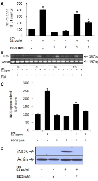
Fig. 1. Effects of EGCG on NO release and iNOS RNA ex- pression. (A) Cells were pretreated with EGCG (1-2uM) for 1 hr followed by treatment with LPS 100 ng/ml for 24 hr. (B) The expression levels of iNOS mRNA were determined in BV-2 microglia by RT-PCR. (C) The ex- pression levels of iNOS mRNA were determined in BV-2 microglia by Real-Time PCR (24 hr). (D) iNOS pro- tein expression level. The iNOS protein level were measured by western blot (24 hr). All values are the mean ±SEM from triplicate. *. p<0.05 compared with LPS treated.
식균작용에 의해 세포 내로 유입된 bead를 관찰하였다.
조건배지를 이용한 SN4741 세포 독성 확인
미세아교세포의 활성에 의한 신경세포의 손상을 확인하기 위해 조건배지를 만들어 SN4741 세포 주에 처리하여 세포 생 존율을 확인하였다. BV-2 미세아교세포에 2 uM EGCG를 처 리한 다음 LPS를 이용해 세포활성을 유도하였다. 4시간 후 EGCG와 LPS가 처리된 배지를 제거한 다음 새로운 배지를 넣어 20시간 동안 배양하여 조건배지를 만들었다. SN4741 도 파민성 신경세포에 조건 배지를 처리하여 24시간 동안 배양한 다음 조건배지에 의한 세포 생존율을 MTT assay로 세포 생존 율을 확인하였다.
결 과
미세아교세포에서 EGCG의 LPS에 의한 NO 생성 억제 미세아교세포는 신경염증반응에 민감한 세포로써 외부 자 극에 의해 pro-inflammatory cytokine이나 NO를 분비하는 세 포로 알려져 있다. EGCG의 NO 생성억제 효능을 확인하기 0.1 μg/ml LPS로 활성화된 미세아교세포에 1, 2 μM EGCG를 처리하여 분비되는 NO 양을 Griess assay로 측정하였다. 0.1 μg/ml의 LPS를 BV-2 미세아교세포에 처리하여 24시간 동안 배양하였을 때 아무것도 처리하지 않은 실험군 대비 4배의 NO가 분비되는 것을 확인하였다. EGCG 1, 2 μM을 1시간 전 처리한 다음 0.1 μg/ml의 LPS를 처리한 실험군에 비해 NO의 분비량이 감소하는 것을 확인할 수 있었다(Fig. 1A). RT-PCR 과 Real-time PCR, Western Blot을 이용해 iNOS 유전자와 단 백질의 발현량을 확인하였다. iNOS는 세포 내에서 NO의 합 성 관여하는 중요한 인자이다. RT-PCR 결과, LPS를 단독 처리 한 실험군에서 6 시간 이후 iNOS RNA 발현이 증가하는 것을 확인하였다. 이에 반해, 2 μM EGCG를 1시간 전 처리 후 LPS 를 처리한 실험군에서는 iNOS 유전자의 발현이 증가되지 않 는 것을 알 수 있었다(Fig. 1B, C). 뿐만 아니라 LPS의해 iNOS 단백질의 발현이 증가하고 EGCG에 의해 억제되는 것을 확인 하였다(Fig. 1D).
EGCG의 LPS에 의한 pro-inflammatory cytokine 생 성 억제 확인
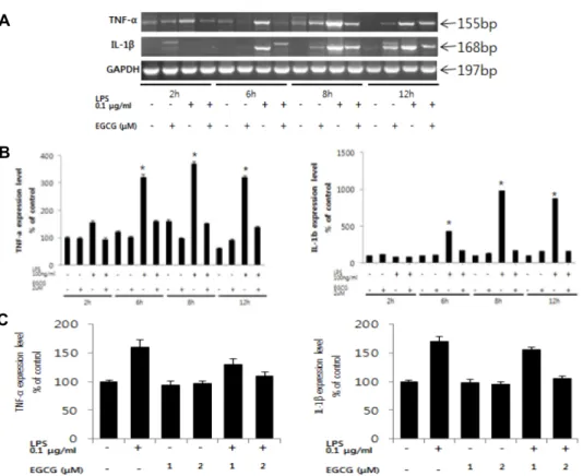
LPS에 의해 유도되는 pro-inflammatory cytokine중에서 대 표적인 TNF-α와 IL-1β의 발현을 RT-PCR을 통해 확인하였다.
0.1 μg/ml LPS를 처리하였을 때 TNF-α 유전자는 LPS 단독 처리 실험군에서 2시간 이후부터 지속적으로 발현량이 증가 하는 것을 확인할 수 있었고, IL-1β 발현량은 LPS 처리 4시간 이후부터 증가하지만, 2 μM EGCG를 전 처리한 실험군에서는 pro-inflammatory cytokine 발현이 감소되는 것을 확인하였다 (Fig. 2).
세포막에서 LPS의 수용체로 작용하는 TLR-4 수용체의 발 현을 RT-PCR을 이용해 확인하였다(Fig. 3). TLR-4 수용체는 미세아교세포의 활성화와 직접적인 관련이 있으며 LPS에 의 한 신경염증반응의 주요 경로로 알려져 있다. 그림 3의 결과와 같이 LPS 단독 처리한 실험군에서 6시간 이후부터 TLR-4 수용 체의 발현이 증가하는 것을 확인 할 수 있었다. 그리고 EGCG 를 1시간 전 처리한 실험군에서는 TLR-4 수용체의 발현량의
A
B
C
Fig. 2. Effects of EGCG on LPS-induced proinflammatory cytokines mRNA expression in BV-2 cells. (A) The expression levels of TNF-α and IL-1β mRNA were determined in LPS-stimulated cells for each time (2, 6, 8, 12 hr) by RT-PCR. (B) Intensity of each band were measured by denditometric analysis and the compared with control. . (C) The expression levels of TNF-α and IL-1β mRNA were determined in BV-2 microglia by Real-Time PCR (8 hr). All values are the mean ±SEM from triplicate.
A
B
Fig. 3. Effects of EGCG on TLR-4 mRNA expression in BV-2 cells. (A) The expression levels of TLR-4 recepter mRNA were determined in LPS-stimulated cells for each time (2, 6, 8, 12 hr) by RT-PCR. (B) Intensity of each band were measured by densitometric analysis and the com- pared with control. All values are the mean ±SEM from triplicate. *. p<0.05 compared with LPS treated.
변화가 없는 것을 확인했다.
EGCG의 미세아교세포의 활성에 따른 phagocytosis 활 성 억제 확인
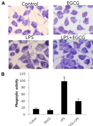
3 nm 직경의 latex bead를 이용해 LPS에 의한 식균활성을 확인하였다. 미세아교세포는 활성화되어 다양한 신경염증반 응물질의 분비와 함께 식균활성을 통해 손상된 신경세포를 제거하는 것으로 알려져 있다. 0.1 μg/ml의 LPS를 처리한 실 험군에서 latex bead 가 세포 내로 다 수 이동하는 것을 확인할 수 있었다(Fig. 4). 그러나 2 μM EGCG를 1시간 전 처리한 다음 LPS를 처리한 실험군에서 leatex bead의 이동이 아무것도 처 리하지 않은 실험군과 비슷한 수준으로 감소하는 것을 확인할 수 있었다.
Conditioned media에 의한 SN4741 세포 독성 확인 EGCG에 의한 미세아교세포의 활성 억제가 신경세포에 미 치는 영향을 확인하기 위해 배양 여액을 이용하여 조건 배지 를 만들어 SN4741 도파민성 신경세포에 처리하였다(Fig. 5).
그 결과 0.1 μg/ml LPS를 단독 처리하여 활성화된 조건배지를 처리한 실험군에서 세포 생존율이 60% 가량으로 감소하는 것 을 확인하였다. 이에 반해 2 μM EGCG를 전 처리한 다음 LPS
A
B
Fig. 4. Effect of EGCG on phagocytotic activity. (A). Micro- graphs indicate that BV2 cells exhibit phagocytotic ac- tivity by engulfment of latex beads. The optical bead density was evaluated after counting 10 fields corre- sponding to at least 200 cells. (B). Phagocytotic ability was evaluated by calculating the mean percentage cells engulfing at least one bead. The phagocytotic capacity was evaluated by estimating the mean number of en- gulfed beads/cell. Results are expressed as mean ± SEM of three independent experiments. *p<0.05, compared with LPS alone. Scale bars indicate 10 μm.
Fig. 5. Effect of EGCG in LPS-stimulated BV-2 microglial con- ditioned medium on neuronal cell viability. SN4741 cells, a murine dopaminergic neuronal cell line, were treated with different BV-2 conditioned media. BV2 cells were pretreated with EGCG (2 μM) stimulated with LPS (0.1 μg/ml) for 24 hr. The viability of SN4741 cultured with BV-2 conditioned media was assessed by MTT assay. All values represent mean ± SEM of three in- dependent experiments. *p<0.05.
로 활성을 유도한 실험군서는 세포 생존율의 변화가 거의 없 는 것을 확인할 수 있었다.
고 찰
미세아교세포의 활성화로 대표될 수 있는 신경염증반응은 신경퇴행성 질환의 주요원인으로 작용한다[4-6, 8, 12, 15]. 녹 차 유래 polyphenol EGCG의 항 산화, 항 노화 효과에 대해서 는 많은 연구가 진행되고 있다[7, 13, 16]. 본 연구에서는 EGCG 를 이용하여 미세아교세포의 활성 억제를 확인하였다. LPS에 의해 활성화된 미세아교세포에서 NO의 생성과 pro-inflam- matory cytokine 유전자의 발현을 확인하였다(Fig. 1, Fig. 2).
그러나 2 μM EGCG를 1시간 전 처리한 미세아교세포에서는 LPS에 의한 활성이 억제되는 것을 알 수 있었다. TLR-4는 세포 막에 존재하는 LPS 특이적 수용체로 자극에 반응해 pro-in- flammatory cytokines의 발현을 유도하는 것으로 알려져 있다 [1, 2]. 본 연구에서 LPS에 의해 TLR-4의 mRNA 발현이 증가 하였고, EGCG를 1 시간 전 처리한 경우 발현이 억제되는 것을 확인할 수 있었다. 이러한 결과를 토대로 BV-2 미세아교세포 에서 EGCG에 의한 NO, pro-inflammatory cytokines의 억제 는 TLR-4 수용체 억제에 의한 것임을 알 수 있었다. TLR-4 수용체의 발현을 제어함으로써 LPS에 의한 신경염증반응을 억제하고 latex bead를 이용한 식균활성 확인 결과가 의미하 는 것과 같이 EGCG에 의한 미세아교세포의 활성이 억제되는 것을 알 수 있었다(Fig. 4).
뿐만 아니라 미세아교세포의 배양여액을 이용한 조건배지 에 대한 신경세포의 생존율을 확인하였을 때, LPS를 처리한 조건 배지에서 신경 세포의 사멸이 유도되고, EGCG 1시간 전 처리한 다음 LPS를 처리한 조건배지에서 신경세포의 생존 율이 회복되는 것을 알 수 있었다. 이러한 결과는 EGCG를 이용해 미세아교세포의 활성과 식균 작용을 제어할 수 있고 더 나아가 신경염증반응에 의한 다양한 신경퇴행성 질환의 진행에 긍정적인 영향을 미칠 수 있으며, 녹차 유래 polyphenol 을 이용한 치료제 개발에 기여할 수 있을 것으로 보인다.
감사의 글
이 논문은 한국연구재단의 기초연구사업(NRF-2015R1D1 A1A01059448)의 지원과 2014학년도 조선대학교 학술연구비 의 지원을 받아 연구되었으며 이에 감사드립니다.
References
1. Beutler, B., Du, X. and Poltorak A. 2001. Identification of Toll-like receptor 4 (Tlr4) as the sole conduit for LPS signal transduction: genetic and evolutionary studies. J. Endotoxin Res. 7, 277-280.
초록:BV-2 미세아교세포의 활성에 대한 녹차 유래 폴리페놀 EGCG의 억제 효과
박으뜸․전홍성*
(조선대학교 자연과학대학 의생명과학과)
본 연구에서는 녹차 유래 polyphenol 중의 하나인 epigallocatechine gallate (EGCG)를 이용한 신경염증 억제 효과를 확인하였다. LPS로 유도된 미세아교세포의 활성화로 분비되는 nitric oxide (NO)와 pro-inflammatory cyto- kine을 포함하여 iNOS, TNF-a와 IL-1b 유전자의 발현과 LPS 수용체인 TLR-4의 활성에 미치는 EGCG의 억제 효능을 확인하였다. Latex beads를 이용한 phagocytotic activity를 확인한 결과 LPS로 유도된 미세아교세포 활성에 의한 식균활성이 EGCG에 의해 억제되는 것을 볼 수 있었다. 뿐만 아니라, BV-2 미세아교세포 조건배지를 이용하여 도파민성 신경세포 SN4741의 세포 사멸확인에서도 EGCG에 의한 보호 효과를 확인하였다. 본 연구 결과는 녹차 유래 polyphenol인 EGCG의 신경염증 반응억제효능과 신경퇴행성 질환 제어 가능성을 확인하였다. 본 연구의 결과 는 녹차 유래 polyphenol인 EGCG의 신경염증 반응과 그로 인한 신경 퇴행성 질환 제어 가능성을 제시하였다.
2. Chen, Z., Jalabi, W., Shpargel, K. B., Farabaugh, K.T., Dutta, R., Yin, X., Kidd, G. J., Bergmann, C. C., Stohlman, S. A.
and Trapp, B. D. 2012. Lipopolysaccharide-induced micro- glial activation and neuroprotection against experimental brain injury is independent of hematogenous TLR4. J.
Neurosci. 32, 11706-11715.
3. Clement, Y. 2009. Can green tea do that? A literature review of the clinical evidence. Prev. Med. 49, 83-87.
4. Collin, M., McGovern, N. and Haniffa, M. 2013. Human dendritic cell subsets. Immunology 140, 22-30.
5. Colton, C. A. 2009. Heterogeneity of microglial activation in the innate immune response in the brain. J. Neuroimmune Pharmacol. 4, 399-418.
6. Fuhrmann, M., Bittner, T., Jung, C. K., Burgold, S., Page, RM., Mitteregger, G., Haass, C., LaFerla, F. M., Kretzschmar, H. and Herms, J. 2010. Microglial Cx3cr1 knockout prevents neuron loss in a mouse model of Alzheimer's disease. Nat.
Neurosci. 13, 411-413.
7. Gonzalez, H., Elgueta, D., Montoya, A. and Pacheco, R.
2014. Neuroimmune regulation of microglial activity in- volved in neuroinflammation and neurodegenerative diseases. J. Neuroimmunol. 274, 1-13.
8. Hirsch, E. C. and Hunot, S. 2009. Neuroinflammation in Parkinson’s disease: a target for neuroprotection? Lancet Neurol. 8, 382-387.
9. Kamon, M., Zhao, R. and Sakamoto, K. 2010. Green tea poly- phenol (−)-epigallocatechin gallate suppressed the differ- entiation of murine osteoblastic MC3T3-E1 cells. Cell Biol.
Int. 34, 109-416.
10. Khandelwal, P. J., Herman, A. M. and Moussa, C. E. 2011.
Inflammation in the early stages of neurodegenerative pathology. J. Neuroimmunol. 238, 1-11.
11. Li, Y., Du, X. F., Liu, C. S., Wen, Z. L. and Du, J. L. 2012.
Reciprocal regulation between resting microglial dynamics and neuronal activity in vivo. Dev. Cell 23, 1189-1202.
12. Maetzler, W., Apel, A., Langkamp, M., Deuschle, C., Dilger,
S. S. and Stirnkorb, J. G., 2014. Comparable auto antibody serum levels against amyloid-and inflammation-associated proteins In Parkinson’s disease patients and controls. PLoS One 9, 88604.
13. Seija, L. 2010. Innate immunity and neuroinflammation in the CNS: The role of microglia in toll-like receptor- medi- ated neuronal injury. GLIA 58, 253-263.
14. Shenouda, S. M. and Vita, J. A. 2007. Effects of flavonoid- containing beverages and EGCG on endothelial function. J.
Am. Coll. Nutr. 26, 366-372.
15. Singh, B. N., Shankar, S. and Srivastava, R. K. 2010. Green tea catechin, epigallocatechin-3-gallate (EGCG): mecha- nisms, perspectives and clinical applications. Biochem. Phar- macol. 82, 1807-1821.
16. Takeuchi, H., Mizoguchi, H., Doi, Y., Jin, S., Noda, M., Liang, J., Li, H., Zhou, Y., Mori, R., Yasuoka, S., Li, E., Parajuli, B., Kawanokuchi, J., Sonobe, Y., Sato, J., Yamanaka, K., Sobue, G., Mizuno, T. and Suzumura, A. 2011. Blockade of gap junction hemichanel suppresses disease progression in mouse models of amyotrophic lateral sclerosis and Alzheimer's disease. PLoS One 6, 21108.
17. Whitney, N. P., Eidem, T. M., Peng, H., Huang, Y. and Zheng, J. C. 2009. Inflammation mediates varying effects in neurogenesis: Relevance to the pathogenesis of brain injury and neurodegenerative disorders. J. Neurochem. 108, 1343-1359.
18. Wilms, H., Rosenstiel, P., Sievers, J., Deuschl, G., Zecca, L.
and Lucius, R. 2003. Activation of microglia by human neu- romelanin is NF-kappaB dependent and involves p38 mi- togen-activated protein kinase: implications for Parkinson’s disease. FASEB J. 17, 500-507.
19. Zecca, L., Wilms, H., Geick, S., Claasen, J. H., Brandenburg, L. O. and Holzknecht, C. 2008. Human neuromelanin in- duces neuroinflammation and neurodegeneration in the rat substantia nigra: implications for Parkinson’s disease. Acta Neuropathol. 116, 47-55.