Printed in the Republic of Korea
Bis[(2-pyridyl)methyl]-2-(2-pyridyl)ethylamine 구리 (II) 착물의 합성 ,
성질 및 X-ray 결정구조
최 기 영*
공주대학교사범대학화학교육과
(2006. 11. 30 접수)
Synthesis, Properties, and X-ray Crystal Structure of Copper(II) Complex with Bis[(2-pyridyl)methyl]-2-(2-pyridyl)ethylamine
Ki-Young Choi*
Department of Chemistry Education, Kongju National University, Kongju 314-701, Korea (Received November 30, 2006)
요 약. 트리포달 구리(II) 착물 [Cu(pmea)(H2O)](ClO4)2·H2O (1) (pmea=bis[(2-pyridyl)methyl]-2-(2-pyridyl)- ethylamine)을합성하고 X-선회절법으로구조를규명하였다. 이착물은삼사정계, 공간군 P-1, a= 9.9362(9) Å, b= 15.7299(17) Å, c= 18.0562(11) Å, α= 68.760(8)°, β= 76.331(6)°, γ= 77.092(9)°, V= 2526.2(4) Å3, Z= 2로결
정화되었다. 각각의구리원자는약간일그러진사각뿔구조를갖는데, 바탕면은 pmea 리간드에있는세개의질소
원자와물분자가포함되어있으며축방향에는피리딘고리의질소원자가포함되어있다. 착물1의순환전압전류그 림은가역일-전자산화 CuIII및환원 CuI과정으로진행됨을보여주었다.
주제어: 결정구조, 구리(II) 착물, 트리포달리간드, 일그러진사각뿔구조, Bis[(2-pyridyl)methyl]-2-(2-pyridyl)- ethylamine
ABSTRACT. The tripodal copper(II) complex [Cu(pmea)(H2O)](ClO4)2·H2O (1) (pmea = bis[(2-pyridyl)methyl]-2-(2- pyridyl)ethylamine) has been synthesized and structurally characterized by X-ray diffraction method. It crystallizes in the triclinic system P-1 with a= 9.9362(9), b= 15.7299(17), c= 18.0562(11) Å, α= 68.760(8), β= 76.331(6), γ= 77.092(9)°, V = 2526.2(4) Å3, Z = 2. Each copper atom reveals a distorted square pyramidal with three nitrogen atoms of the pmea ligand and water molecule occupying the basal plane and one nitrogen atom from the pyridine ring according the axial position. The cyclic voltammogram of 1 undergoes reversible one-electron oxidation to the CuIII and reversible one-elec- tron reduction to the CuI.
Keywords: Crystal Structure, Copper(II) Complex, Tripodal Ligand, Distorted Square Pyramidal Geometry, Bis[(2- pyridyl)methyl]-2-(2-pyridyl)ethylamine
INTRODUCTION
Copper complexes with tripodal ligands have attracted considerable interest due to their impor- tance in a variety of synthetic, industrial, and bio- logical processes.1-3 Several authors have systematically investigated copper complexes of tripodal ligands
by appropriate ligand design and use of low tem- perature synthesis, handling and characterization to provide insight into the kinetics and thermodynam- ics of their formation, possible structures and spec- troscopy and physicochemical properties.4-12 For example, the tripodal copper(II) complexes [Cu- (tmpa)Cl]PF6 (tmpa=tris(2-pyridylmethyl)amine) and
[Cu(tepa)Cl]PF6 (tepa = tris[2-(2-pyridyl)ethyl]amine) exhibit a distorted square-pyramidal geometry, with N3Cl basal plane and one nitrogen atom of the axial pyridiyl group.8 In a previous paper, Karlin et al.9 report the synthesis and characterization of [Cu (pmea)]PF6 (pmea = bis[(2-pyridyl)methyl]-2-(2- pyridyl)ethylamine) in which the geometry about copper atom is best described as pyramidal with the amine nitrogen occupying the axial position and three pyridyl nitrogens in the trigonal plane. In con- trast, the crystal structure of [Cu(pmea)Cl]ClO4
·H2O reveals a five coordinate distorted square pyramidal CuN4Cl environment.9 It seems that the chlorine anion could play a role here to stabilize the copper(II)-pmea complex. In order to better under- stand some aspects of the different molecular topol- ogies, we report the synthesis and crystal structure of tripodal copper(II) complex [Cu(pmea)(H2O)]
(ClO4)2·H2O (
1
). By metathesis of uncoordinated chlo- ride with perchlorate anions, we could obtain the single crystals of title complex, in which this com- plex exhibits some uncommon feature.EXPERIMENTAL
Materials and physical measurements.
The bis[(2-pyridyl)methyl]-2-(2-pyridyl)ethylamine (pmea) was synthesized according to the literature method.13 IR spectra were recorded as KBr pellets on a Perkin-Elmer Paragon 1000 FT-IR spectrome- ter. Solution and solid state electronic spectra were obtained on a Jasco Uvidec-610 spectrophotome- ter. Elemental analysis (C, H, N) were performed on a Perkin Elmer CHN-2400 analyzer. Electro- chemical measurements were accomplished with a three electrode potentiostat BAS-100BW system. A 3-mm Pt disk was used as the working electrode.The counter electrode was a coiled Pt wire and a Ag/AgCl electrode was used as a reference elec-
trode. Cyclic voltametric data were obtained in DMSO solution using 0.10 M tetraethylammonium perchlorate (TEAP) as supporting electrolyte at 20.0±0.1 °C. The solution was degassed with high purity N2 prior to carrying out the electrochemical measurements.
Synthesis of [Cu(pmea)(H
2O)](ClO
4)
2·H
2O (1).
To a methanol solution (20 mL) of Cu(ClO4)2·6H2O (185 mg, 0.5 mmol) was added pmea (152 mg, 0.5 mmol) The mixture was heated to reflux for 1 h and then cooled to room temperature. The solution was filtered and left at room temperature until blue crys- tals formed. The product was filtered out and one of them was subjected to the X-ray analysis. Yield:
72%. Calc. (found) for C38H48Cl4Cu2N8O20: C, 37.85 (37.76); H, 4.01 (4.11); N, 9.29 (9.17)%. IR (KBr;
cm-1): 3484(m), 3422(m), 3064(w), 1608(m), 1568(m), 1482(m), 1443(m), 1309(w), 1289(w), 1144(s), 1116(s), 1088(s), 1029(w), 1004(w), 942(w), 808(w), 766(m), 629(m), 545(w), 422(w). UV-Vis in DMSO [λmax, nm (ε, M-1cm-1)] 261(1.43×10⁴), 641(134); in diffuse reflectance spectrum (λmax, nm): 261, 645.
X-ray crystallography.
Intensity data for the compounds were measured on an Enraf-Nonius CAD4 diffractometer using graphite-monochromated Mo-Kα radiation in the ω-2θ scan mode. Accurate cell parameters and an orientation matrix were determined by least-squares fit of 25 reflections.The intensity data were collected for Lorentz and polarization effects. An empirical absorption cor- rection bases on ϕ-scan was applied. The structure was solved direct methods14 and the least-squares refinement of the structure was performed by the program SHELXL-97.15 All atoms except all hydro- gen atoms, O(1), O(4), O(6), O(9), O(11), O(12), O(16), Ow(4), C(10), C(11), C(13), and C(17) were refined anisotropically. The hydrogen atoms were placed in calculated positions allowing to ride on their parent C atoms with Uiso(H)=1.2Ueq(C or N).
The hydrogen atoms of Ow(1), Ow(2), Ow(3), and Ow(4) were not found. The rather higher R1 and
wR2 values may be attributed mainly to the bad quality of the sample compound. Crystal parame- ters and details of the data collections and refine- ment are listed in Table 1.
RESULTS AND DISCUSSION
Structural description.
An ORTEP drawing16 of1
with the atomic numbering scheme is shown inFig. 1. Selected bond distances and angles are listed in Table 2. Two crystallographically independent but chemically identical [Cu(pmea)(H2O)]+ cations exist in the asymmetric unit. Each copper(II) atom in the cations is pentacoordinated structure with three pyridyl nitrogens, one aliphatic amine nitrogen and
a water molecule. The Cu(1) atom reveals a CuN4O coordination environment with three nitrogen atoms of the pmea ligand and water molecule occupying the basal plane [Cu(1)-N(1) 1.997(8), Cu(1)-N(2) 2.100(9), Cu(1)-N(4) 2.010(8), Cu(1)-Ow(1) 2.087(7) Å] and one nitrogen atom from the pyridine ring according the axial position [Cu(1)-N(3) 2.226(8) Å], which can be described as a distorted square pyramidal with a τ value of 0.18 (values of 0 and 1 are indicative of idealized square-pyramidal and
Table 1. Crystallographic Data Chemical formula
Formula weight Temperature Crystal system Space group Unit cell dimensions
a (Å)
b (Å)
c (Å)
α (°)
β (°)
γ (°) Volume (Å3)
ZDensity (calculated, mg/m3) Absorption coefficient (mm−1) Diffractometer/scan Radiation/wavelength
F(000)
Crystal size (mm3)
θ range for data collection (°) Index ranges
Reflections collected/unique Absorption correction
Maximum and minimum transmission Refinement method
Data/restraints/parameters Goodness of fit on F2 Final R indices [I > 2σ (I)]
R1
wR2
R indices (all data)
R1
wR2
Weighting scheme
Largest difference peak and hole
C38H48Cl4Cu2N8O20
1205.72 293(2) K Triclinic
P-1 9.9362(9) 15.7299(17) 18.0562(11) 68.760(8) 76.331(6) 77.092(9) 2526.2(4) 21.585 1.136
Enraf-Nonius/ω-2θ
Mo-Kα/0.71073 12360.40 × 0.30 × 0.20 1.23 to 24.96
–11≤h ≤11, –17≤k ≤18, 0≤l ≤21 8915/8613 (Rint = 0.0320)
ϕ-scan
0.8047 and 0.6594
full-matrix least-squares on F2 8613/0/589
1.038 0.0902 0.2398 0.1950 0.2981
w = 1/[σ2(Fo2) + (0.1630P)2 + 3.0031P] with P = (Fo2 + 2Fc2)/3
1.397 and –0.858 eÅ-3
Note. R1 = ∑||Fo| −|Fc||/|Fo|. wR2= {∑[w(Fo2 −Fc2)2]/∑[w(Fo2)2]}1/2.
trigonal bipyramidal geometries, respectively).17 The Cu(2) atom presents a similar coordination environ- ment [Cu(2)-N(5) 2.021(8), Cu(2)-N(6) 2.015(9), Cu(2)-N(7) 2.012(9), Cu(2)-Ow(2) 2.036(7), Cu(2)- N(8) 2.246(8) Å], but in this case, the τ value is 0.05. The Cu(1) and Cu(2) atoms are displaced
0.188(5) and 0.274(4) Å from the least-squares plane defined by the N3O basal plane toward the pyridyl nitrogen atoms N(3) and N(8). The axial Cu-Npy bond distances of Cu(1)-N(3) and Cu(2)-N(8) are ca. 0.22 Å longer than the equatorial Cu-Npy
bond distances. Similar results are reported on the related complexes [Cu(tepa)Cl]PF6 and [Cu(tmpa)Cl]PF6, which indicates the distorted square-pyramidal geometry.8 The average Cu-Npy and Cu-Namine bond distances are similar to those found in the chloride derivative [Cu(pmea)Cl]ClO4·H2O (2.092(2) Å and 2.055(2) Å],9 which exhibits a distorted square pyramidal geometry (τ= 0.12 and 0.14) with N3Cl basal plane and one of the pyridines in the axial position. The N(1)-Cu(1)-N(2), N(2)-Cu(I)-N(3), N(5)-Cu(2)-N(6) and N(6)-Cu(2)-N(7) bite angles of the five-membered chelate rings [81.4(4)º, 87.2(4)º, 83.3(4)º and 84.6(4)º] are larger than the N(2)- Cu(1)-N(4) and N(6)-Cu(2)-N(8) bite angles of the six-membered chelate rings [90.9(4)º and 96.1(4) º].
The axial Cu(1)-N(3) and Cu(2)-N(8) linkages are bent slightly off the perpendicular to CuN3O basal plane by 2.1-13.4º and 6.1-9.0º, respectively.
Fig. 1. An ORTEP drawing of 1 showing the atomic numbering scheme (30% probability ellipsoids). The hydrogen atoms and perchlorate anions are omitted for clarity.
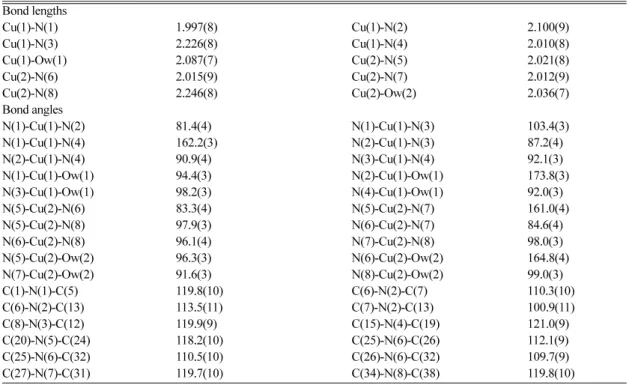
Table 2. Selected Bond Lengths (Å) and Angles (°) Bond lengths
Cu(1)-N(1) Cu(1)-N(3) Cu(1)-Ow(1) Cu(2)-N(6) Cu(2)-N(8) Bond angles N(1)-Cu(1)-N(2) N(1)-Cu(1)-N(4) N(2)-Cu(1)-N(4) N(1)-Cu(1)-Ow(1) N(3)-Cu(1)-Ow(1) N(5)-Cu(2)-N(6) N(5)-Cu(2)-N(8) N(6)-Cu(2)-N(8) N(5)-Cu(2)-Ow(2) N(7)-Cu(2)-Ow(2) C(1)-N(1)-C(5) C(6)-N(2)-C(13) C(8)-N(3)-C(12) C(20)-N(5)-C(24) C(25)-N(6)-C(32) C(27)-N(7)-C(31)
1.997(8) 2.226(8) 2.087(7) 2.015(9) 2.246(8) 81.4(4) 162.2(3) 90.9(4) 94.4(3) 98.2(3) 83.3(4) 97.9(3) 96.1(4) 96.3(3) 91.6(3) 119.8(10) 113.5(11) 119.9(9) 118.2(10) 110.5(10) 119.7(10)
Cu(1)-N(2) Cu(1)-N(4) Cu(2)-N(5) Cu(2)-N(7) Cu(2)-Ow(2) N(1)-Cu(1)-N(3) N(2)-Cu(1)-N(3) N(3)-Cu(1)-N(4) N(2)-Cu(1)-Ow(1) N(4)-Cu(1)-Ow(1) N(5)-Cu(2)-N(7) N(6)-Cu(2)-N(7) N(7)-Cu(2)-N(8) N(6)-Cu(2)-Ow(2) N(8)-Cu(2)-Ow(2) C(6)-N(2)-C(7) C(7)-N(2)-C(13) C(15)-N(4)-C(19) C(25)-N(6)-C(26) C(26)-N(6)-C(32) C(34)-N(8)-C(38)
2.100(9) 2.010(8) 2.021(8) 2.012(9) 2.036(7) 103.4(3) 87.2(4) 92.1(3) 173.8(3) 92.0(3) 161.0(4) 84.6(4) 98.0(3) 164.8(4) 99.0(3) 110.3(10) 100.9(11) 121.0(9) 112.1(9) 109.7(9) 119.8(10)
Chemical properties.
The infrared spectrum of complex1
exhibits bands at 1443-1608 cm-1 associ- ated with pyridine skeleton. The strong bands at 1088 cm-1 was also assigned to the ν(Cl-O). The visible spectra of1
in DMSO solution and the solid state show d-d transition bands at 641 and 645 nm, which is typical of a square pyramidal Cu(II) com- plex.18 Similar absorption bands (approximately 650 nm) were also found in other Cu(II) complexes of tripodal ligands.8,9 Cyclic voltammetric data for the copper(II) complexes in 0.10 M TEAP-DMSO solu- tion are listed in Table 3. The cyclic voltammogram of1
is shown in Fig. 2. The oxidation and reduc- tion potentials of1
gives the reversible one-electron processes at +0.23 V and -0.22 V vs the Ag/AgCl ref- erence electrode, assigned to the CuII/CuIII and CuII/CuI couples, respectively. The redox potential for1
is slightly more negative than that of the square-pyra- midal complex[Cu(Hdpa)]Cl2.19 This can be attrib- uted the more serve steric crowding caused by the presence of the N-coordinated pyridine group in which the complex1
makes the oxidation of Cu(II)to Cu(III) easier and the reduction to Cu(I) difficult.
Supplementary Material.
Atomic coordinates, bond lengths and angles, and thermal parameters for1
are available from author K.-Y. Choi on request.Acknowledgement.
This work was supported by grant No. R05-2003-000-10536-0 from the Basic Research Program of the Korea Science & Engi- neering Foundation.REFERENCES
1. Kitajima, N.; Morook, Y. Chem. Rev. 1994, 94, 737.
2. Karlin, K. D.; Tyeklar, Z. Adv. Inorg. Biochem.1994, 9, 3. Harata, M.; Jitsukawa, K.; Masuda, H.; Einaga, H. 123. J.
Am. Chem. Soc. 1994, 116, 10817.
4. Karlin, K. D.; Kaderli, S.; Zuberbühler, A. D. Acc.
Chem. Res.1997, 30, 139.
5. Karlin, K. D.; Lee, D.-H., Kaderli, S.; Zuberbühler, A.
D. Chem. Commun.1997, 745.
6. Wei, N.; Murthy, N.; Chen, Q.; Zubieta, Z.; Karlin, K.
D. Inorg. Chem. 1994, 33, 1953.
7. Karlin, K. D.; Wei, N.; Jung, B.; Kaderli, S.; Niklaus, P.; Zuberbühler, A. D. J. Am. Chem. Soc.1993, 115, 9506.
8. Karlin, K. D.; Hayes, J. C.; Juen, S.; Hutchinson, J. P.;
Zubieta, J. Inorg. Chem. 1982, 21, 4108.
9. Schatz, M.; Becker, M.; Thaler, F.; Hampel, F.; Schin- dler, S.; Jacobson, R. R.; Tyeklar, Z.; Murthy, N. N.;
Ghosh, P.; Chen, Q.; Zubieta, J.; Karlin, K. D. Inorg.
Chem. 2001, 40, 2312.
10. Kobayashi, T.; Ito, S.; Hamazaki, H.; Ohba, S.; Nishida, Y. Chem. Lett.1996, 347.
11. So, K. W.; Yang, C.-T.; Vittal, J. J.; Ranford, J. D. Inorg.
Chim. Acta. 2003, 349, 135.
12. Yang, G. J. Chem. Crystallogr. 2004, 34, 269.
13. Oki, A.; Glerup, J.; Hodgson, D. J. Inorg. Chem. 1990, 29, 2435.
14. Sheldrick, G. M. Acta Crystallogr. 1990, A46, 467.
15. Sheldrick, G. M. SHELXL97, Program for Crystal Struc- ture Refinement; University of Göttingen: Germany, 1997.
16. Farrugia, L. J. J. Appl. Crystallogr. 1997, 30, 565.
17. Addison, A. W.; Rao, T. N.; Reedijk, J.; van Rijn, J.;
Verschoor, G. C. J. Chem. Soc., Dalton Trans. 1984, 1349.
18. Lever, A. B. P. Inorganic Electronic Spectroscopy; Elsevier: Amsterdam, 1984.
19. Choi, K.-Y.; Ryu, H.; Sung, N.-D.; Suh, M. J. Chem.
Crystallogr. 2003, 33, 947.
Table 3. Cyclic Voltammetric Data
Potentials (V) versus Ag/AgCl
Complex Cu(II)/Cu(III) Cu(II)/Cu(I)
[Cu(Hdpa)]Cl2b
[Cu(pmea)(H2O)](ClO4)2·H2O(1) +0.25
+0.23 −0.17
−0.22
aMeasured in 0.10 M TEAP-DMSO solution at 20.0±0.1°C.
bRef. 19.
Fig. 2. Cyclic voltammogram of 1 in 0.10 M TEAP-DMSO solution at 20.0±0.1°C. The scan rate is 50 mV/s.
![Fig . 1. Selected bond distances and angles are listed in Table 2. Two crystallographically independent but chemically identical [Cu(pmea)(H 2 O)] + cations exist in the asymmetric unit](https://thumb-ap.123doks.com/thumbv2/123dokinfo/5300142.155028/3.892.128.762.224.837/selected-distances-crystallographically-independent-chemically-identical-cations-asymmetric.webp)