http://dx.doi.org/10.5369/JSST.2019.28.4.225 pISSN 1225-5475/eISSN 2093-7563
Synthesis of Au@TiO 2 Core-shell Nanoparticle-decorated rGO Nanocomposite and its NO 2 Sensing Properties
Gautam Kumar Naik and Yeon Tae Yu
+Abstract
Au@TiO
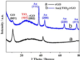
2core-shell decorated rGO nanocomposite (NC) was prepared using a simple solvothermal method followed by heat treat- ment for gas sensor application. The crystal structure and morphology of the composites were characterized by X-ray powder diffraction and transmission electron microscopy, respectively. The NO
2sensing response of the Au@TiO
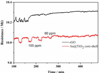
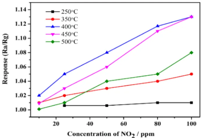
2/rGO NC was tested at operating tem- peratures from 250°C to 500°C, and was compared with those of the bare rGO and Au@TiO
2core-shell NPs. The Au@TiO
2/rGO NC- based sensor showed a far higher response than the rGO or Au@TiO
2core-shell based sensors, with the maximum response detected when the operating temperature was 400°C. This improved response was due to the high rGO gas absorption capability for NO
2gas and the catalytic effect of Au@TiO
2core-shell NPs in oxidizing NO
2to NO
3.
Keywords: Au@TiO
2core-shell NPs, NO
2, gas sensing response, rGO, nanocomposite
1. INTRODUCTION
Rapid industrialization can cause serious air pollution. Gases such as CO and NO
x(NO
2, N
2O, and NO) are often prime emission constituents, and, being highly carcinogenic in nature, can cause serious human health problems [1-4] which has led to a high demand for gas sensors which can detect NO
2and other nitrogen oxide gases. Among various gas sensors, metal oxide semiconductors (MOS) have been widely used based on their simple sensing mechanism, low cost, and very small size. Various studies have been undertaken to develop MOS-based sensors with enhanced selectivity and sensitivity [5,6]. The efficiency of a sensor material generally depends on its physical (surface area and thermal stability) and electrical properties, and many strategies, such as surface modification, doping of different noble metals and formation of composites, have been studied in an effort to enhance their sensing performance [7-10].
Reports have been published on resistant type, TiO
2based gas
sensors, for NO
2and CO gas sensing [11-13]. Our recent study found that Au@TiO
2core-shell nanoparticles showed very good response and selectivity for CO gases, at an operating temperature of 600°C, however there have been very few reports on use of a TiO
2based sensor for NO
2gas sensing [14]. In addition, the available reports only showed the sensitivity of TiO
2in targeting gases like CO and NO
2operating temperature > 500°C. This sensitivity can be increased by adding Au nanoparticles (NPs) to the TiO
2surface, and detailed studies have shown that insertion of Au NPs facilitated oxidation of target gases such as CO and ethanol, which resulted in improved sensitivity [15-17]. This showed that there was scope to develop TiO
2based sensing materials for NO
2sensing at low temperature, by forming composites with nanomaterials that contributed catalytic and electrical effects.
Among developed nanostructured mateials, it has been established that reduced graphene oxide (rGO) has a relatively high surface area and electrical conductivity, indicating that rGO had potential to improve MOS gas sensing properties. Early reports have stated that the working principle of an rGO-based gas sensor relies on charge transfer between adsorbed gas molecules and the rGO sheet [18].
In our work, in order to develop improved NO
2gas sensing ability at low operating temperatures, we synthesized nanocomposite (NC) consisting of Au@TiO
2core-shell NPs with rGO, using a microwave-assisted hydrothermal method, and investigated their gas sensing properties toward NO
2gas across concentrations Division of Advanced Materials Engineering and Research Centre for
Advanced Materials Development, Chonbuk National University, Jeonju 54896, South Korea
+