447
서 론
Helicobacter pylori (H.pylori)는 현재 만성위염, 소화성궤양의 원인뿐 아니라 위선암 및 위림프종 발생의 중요 위험인자로 인식되 고 있다. 따라서 이 세균의 감염을 정확하고 예민하게 진단하는 방 법이 연구되어 왔다[1-6]. 최근 H. pylori에 대한 항체반응이 보고되었고 이후 H. pylori 감 염을 비침습적으로 진단할 수 있는 상품화된 혈청학적 kit에 대한 관심이 높아져 현재 국내에도 다양한 제품이 소개되어 있다. 그러나 아직 H. pylori에 대한 항체를 검사하는 표준 방법은 결정되지 않은 상태이다. 이러한 kit들은 각각 다른 항원정제과정과 검사원리를 적 용하고 있어 서로 다른 결과를 보일 수 있으리라 생각되었다. 본 연구에서는 본원 내과 소화기병연구소에서 위내시경을 시행 받은 환자에게서 H. pylori 감염을 확인하였고 동시에 이들의 혈청
혈청학적
Helicobacter pylori IgG 항체 면역검사법의 임상적 유용성 :
6 가지 kit의 비교 평가
용동은․이혁민․김현숙․이준구*․이용찬*
연세대학교 의과대학 임상병리과학교실, 내과학교실*
Clinical Usefulness of
Helicobacter pylori IgG Ab Assay :
Comparison of Six Commercial Kits
Dongeun Yong, M.D., Hyukmin Lee, M.D., Hyon-Suk Kim, M.D., Joon Gu Lee, M.D.,* and Yong Chan Lee, M.D.*
Department of Clinical Pathology and Internal Medicine*, Yonsei University College of Medicine, Seoul, KoreaBackground : The diagnostic significance of the serological detection of antibodies to Heli-cobacter pylori (H. pylori) has been reported by many investigators. But the comparison data between the various serological kits were not established in Korea.
Methods : Forty nine patients with upper gastrointestinal symptoms were studied from June 1997 to September 1997 in Yonsei University College of Medicine, Severance hospital. Endos-copic gastric biopsy specimens were obtained for microsEndos-copic examination of the bacteria and rapid urease test (CLO test). The sera of these patients were obtained for the serological test at the same time. The six commercial kits (Cobas Core II, G.A.P. test IgG, PYLORAGEN, Quick-Vue, BIOCARD Helicobacter pylori IgG, EZ-H.P.) for the detection H. pylori antibodies were evaluated for diagnosis and screening of H. pylori infection.
Results : Sensitivities for the six kits were from 71% to 96%, specificities were from 24% to 71%, positive predictive values were from 68% to 81%, negative predictive values were from 60% to 80%, respectively. There were statistically significant differences in four groups, between G.A.P. test and Cobas Core, G.A.P. test and PYLORAGEN, QuickVue and Cobas Core, QuickVue and PYLORAGEN.
Conclusions : Sensitivities and specificities obtained in different studies revealed as great differences in the results with the same kits as between the results obtained with different kits in the same study. So, the serologic method alone for the diagnosis of H. pylori infection is not recommended. But in the screening of H. pylori infection, it can be used, because sensi-tivities and negative predictive values are relatively high. (Korean J Clin Pathol 1998; 18: 447-51)
Key words : Helicobacter pylori (H. pylori), serologic detection, IgG Ab, Commercial kits
접 수 : 1998년 5월 22일 접수번호: KJCP1168 수정본접수 : 1998년 6월 9일 교 신 저 자 : 김 현 숙 우 120-752 서울시 서대문구 신촌동 134 연세대학교 의과대학 임상병리과학교실 전화 : 02-3497-3531, Fax : 02-3462-9493
을 대상으로 6가지 commercial kit를 이용한 혈청학적 방법의 연관 성을 조사하고 각 kit의 특징을 비교하고자 하였다.
대상 및 방법
1. 대상 소화기계 증상을 주소로 1997년 6월부터 1997년 9월까지 연세의 대 세브란스 병원에서 위내시경 및 혈청학적 비교검사를 시행한 49 명을 대상으로 하였다. 전혀 약을 쓰지않은 신환을 대상으로 함을 원칙으로 하였으며 이전에 위수술을 받았거나 최근 3개월내 제산 제나 위산 분비 억제제를 복용한 적이 있는 환자는 연구 대상에서 제외하였다. 2. 방법 1) 조직학적 검사 내시경 검사를 시행한 환자들로부터 2곳 이상에서 위점막을 채 취하여 한조각은 배양용으로, 한조각은 신속 요소분해 효소 검사 (Campylobacter-like organism test, CLO)에, 나머지 한조각은 조직학적 도말 염색에 사용하였다. 위생검 조직 표본을 멸균 식염 수 2 mL에 담아 수송하였으며 그람염색하여 S자형의 만곡된 그람 음성 간균이 나타났을 때를 양성으로 판정하였다.2) 신속 요소분해 효소법
CLO test (Delta West Ltd, Bentley, Western, Australia)를 사용하여 2 분안에 분홍색 혹은 짙은 노랑색으로의 색깔변화가 있 으면 양성, 그렇지 않으면 음성으로 판독하였다. 1)에서 양성인 경우 H. pylori 감염양성으로 판정하였고 1)과 2) 에서 모두 음성인 감염 음성으로 판정하였다. 그리고 1)에서 음성 이었으나 2)에서 양성이면 감염 미상으로 분류하여 이 경우 결과 분석에서 제외하였다[15]. 3) 혈청학적 검사 내시경 검사 시행 직후 채혈하여 분리한 혈청을 분주하여 -70 ℃에서 보관하였으며, 혈청내에 존재하는 H. pylori에 대한 IgG 항 체를 검출하기 위하여 Cobas Core II (Roche, Basel, Switzer-land), G.A.P. test IgG (BIO-RAD, Richmond, CA, USA), PYLORAGEN (HYCOR Biromedical Inc. California, USA), QuickVue (QUIDEL, San Diego, USA), BIOCARD Helicobac-ter pylori IgG (ANI Biotech OY, Helsinki, FINLAND), EZ- H.P. (LG Chem. Ltd. Korea)의 상품화된 5개 kit를 사용하였으 며, 각 kit마다 지시하는 방법대로 검사하였다(Table 1). 정량검사 인 Cobas Core II, G.A.P. test IgG, PYLORAGEN은 각각의 kit 별로 포함된 cut off 치에 따라서, 정성검사인 QuickVue, BIO-CARD Helicobacter pylori IgG, EZ-H.P.은 발색유무에 따라 그 결과를 판정하여 민감도, 특이도, 그리고 양성 및 음성 예측치를 구 하였다.
검사간의 차이는 비모수 통계분석 중 two-sided McNemar test 를 사용하여 비교하였다.
결 과
전체 49명 환자 중 조직학적검사는 음성이나 CLO검사 양성인 4명은 감염미상으로 제외하고 45명의 환자를 분석하였다. 이들 45 명에서 연령평균은 51세, 연령 중간값은 53세, 남녀 비율은 1:1.1이 었다. 조직학적 확인검사상 H. pylori 양성은 28명, 음성은 17명이 었고 유병율은 62.2%이었다(Table2).Table 1. Instructions of the six commercial H.pylori IgG antibody detection kits
Cobas Core II G.A.P. test PYLORAGEN Quickvue BIOCARDTM EZ-H.P.TM
제조원 Roche BIO-RAD HYCOR QUIDEL ANI BIOTECH LG CHEM
원리 효소면역법 (bead) 효소면역법 (microplate) 효소면역법 (microplate) 효소면역법 (membrane) Chromatographic absorbent (membrane) Chromatographic absorbent (membrane) 특징 정량, 정성 정량, 정성 정량, 정성 정성 정성 정성
항원 FPLC fraction Purified Ag Purified Ag Purified Ag Purified Ag Purified Ag
검체 희석 배수 1: 40 1: 200 1: 100 1: 6 1: 200 1: 200 필요 검체 (양) serum (10µL) serum plasm (25µL) serum (10µL) serum plasma whole blood (120µL) serum whole blood (5µL) serum plasma whole blood (5µL)
효소 POD POD POD ALP -
-총 반응시간 45분 140분 60분 10분 2분 10분
반응 온도 37℃ 25℃ 실온 실온 실온 실온
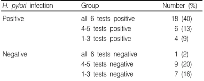
45명중 만성 표재성위염이 35명(78%)로 가장 많았고 그외 소화 성 궤양이 2명, 악성 위종양이 6명, 유두종이 1명, 정상이 1명이었다 (Table 3). 각각의 질환군에서 H. pylori 감염으로 판단된 환자의 비율은 60%에서 100%로 다양하였으나 환자수가 작아 의의를 부 여할 수는 없었다. 이들 45명에서 각각 6가지 혈청학적 검사를 시행하였다. 민감 도는 71%에서 96%, 특이도는 24%에서 71%, 양성예측치는 68%에서 81%, 음성예측치는 60%에서 80%로 다양한 결과를 나 타내었다(Table 4). 검사간 two-sided McNemar test를 시행한 결과 4가지 군, 즉 G.A.P. test와 Cobas Core II, G.A.P. test와 PYLORAGEN, QuickVue와 Cobas Core II 그리고 QuickVue 와 PYLORAGEN간에 유의한 차이를 보이는 것으로 나타났다 (P<0.025). H. pylori 감염양성인 환자중 모든 혈청학적 검사에 양성인 경우 는 18명이었고 감염음성인 환자중 모든 혈청학적 검사에 양성인 경우는 1명이었다. 나머지 26명 환자(58%)는 적어도 한 개 이상 검사간의 불일치가 있었다. 감염양성 환자 중 4-5가지 혈청학적 검 사간의 일치를 보인 환자는 6명이고 1-3가지 검사만의 일치를 보인 환자는 4명이었다. 감염음성 환자중 4-5가지 혈청학적 검사간의 일 치를 보인 환자는 9명이고 1-3가지 검사만의 일치를 보인 환자는 7명이었다(Table 5).
고 찰
H. pylori는 1984년 Marshall과 Warren이 만성위염환자의 위생 검 조직에서 나선모양의 그람음성 간균을 처음 분리하고 만성 표피 성위궤양(chronic superficial gastritis)과 연관이 있음을 주장하였 다. 현재 H. pylori 감염은 만성위염, 소화성궤양의 원인과 위선암
및 위림프증 발생의 중요 위험인자로 인식되고 있고 H. pylori를
제거할 경우 위궤양의 재발이 현저히 낮아짐이 알려져 있다. 따라 서 이 세균의 감염을 정확하고 예민하게 진단하는 방법이 연구되어 왔다. 침습적 검사로는 위장관 내시경으로 얻은 생검조직으로 염색 하여 조직학적으로 검출하거나 rapid urease test, 세균 배양법이 있고, 비침습적 검사로는 요소호흡 검사나 혈청학적 검사가 있다.
조직학적 방법은 위내시경을 필요로하고 가격이 비싸고 위생검 Table 2. Prevalence of H.pylori infection in 49 subjects tested by
Hematoxylin-Eosin stain on histology and rapid urease test (CLO) of the gastric tissue
Histology
+
-CLO + 22 4
- 6 17
Total 28 21
Table 3. Characteristics of the patients with dyspeptic symptoms included in this study
Endoscopic diagnosis No. of patients Histology positive (%) CLO positive (%) H. pylori infection (%)
+
-Chronic superficial gastritis
Benign peptic ulcer Stomach Carcinoma Papilloma Normal Total 35 2 6 1 1 45 21 (60) 2 (100) 4 (67) 1 0 28 (62) 18 (51) 2 (100) 1 (17) 1 0 22 (49) 21 (60) 2 (100) 4 (67) 1 0 28 (62) 14 (40) 0 (0) 2 (33) 0 1 17 (38)
Table 4. Sensitivity, specificity and predictive values of the six commercial kits Seropositive of 28 H. pylori positive Seronegative of 27 H. pylori negative Sensitivity % Specificity % Positive predictive value % Negative predictive value % Efficiency % Cobas Core II
G.A.P. test IgG PYLORAGEN QuickVue BIOCARDTM EZ-H.P.TM 25 22 27 20 24 25 8 12 4 12 7 8 89 78 96 71 86 89 47 71 24 71 41 47 74 81 68 80 71 74 73 67 80 60 64 73 73 53 69 71 69 73 Table 5. Detailed results from six commercial kits of the subjects
H. pylori infection Group Number (%) Positive all 6 tests positive
4-5 tests positive 1-3 tests positive
18 (40) 6 (13) 4 (9) Negative all 6 tests negative
4-5 tests negative 1-3 tests negative
1 (2) 9 (20) 7 (16)
술식에 의존적인 단점이 있고[7, 8] 균주를 증명하기 위한 gold standard인 세균배양은 검체수송, 배지선택, 배양상태에 영향받고 시간이 오래 걸리는 단점이 있다[9]. 비침습적 방법중 요소 호흡법은 매우 정확하고 전체 위점막 상태 를 반영하는 장점이 있으나 가격, 소요시간, 방사선 노출 위험등으 로 인해 일반 검사실 검사로는 부적절하다[10]. 이에 반해 혈청학적 검사는 일반 검사실에서 시행가능하고 경제 적이고 시간이 적게 걸리기 때문에 약물투여후의 반복 추적검사나 대규모의 선별검사에 유용할 것으로 생각된다[11, 12]. H. pylori 항체를 측정하는 혈청학적인 방법은 항원 정제방법에 따라 분류할 수 있다[13]. 민감도는 높으나 특이도가 낮은 전체세포항원이용법 이나 초음파 처리법[14], 그리고 부분적인 항원정제법으로 외막항 원만을 쓰거나[15], acid glycine 법 등이 있고[16] 특이도는 높으 나 민감도가 낮은 고도정제항원법으로 high molecular weight cell-associated protein 등[17]이 있다. 또 항체측정방법에 따라 세균 응집(bacterial agglutination), 보 체 결합(complement fixation) 그리고 효소면역법 등으로 분류할 수 있는데 효소면역법이 가장 민감한 것으로 알려져 있다[18]. 본 연구에서의 H. pylori의 유병율은 62%로서 1996년도 Malaty 등이 보고한 우리나라 성인에서의 75%의 유병율보다 낮게 측정되 었다[19]. 그러나 세균배양과 Silver stain을 통한 조직검사를 병행 하면 유병율은 더 높아질 것으로 생각된다. 본 연구에서는 현재 국 내에 소개되어있는 고도정제항원을 사용한 효소면역법 kit 4개와 chromatographic absorbent법 kit 2개를 사용하여 검사하고 각 kit 의 결과를 비교하였고 기존 보고와 비교해 보았다.
1992년 Goossens 등이 Cobas Core II (Roche, Basel, Switzer-land)로 민감도, 특이도, 양성 및 음성 예측치를 93%, 91%, 94%, 및 94%로 보고하였고[20] Feldman 등은 1995년 99.3%와 86.5% 의 민감도와 특이도를 보고하였다[21]. 본 연구에서는 민감도 89%, 특이도 47%, 그리고 양성 및 음성 예측치는 모두 74%를 보 였는데 그외의 혈청학적 kit를 사용한 결과도 기존 보고와 비교해 볼 때 다양한 양상을 보이는 것을 알 수 있었다[22-25]. 이와같은 연구자간의 차이는 각 연구대상군과 기준검사의 정의 차이때문이 라고 해석할 수 있겠다. 이와 같이 혈청학적 검사가 민감도에 비해 특이도가 낮은 이유는 H.pylori가 치료된 후에도 계속 항체가 존재하여 검사상 위양성을 보이거나[26] 다른 균과의 교차반응을 가질 수 있기 때문이라고 한다[27]. 그외에도 세계적으로 각 지역마다 분포하는 H. pylori의 항원성이 다르다라는 보고가 있어[28] 같은 kit를 사용하여도 검출 율은 차이를 보일 수 있으리라 생각된다. 같은 환자의 혈청으로 검 사해도 kit 간에 차이를 보이는 이유는 각 kit의 항원제조과정과 항 원종류가 다르기 때문이라고 해석할 수 있겠다. 1988년 Megraud 등은 조직학적 검사는 CLO 검사에 비해 혈청 학적 검사의 민감도와 특이도가 낮아 진단검사로서 가치가 떨어진 다고 하였는데[29], 본 연구결과로 보아 혈청학적 검사를 단일 진 단 검사로서 이용하기는 아직도 어려울 것으로 생각된다. 이상의 결과를 요약하면 H. pylori의 혈청학적 검사는 아직도 특 이도가 낮아서 진단검사로서 이용하기 위해서는 개선되어야할 점 이 남아있으나, 민감도와 음성예측치가 특이도보다 상대적으로 높 으므로 선별검사로서는 유용한 것으로 사료되었다.
요 약
배경 : Helicobacter pylori는 그람음성, 편모를 가진 S형 세균으 로 만성위염, 소화성 궤양과 악성 위종양의 위험인자로 보고되었 다. 따라서 비침습적 방법으로 이 세균의 감염을 진단, 선별할 수 있는 혈청학적 검사에 대해 많은 연구가 있어 왔다. 본 연구에서는 현재 국내에 소개된 상품화된 6종류의 혈청학적 검사 kit 간 비교하 고자 하였다. 방법 : 1997년 6월에서 9월까지 연세의대 세브란스병원에서 상 부소화기계 증상을 주소로 위내시경을 시행한 49명 환자 중 45명을 대상으로 조직학적 검사, CLO 검사와 혈청학적 검사를 시행하였 다. 혈청학적 검사는 상품화된 6개 kit인 Cobas Core II, G.A.P. test IgG, PYLORAGEN, QuickVue, BIOCARD Helicobacter pylori IgG, 및 EZ-H.P.로 시행하였다.결과 : 민감도는 71%에서 96%로, 특이도는 24%에서 71%로, 양성예측치는 68%에서 81%로, 음성예측치는 60%에서 80%로 다 양하였다. 검사결과간 two-sided McNemar test를 시행하여 네가 지 군, 즉 G.A.P. test IgG와 Cobas Core, G.A.P. test IgG와 PYLORAGEN, QuickVue와 Cobas Core 그리고 QuickVue와 PYLORAGEN간에 유의한 차이를 보이는 것을 알 수 있었다(P< 0.025). 결론 : 본 연구결과와 이미 보고된 결과와 비교해 볼 때 민감도와 특이도가 차이를 보였고 같은 환자군을 대상으로한 여러 검사 kit 간에도 통계적으로 유의한 차이를 보였다. 이로보아 단일 진단검사 로서 혈청학적검사를 사용하기에는 아직 개선해야할 점이 남아있 다고 할 수 있으나 민감도와 음성예측치가 상대적으로 특이도보다 높으므로 선별검사로서는 유용한 것으로 사료되었다.
참고문헌
1. Graham DY and Go MF. Helicobacter pylori current status.
Gas-troenterology 1993; 105: 279-82.
2. Parsonnet GD, Fredman FD, Vandesteen DP, Chang Y, Vogel-man JH, et al. Helicobacter pylori infection and the risk of gastric
carcinoma. N Engl J Med 1991; 325: 1127-31.
3. Tompkins LS and Falkow S. The new path to preventing ulcers.
Science 1995; 267: 1621-2.
4. Marshall BJ and Warren JR. Unidentified curved bacilli in the
stomach of patients with gastritis and peptic ulceration. Lancet 1984; 4: 1311-5.
5. Nomura A, Stemmermann GN, Chyou PH, Kato I, Perez-Perez
GI, Blaser MJ. Helicobacter pyori infection and gastric carcinoma
1132-6.
6. Correa P. Human gastric carcinogenesis: a multistep and
multifac-torial process-First American Cancer Society Award Lecture on Can-cer Epidemiology and prevention. CanCan-cer Res 1992; 52: 6735-40.
7. Barthel JS and Everett ED. Diagnosis of Campylobacter pylori
infec-tions: the "gold standard" and the alternatives. Reviews of Infectious Disease 1990; 12: S107-14.
8. Loffeld RJ, Stobberingh E, Arends JW. A review of diagnostic
techniques for Helicobacter pylori infection. Dig Disease 1993; 11: 173-80.
9. Jerris RC. Helicobacter. In: Murray PR, ed. Manual of clinical
mi-crobilogy. 6th ed. Washington D.C.: ASM Press, 1995: 492-8.
10. Von Wulffen H, Gatermann S, Windler E, Gabbe E, Heinrich HC. Performance of Helicobacter pylori acid extract and ureased
enzyme-linked immunosorbent assays in relation of 14C-urea breath test. Int J Med Microbiol Virol Parasitol Infect Dis 1993; 280: 203-13.
11. Marchildon PA, Ciota LM, Zamaniyan FZ, Peacock JS, Graham DY. Evaluation of three commercial enzyme immunoasays compare
with the 13C urea breath test for detection of Helicobacter pylori infection. J Clin Microbiol 1996; 34: 1147-52.
12. Cutler A, Schubert A, Schubert T. Role of Helicobacter pylori
serol-ogy in evaluation treatment success. Digestive Disease & Sciences 1993; 38: 2262-6.
13. 이우진, 김재규, 김용태, 최상운, 정현채, 송인성, 등. Helicobacter
pylori 감염 진단에서 혈청학적 검사의 타당성. 대한소화기병학회지 1994; 26: 631-6.
14. Kaldor J, Tee W, McCarthy P, Dwyer B. Immune response of
Campylobacter pyloridis in patients with peptic ulceration. Lancet 1985; 1: 921.
15. Czin S, Carr S, Sheffler L, Aronoff S. Serum IgG antibody to the
outer membrane proteins of Campylobacter pylori in children with gastroduodenal disease. J Infect Dis 1989; 159: 586-9.
16. Goodwin CS, Blincow E, Peterson G, Sanderson C, Cheng W, marshall B, et al. Enzyme-linked immunosorbent assay for
Campylo-bacter pyloridis: Correlation with presence of C. pyloridis in the gastric mucosa. J Infect Dis 1987; 155: 488-92.
17. Evans DJ, Evans DG, Graham DY, Klein PD. A sensitive and
specific serologic test for detection of Campylobacter pylori infection. Gastroenterology 1989; 96: 1004-8.
18. Blecker U, Hauser B, Lanciers S, Peeters S, Suys B, Vandenplas Y. The prevalence of Helicobacter pylori-positive serology in
asymp-tomatic children. J Pediatr Gastroenterol & Nutr 1993; 16: 252-6.
19. Malaty HM, Kim JG, Kim SD, Graham DY. Prevalence of
Heli-cobacter pylori infection in Korean children: inverse relation to socio-economic status despite a uniformly high prevalence in adults. Am J Epidemiol 1996; 143: 257-62.
20. Goossens H, Glupczynski Y, Burette A, van den Borre C, But-zler J. Evaluation of a commercially available second-generation
im-munoglobulin G enzyme immunoassay for detection of Helicobacter pylori infection. J Clin Microbiol 1992; 30: 176-80.
21. Feldman RA, Deeks JJ, Evans SJ. Multi-laboratory comparison of
eight commercially available Helicobacter pylori serology kits. Eur J Clin Microbiol Infect Dis 1995; 14: 428-33.
22. Van den Oeyer H, Loffeld RJ, Stobberingh EE. Usefulness of a
new serological test (Bio-Rad) to diagnose Helicobacter pylori-asso-ciated gastritis. J Clin Microbiol 1991; 29: 283-6.
23. 김준명, 임새중, 김응. 위염 및 소화성 궤양에서 간접형광 항체법 에 의한 Campylobacter pylori 항체의 검출. 대한내과학회지 1990; 38: 463-9. 24. 이정호, 박성진, 구정완, 김도헌, 양창헌, 김성철 등. Helicobacter pylori 감염의 진단을 위한 혈청 IgG 항체가의 유용성. 대한소화기 병학회지 1994; 26: 39-46.
25. Graham DY, Evans DJ, Peacock J, Baker JT, Schrier WH. Com-parison of rapid serological tests (FlexSure HP and QukiVue) with conventional ELISA for detection of Helicobacter pylori infection. Am J Gastroenterol 1996; 91: 942-8.
26. Meyer B, Werth B, Beglinger C, Dill S, Drewe J, Vischer WA, et al. Helicobacter pylori infection in healthy people: a dynamic
process? Gut 1991; 32: 347-50.
27. Newell DG. Identificaton of outer membrane proteins of
Campylo-bacter pyloridis and antigenic cross reactivity between CampyloCampylo-bacter pyoridis and Campylobacter jejuni J General Microbiol 1987; 133: 163-70.
28. Johanna HN, Perez-Perez GI, Martin JB. Antigenic characterizaion
of Helicobacter pylori strains form different parts of the world. Clinical and Diagnostic Laboratory Immunology 1997; 4: 592-7.
29. Megraud F. Comparison of different tests for Campylobacter pylori.