저작자표시-비영리-변경금지 2.0 대한민국 이용자는 아래의 조건을 따르는 경우에 한하여 자유롭게 l 이 저작물을 복제, 배포, 전송, 전시, 공연 및 방송할 수 있습니다. 다음과 같은 조건을 따라야 합니다: l 귀하는, 이 저작물의 재이용이나 배포의 경우, 이 저작물에 적용된 이용허락조건 을 명확하게 나타내어야 합니다. l 저작권자로부터 별도의 허가를 받으면 이러한 조건들은 적용되지 않습니다. 저작권법에 따른 이용자의 권리는 위의 내용에 의하여 영향을 받지 않습니다. 이것은 이용허락규약(Legal Code)을 이해하기 쉽게 요약한 것입니다. Disclaimer 저작자표시. 귀하는 원저작자를 표시하여야 합니다. 비영리. 귀하는 이 저작물을 영리 목적으로 이용할 수 없습니다. 변경금지. 귀하는 이 저작물을 개작, 변형 또는 가공할 수 없습니다.
Significance of Salivary Gland Radioiodine
Retention on Post-ablation
131I Scintigraphy as a
Predictor of Salivary Gland Dysfunction in
Patients with Differentiated Thyroid Carcinoma
by
Kyung Sook Jo
Major in Medicine
Department of Nuclear Medicine & Molecular Imaging
The Graduate School, Ajou University
Significance of Salivary Gland Radioiodine
Retention on Post-ablation
131I Scintigraphy as a
Predictor of Salivary Gland Dysfunction in
Patients with Differentiated Thyroid Carcinoma
by
Kyung Sook Jo
A Dissertation Submitted to The Graduate School of
Ajou University in Partial Fulfillment of the Requirements for
The Degree of Master of Medicine
Supervised by
Joon-Kee Yoon, M.D., Ph.D.
Major in Medicine
Department of Nuclear Medicine & Molecular Imaging
The Graduate School, Ajou University
This certifies that the dissertation
of Kyung Sook Jo is approved.
SUPERVISORY COMMITTEE
Joon-Kee Yoon
Young Sil An
Su Jin Lee
The Graduate School, Ajou University
June, 2nd, 2014
ABSTRACT
-Si
gni
f
i
canceofSal
i
var
y Gl
andRadi
oi
odi
neRet
ent
i
on on
Post
-abl
at
i
on
131ISci
nt
i
gr
aphy asaPr
edi
ct
orof
Sal
i
var
y Gl
andDysf
unct
i
on i
n Pat
i
ent
swi
t
h
Di
f
f
er
ent
i
at
edThyr
oi
dCar
ci
noma
Purpose:Weinvestigatedwhether131Iwhole-body scintigraphy couldpredict functionalchangesinsalivaryglandsafterradioiodinetherapy.
M ethods:We evaluated 90 patients who received initialhigh-dose (≥3.7 GBq) radioiodine therapy after totalthyroidectomy.Allpatients underwent diagnostic (DWS) and post-ablation (TWS)131I whole-body scintigraphy. Visualassessmentofsalivary radioiodine retention on DWS and TWS was usedtodividethepatientsintotwotypesofgroups:aDWS+orDWS-group and a TWS+orTWS-group.Salivary gland scintigraphy wasalso performed beforeDWS andatthefirstfollow-upvisit.Peakuptakeand%washoutwere calculated in ROIsofeach gland.Functionalchanges(ΔuptakeorΔwashout) ofsalivary glandsafterradioiodinetherapy werecompared between thetwo groups.
Results:Both peak uptake and the %washoutofthe parotid glands were significantly lower after radioiodine therapy (allp values <0.001),whereas only the %washoutwere significantly reduced in the submandibularglands (allp values<0.05).Fortheparotid glands,theTWS+ group showed larger Δuptake and Δwashoutafterradioiodine therapy than did the TWS- group (all p values <0.01). In contrast, the Δuptake and Δwashout of the
submandibular glands did not significantly differ between the TWS+ and TWS- groups (allp values >0.05).Likewise,no differences in Δuptake or Δwashoutwereapparentbetween theDWS+ andDWS- groupsin eitherthe parotidorsubmandibularglands(allpvalues>0.05).
Conclusion: Salivary gland radioiodine retention on post-ablation 131I scintigraphyisagoodpredictoroffunctionalimpairmentoftheparotidglands afterhigh-doseradioiodinetherapy.
Keyword: Radioiodine therapy, Salivary gland dysfunction, Differentiated thyroid cancer,131Iscintigraphy,Salivaryglandscintigraphy.
TABLE OF CONTENTS
ABSTRACT···ⅰ TABLE OF CONTENTS ···ⅲ LIST OF FIGURES ···ⅳ LIST OF TABLES ···ⅴ
Ⅰ.Introduction···1
Ⅱ.MaterialsandMethods···4
A.Subjects···4
B.RadioiodineTreatmentProtocol···5
C.AcquisitionandImageAnalysisofSalivaryGlandScintigraphy···6
D.AcquisitionandImageAnalysisof131IWhole-BodyScintigraphy···8
E.StatisticalAnalysis···9
Ⅲ.Results···10
A.CharacteristicsofPatients···10
B.SalivaryGlandFunction···11
C.RadioiodineRetentionon131IWhole-BodyScintigraphy···12
D.RelationbetweenRadioiodineRetentionandSalivaryGlandFunction··13 Ⅳ.Discussion···16
V.Conclusion···22
References···23
LI
ST OF FI
GURES
Fig.1.Flow diagram showingtheprocessofradioiodinetherapy···6
Fig.2.Polygonalregion ofinterests(ROIs)weredrawn overbilateralpaotid and submandibularglands.A circularROIwasplaced overtheskull forthecorrectionofbackgroundactivity.···8
LI
ST OF TABLES
Table1.Characteristicsofpatients···10
Table2.Salivaryglandfunctionbeforeandafter131Iablationtherapy···12
Table3.Salivaryglandradioiodineretentionondiagnosticorpost-ablation
131Iwhole-bodyscintigraphy········································································13
Table4.Relationbetweentheradioiodineretentionondiagnostic131Iwhole -bodyscintigraphyandthefunctionalchangeofsalivarygland···15
Table5.Relationbetweentheradioiodineretentiononpost-ablation131Iwhole -bodyscintigraphyandthefunctionalchangeofsalivarygland···15
Ⅰ.I
nt
r
oduct
i
on
Themanagementofpatientswith well-differentiatedthyroidcarcinomas includes surgicalremovalofalltumortissue followed by radioactive iodine
131Itherapy.Postoperative131Itherapy is used to destroy remnant thyroid
tissue as well as microscopic metastases, and affords the benefits of significantreductionsinrecurrenceandcancermortalityratesinpatientswith papillary or follicular thyroid cancer(Mazzaferri and Jhiang, 1994; Schlumberger,1998).Ithasalsobeen reported that131Itherapy improved the 5-year recurrence-free survival rates even of patients with papillary microcarcinoma (Creach et al, 2012). Furthermore, administration of a therapeutic dose of131Ienables visualization ofpersistenttumortissue,via highly sensitive whole-body scanning,in patients with elevated levels of serum thyroglobulin(Lind,1999).
However,although high-dose131Itherapy prolongs survival,some side effectsareapparent.Alexanderetal.reported that~77% ofpatientsreported intermediate orlong-term complaints including sialadenitis,transientloss of taste or smell,transient alopecia,chronic/recurrent conjunctivitis,increased frequencyofinfluenzainfectionandhematologicabnormalities(Alexanderetal, 1998).Apartfrom thesesideeffects,high-dose131Itherapy can impairdental health by increasing the risk of caries and the need for tooth extractions(Walter.1998).Of the problems listed above,sialadenitis is the mostfrequentcomplication,associated with radiation damage caused during the transportofradioiodine into the salivary glands,which have an iodide uptake mechanism similarto thatofthe thyroid,and concentrate iodine to levels 20–100-fold thatin serum.The frequency ofintermediate sialadenitis was initially ~33% and reached 43% 1 yearaftertreatment(Allweiss etal,
2
-1984;Cavalieri,1997;Alexanderetal,1998;MandelS.JandMandelL.,2003). The frequency of radiation-induced sialadenitis is dose-dependent, being proportionalto the cumulative131Idose administered(Spitzweg et al,2001; Hyeretal,2007;Kim etal,2007;Macioszek etal,2008).However,in more than halfofallcases,reduced salivary gland function was notobviously associated with clinically evident xerostomia or post-therapeutic sialadenitis(Alexander et al,1998).Therefore,predicting the occurrence of salivary gland dysfunction is of great importance in the management of patientswithdifferentiatedthyroidcancer.
Salivary gland scintigraphy using 99mTc-pertechnetate can assess functionaldamage to the glands following radioiodine therapy(Malpanietal, 1996; Caglar et al, 2002). Intravenously administered 99mTc-pertechnetate gradually accumulates in the salivary glands over20min post-administration and is then excreted as saliva production is enhanced. This type of scintigraphy yields semiquantitative data on uptake ratios and %excretion values(Raza et al,2006;Kang et al,2011;An et al,2013).However,no reliable method has yetbeen developed to predictfunctionalimpairmentof salivaryglandsatthetimeof131Iablationtherapy.
Diagnostic131Iwhole-body scintigraphy (DWS)can beused to acquire informationonthepresenceofiodine-avidremnantthyroidtissueandalsoto detectlocoregionalordistantmetastases(Cooperetal,2009).Post-ablation131I whole-body scintigraphy (TWS)isalsousefulin thiscontext,particularly in patients with elevated serum thyroglobulin levels,because TWS is more sensitivethanDWS whenusedtodetectmetastaticdisease(Lind,1999;Chong etal,2010).Ourclinicalexperiencehastaughtusthatsomepatientsexhibit
131Iretention in the salivary glands on eitherDWS orTWS.We conducted
whole-body scintigraphy atthe time of131Iablation could predictfunctional impairmentofthesalivaryglandsinpatientswithpapillarythyroidcancer.
4
-Ⅱ.Mat
er
i
al
sandMet
hods
A.Subj
ect
s
Study subjectswererecruited by review ofmedicalrecordsfrom June 2010toOctober2011.Duringthisperiod,atotalof229patientswerereferred for131I ablation therapy to treat differentiated thyroid cancer. Of these patients,those who metthe following criteria were selected:(1) the first course of radioiodine therapy was given after total thyroidectomy; (2) treatment featured high-dose131I therapy (≥3.7GBq); (3) both DWS and TWS were performed;(4)salivary gland scintigraphy was performed both before and afterradioiodine therapy.We limited oursubjects using criterion (1) above because some patients already had salivary gland dysfunctions, caused by previous therapy, at the time when the second course of radioiodine therapy was given, or thereafter. Eighteen patients who had undergonelow-dose(1.1GBq)131Itherapy werealsoexcluded from thestudy because radiation-induced sialadenitis rarely occurs in such patients. Furthermore,DWS was frequently notperformed on such patients.Another fourpatientsexcluded from thestudy had undergonehigh-doseradiotherapy (5.5or7.4GBq)withoutperformanceofDWS.Initially,they werescheduled for low-dose radioiodine therapy, but the doses were raised because of elevationsinserum thyroglobulinconcentrations.
Information on patients’characteristics,including symptoms of salivary dysfunction and the details of radioiodine ablation therapy,was collected retrospectively via medicalrecord review.Pain and/orswelling in a salivary gland, and a dry mouth, were considered to be symptoms of salivary dysfunction(Hyeretal,2007).
B.Radi
oi
odi
neTr
eat
mentPr
ot
ocol
The radioiodine ablation protocolis shown in Figure 1.Four weeks beforeadministering131I,patientswererequestedtostoptaking levothyroxine (T4)and to switch to triiodothyronine (T3).Any form ofthyroid hormone supplementation was prohibited from 2 weeks prior to radioiodine therapy, and,atthesametime,patientswereinstructed to starttaking a low-iodine diet. Eight days before radioiodine therapy, blood was withdrawn for measurementofthyroid-stimulating hormoneand thyroglobulin levelsand,on thesameday,baselinesalivary glandscintigraphy wasperformed.DWS was acquired2daysafteradministration of74MBq131I.131Iablation therapy was performedon thefirstday ofhospitalization andpatientswereencouraged to drink2L ofwaterdaily andtotakesialogoguescontaining vitamin C.TWS was obtained 1 week after radioiodine therapy. All patients underwent post-treatmentsalivary gland scintigraphy attheirfirstfollow-upvisitsafter discharge.
6
-Fig.1.Flow diagram showing theprocessofradioiodinetherapy
C.Acqui
si
t
i
on and I
mage Anal
ysi
s of Sal
i
var
y Gl
and
Sci
nt
i
gr
aphy
Salivary gland scintigraphieswereacquired using dual-head γ-cameras (Infinia Hawkeye 4,GE Healthcare,Milwaukee,WI; or Varicam,Elscint, Haifa,Israel) equipped with low-energy high-resolution collimators.In the supineposition,each patientreceived an intravenousinjection of185MBqof
of the head-and–neck region were acquired 5,10,15,and 20 min later. Immediately afteracquisition ofthe 20-min images,200 mloforange juice wasgivenorallytostimulatesalivaproduction,and40-minimageswerelater acquired. Each image was stored in a 128×128 matrix for 1 min. For quantitative analysis,polygonalregions ofinterest(ROIs)were drawn over theparotidandsubmandibularglandsvisibleonanteriorimages(Figure2).On the same images,a circular ROIwas placed over the skull,to allow of backgroundcorrection.99mTc-pertechnetateuptakewascalculatedastheratios oftheaverageradioactivity ofthesalivary gland to thatofthebackground. Peakuptakewasdefinedasthehighestsuchvaluenotedonthe5-to-20-min images. Percentage washout of salivary glands was calculated as the proportionsofcleared radioactivity (peak radioactivity minustheradioactivity at40min)divided by peak radioactivity.The lateraldifference was assessed by comparing peak uptake and %washout between right and left paroid glands and between rightand leftsubmandibular glands.To evaluate the effect of radioiodine ablation on salivary gland function,peak uptake and %washout of each gland were compared between baseline and follow-up scintigraphies.Functionalchangesin salivary glandsafterradioiodinetherapy werequantitatedasΔpeakorΔwashout:
Δpeak= peak uptake upon follow-up salivarygland scintigraphyminuspeak uptakeuponbaselinesalivaryglandscintigraphy
Δwashout = %washout upon follow-up salivary gland scintigraphy minus %washoutuponbaselinesalivaryglandscintigraphy
8
-Fig.2.Polygonalregion ofinterests (ROIs)were drawn overbilateralpaotid and submandibular glands.A circular ROI was placed over the skullfor the correctionofbackgroundactivity.
D.Acqui
si
t
i
on and I
mage Anal
ysi
s of
131IWhol
e-Body
Sci
nt
i
gr
aphy
(Infinia Hawkeye 4,GE Healthcare,Milwaukee,WI; or Varicam,Elscint, Haifa, Israel) equipped with medium-energy collimators (peak, 364 keV; window,15%).Both anteriorand posteriorplanarimages,from thevertex to theknee,wereobtainedandstoredin 256×1,024matrices,using ascanspeed of9 cm/min.Scintigraphies were retrospectively reviewed by two nuclear medicine physicians (J.K.Y and K.S.C) blinded to patient data, and the presence or absence of radioiodine retention in salivary glands was determined visually by consensus.Using the scintigraphic data,each gland wasclassifiedbyradioiodineretentionstatusaseitherDWS+orDWS-group and eitherTWS+ orTWS- group.TheΔpeak and Δwashoutofeach gland were compared between the two groups to evaluate the significance of radioiodine retention on functional changes to the salivary glands after radioiodinetherapy.
E.St
at
i
st
i
calAnal
ysi
s
Thet-testwasusedtocomparepeakuptake,%washout,Δpeak,andΔ washoutbefore and afterradioiodine therapy,and between the two groups. The chi-squared test was used to compare the frequency of radioiodine retention on DWS and TWS.Allstatisticalanalyses were performed using the MedCalc software (version 12.3.0; MedCalc, Mariakerke, Belgium). P valueslessthan0.05wereconsideredtoreflectsignificance.
10
-Ⅲ.Resul
t
s
A.Char
act
er
i
st
i
csofPat
i
ent
s
Ourfinalstudy population contained 90 patients with papillary thyroid cancer(23 males,67 females;mean age48 years;range,23-70 years).Half (n=45)ofallpatientscomplainedofatleastonesalivarysymptom atthetime of follow-up salivary scintigraphy.Most patients (93.3 %,84/90) received either3.7or5.6GBqofradioiodine(meandose5.0GBq;range,3.7-9.3GBq). Themean intervalfrom baselineto follow-up salivary scintigraphy was223 days,and the intervalfor mostpatients (87.8 %,79/90) was less than 9 months.Table1detailsthecharacteristicsofpatients.
Variables Values
Totalnumberofpatients 90
Male/female 23/67
Age(years) 48±12(23-70)
Duration(days)
From baselinetofollow-upsalivaryglandscintigraphy 223±55(164-421) From diagnostic131Iwhole-bodyscintigraphyto131Iablationtherapy 6±1(4-12) From131Iablationtherapytopost-ablation131Iwhole-bodyscintigraphy 7±1(2-9)
Symptomsofsalivaryglanddysfunction(pain,swellingordrymouth) 50% (45/90) Doseof131Iablationtherapy
Mean 5.0±1.3(GBq)
3.7/5.6/7.4/9.3(GBq) 42/51/3/3% Mean±SD;GBq,gigabecquerel
B.Sal
i
var
y Gl
andFunct
i
on
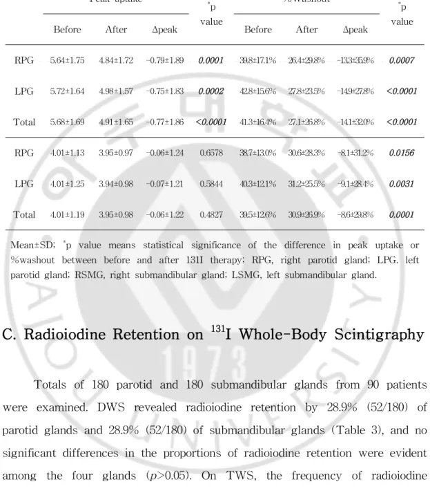
Allsalivary glands showed99mTc-pertechnetate uptake peaks ateither 15 or 20 min (data notshown).Table 2 presents data on salivary gland functionevaluatedby salivaryglandscintigraphybeforeandafter131Iablation therapy.There was no lateraldifference in the peak uptake ofeither the parotid or submandibular glands on either baseline or follow-up salivary gland scintigraphy (allp values >0.05).However,%washout was slightly lowerin right-sided than in left-sided glands(forparotid glands:39.8±17.1% vs. 42.8±15.6%, p=0.0013; and for submandibular glands: 38.7±13.0% vs. 40.3±12.1%,p=0.0156).However,after131Iablation therapy,these differences werenolongerapparent(allpvalues>0.05).
After high-dose 131I ablation therapy, the parotid glands showed significantreductions in both peak uptake (5.68±1.69 → 4.91±1.65,p<0.0001) and %washout (41.3±16.4% → 27.1±26.8%, p<0.0001), whereas, for submandibularglands,only %washoutwas significantly reduced (39.5±12.6% → 30.9±26.9%,p=0.0001).Unlike the parotid glands,the peak uptake ofthe submandibularglands was notsignificantly reduced by131Iablation therapy (4.01±1.19 → 3.95±0.98, p=0.4827). Consequently, larger decrease in peak uptake was observed in the parotid than the submandibular glands (Δpeak=-0.77±1.86 vs.-0.06±1.22,p<0.0001),while the extent of functional decrease in %washoutwas similar between the parotid and submandibular glands(Δwashout=-14.1±32.0% vs.-8.6±29.8%,p=0.091).
12 -Peakuptake * p value %Washout * p value Before After Δpeak Before After Δpeak RPG 5.64±1.75 4.84±1.72 -0.79±1.89 0.0001 39.8±17.1% 26.4±29.8% -13.3±35.9% 0.0007 LPG 5.72±1.64 4.98±1.57 -0.75±1.83 0.0002 42.8±15.6% 27.8±23.5% -14.9±27.8% <0.0001 Total 5.68±1.69 4.91±1.65 -0.77±1.86 <0.0001 41.3±16.4% 27.1±26.8% -14.1±32.0% <0.0001 RPG 4.01±1.13 3.95±0.97 -0.06±1.24 0.6578 38.7±13.0% 30.6±28.3% -8.1±31.2% 0.0156 LPG 4.01±1.25 3.94±0.98 -0.07±1.21 0.5844 40.3±12.1% 31.2±25.5% -9.1±28.4% 0.0031 Total 4.01±1.19 3.95±0.98 -0.06±1.22 0.4827 39.5±12.6% 30.9±26.9% -8.6±29.8% 0.0001
Mean±SD;*p value means statisticalsignificance of the difference in peak uptake or
%washout between before and after 131I therapy;RPG,right parotid gland;LPG.left parotidgland;RSMG,rightsubmandibulargland;LSMG,leftsubmandibulargland.
Table2.Salivary glandfunction beforeand after131Iablation therapy
C.Radi
oi
odi
neRet
ent
i
onon
131IWhol
e-BodySci
nt
i
gr
aphy
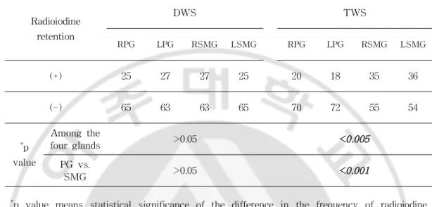
Totals of180 parotid and 180 submandibularglands from 90 patients were examined.DWS revealed radioiodine retention by 28.9% (52/180) of parotidglandsand28.9% (52/180)ofsubmandibularglands(Table3),andno significantdifferencesin theproportionsofradioiodineretention wereevident among the four glands (p>0.05).On TWS,the frequency of radioiodine retention was different among the four glands (p<0.005) where it was observed more frequently in submandibular than parotid glands (39.4% vs. 20.0%,p<0.001).Thus,each gland was identified as DWS+ orDWS- and TWS+orTWS-.
Radioiodine retention DWS TWS RPG LPG RSMG LSMG RPG LPG RSMG LSMG (+) 25 27 27 25 20 18 35 36 (-) 65 63 63 65 70 72 55 54 * p value Amongthe fourglands >0.05 <0.005 PG vs. SMG >0.05 <0.001 *
p value means statisticalsignificance ofthe difference in the frequency ofradioiodine retention.Thefrequency ofradioiodineretention wascomparedamong thefourglands,and itwasthen compared between theparotid and submandibularglands.DWS,diagnostic131I whole-body scintigraphy; TWS,post-ablation 131I whole-body scintigraphy; RPG,right parotid gland;LPG,left parotid gland;RSMG,right submandibular gland;LSMG,left submandibulargland;PG,parotidgland;SMG,submandibulargland
Table 3.Salivary gland radioiodine retention on diagnostic or post-ablation
131Iwhole-body scintigraphy
D.Rel
at
i
on bet
ween Radi
oi
odi
ne Ret
ent
i
on and Sal
i
var
y
Gl
andFunct
i
on
Afterclassification intothesubgroupsdescribed above,itwasapparent thatthe peak uptake ofthe parotid gland was decreased by131Itherapy in both theDWS+ andDWS- groups,buttheΔpeak didnotsignificantly differ betweenthetwogroups(1.02±2.26vs.0.67±1.67,p=0.3207,Table4).Likewise, thefallsin %washoutofboth theparotid and submandibularglandsdid not differbetween theDWS+ and DWS- subgroups(allp values>0.05).On the
14
-otherhand,the peak uptake ofthe submandibulargland changed minimally after131I therapy, and no significant difference in Δpeak was apparent between the DWS+ and DWS- groups (p=0.0586).Therefore,neither peak uptake nor %washoutcorrelated with radioiodine retention,as revealed by DWS,byeithertheparotidorsubmandibularglands.
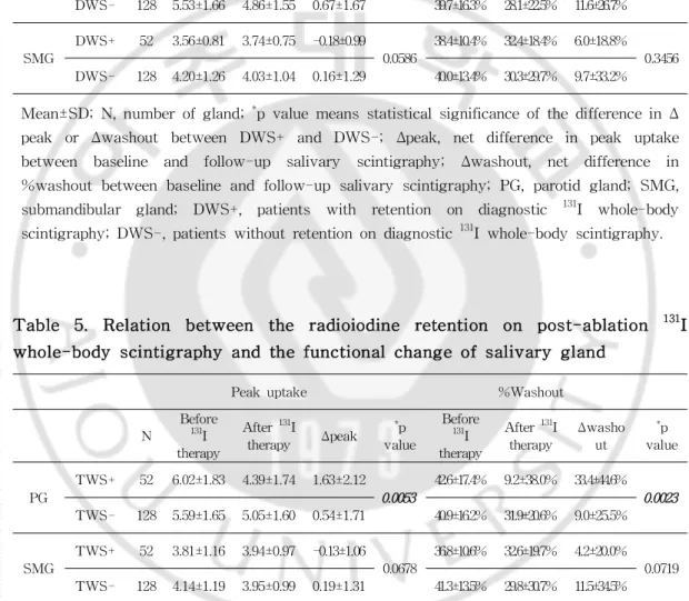
In contrast,after131Itherapy,the fallin peak uptake ofthe parotid gland was greaterin theTWS+ than in the TWS- group (Δpeak=1.63±2.12 vs.0.54±1.71,p=0.0053,Table5).Similarly,the%washoutoftheparotidgland decreased to a greater extent in the TWS+ than the TWS-group (Δwashout=33.4±44.6% vs.9.0±25.5%,p=0.0023).However,aswasnotedwhen DWS data were examined,the submandibular gland exhibited a minimal decrease in peak uptake that was not accompanied by any difference in Δpeak (-0.13±1.06 vs.0.19±1.31,p=0.0678).In addition,Δwashout did not differ between the TWS+ and TWS-groups (4.2±20.0% vs. 11.5±34.5%, p=0.0719).Therefore,radioiodine retention as revealed by TWS was closely related to functional changes in the parotid,but not the submandibular, glands.
Peakuptake %Washout N Before 131 I therapy After131I therapy Δpeak * p value Before 131 I therapy After131I therapy Δwasho ut * p value PG DWS+ 52 6.05±1.73 5.03±1.87 1.02±2.26 0.3207 45.0±16.3% 24.7±35.3% 20.3±42.0% 0.1693 DWS- 128 5.53±1.66 4.86±1.55 0.67±1.67 39.7±16.3% 28.1±22.5% 11.6±26.7% SMG DWS+ 52 3.56±0.81 3.74±0.75 -0.18±0.99 0.0586 38.4±10.4% 32.4±18.4% 6.0±18.8% 0.3456 DWS- 128 4.20±1.26 4.03±1.04 0.16±1.29 40.0±13.4% 30.3±29.7% 9.7±33.2%
Mean±SD;N,numberofgland;*pvaluemeansstatisticalsignificanceofthedifferenceinΔ peak or Δwashout between DWS+ and DWS-; Δpeak,net difference in peak uptake between baseline and follow-up salivary scintigraphy; Δwashout, net difference in %washoutbetween baselineand follow-up salivary scintigraphy;PG,parotid gland;SMG, submandibular gland; DWS+, patients with retention on diagnostic 131I whole-body scintigraphy;DWS-,patientswithoutretentionondiagnostic131Iwhole-bodyscintigraphy.
Table 4. Relation between the radioiodine retention on diagnostic 131I whole-body scintigraphy and thefunctionalchangeofsalivary gland
Peakuptake %Washout N Before 131I therapy After131I therapy Δpeak *p value Before 131I therapy After131I therapy Δwasho ut *p value PG TWS+ 52 6.02±1.83 4.39±1.74 1.63±2.12 0.0053 42.6±17.4% 9.2±38.0% 33.4±44.6% 0.0023 TWS- 128 5.59±1.65 5.05±1.60 0.54±1.71 40.9±16.2% 31.9±20.6% 9.0±25.5% SMG TWS+ 52 3.81±1.16 3.94±0.97 -0.13±1.06 0.0678 36.8±10.6% 32.6±19.7% 4.2±20.0% 0.0719 TWS- 128 4.14±1.19 3.95±0.99 0.19±1.31 41.3±13.5% 29.8±30.7% 11.5±34.5%
Mean±SD;N,numberofgland;*pvaluemeansstatisticalsignificanceofthedifferenceinΔ peak or Δwashout between TWS+ and TWS-; Δpeak,net difference in peak uptake between baseline and follow-up salivary scintigraphy; Δwashout, net difference in %washoutbetween baselineand follow-up salivary scintigraphy;PG,parotid gland;SMG, submandibular gland; TWS+, patients with retention on postablation 131I whole-body
scintigraphy; TWS-, patients without retention on post-ablation 131I whole-body scintigraphy.
Table 5. Relation between the radioiodine retention on post-ablation 131I whole-body scintigraphy and thefunctionalchangeofsalivary gland
16
-Ⅳ.Di
scussi
on
Sialadenitisisthemostprevalentcomplication in patientswhoundergo radioiodine therapy to treat differentiated thyroid carcinoma. Most such conditions can be relieved by conservative treatment featuring adequate hydration; salivary flow stimulation,external compression,and medication including NSAIDs, anticholinergics, or steroids. However, in some cases, invasive procedures are required, for example, sialendoscopy or surgical removalofa salivary gland,and permanentdamage to the salivary glands results(Razaetal,2006;Kim etal,2007;Prendesetal,2013).Salivaryglands cause pain and swelling as early as a few days after radioiodine therapy. However, such early symptoms are frequently transient and subside spontaneously;they are thus not reliable predictors of permanent salivary gland dysfunction(MandelS.Jand MandelL.,2003).In a previous study by Macioszek etal.,theacutesialadenitissymptomsdeveloping afterradioiodine therapy were not correlated with subsequent salivary gland dysfunction(Macioszek et al,2008).However,prediction of salivary gland dysfunction is crucialto selectpatients who require close observation after treatmentand to reduce the extentofsuch dysfunction.In this study,we investigated whethersalivary gland radioiodine retention,as revealed by131I whole-body scintigraphy,could be used to predictfunctionalsalivary gland impairmentcausedbyradioiodinetherapy.
Ourdatashow thattheextentoffunctionalchangesintheparotidglands afterradioiodinetherapy wasgreaterin theTWS+ than in theTWS- group in terms of both peak uptake and %washout.This indicates that parotid glandradioiodineretentionon TWS isclosely associatedwithdevelopmentof latersalivarydysfunction,andsuchpatientsthereforerequiremoreaggressive
management,andcloseobservationtoprotectthesalivaryglands.Incontrast, neitherthe peak uptake nor%washoutofthe submandibularglands differed significantly between the TWS+ and TWS- groups. This implies that submandibulargland dysfunction cannotbepredicted by radioiodineretention revealed by TWS.A possibleexplanation forthedifferencesin theseresults isthefactthatthehistologyofthesalivaryglandsdiffers.Parotidglandsare serous cell-dominantand are affected more by radiation than are mucous cells.However,thesubmandibularglandsarecomposed ofboth mucousand serous cells.In addition the parotid gland transports more radioiodine than doesthesubmandibulargland.Therefore,itisunderstandablethattheparotid gland should be more susceptible to radiation-induced damage than the submandibular gland,in patients receiving 131I ablation therapy(Rigler and Scanlon,1955;Malpanietal,1996;MandelS.JandMandelL.,2003).Esfahani etal.reported thatthedecrease in theproportion ofsaliva secreted by the parotid glands was greater than that from the submandibular glands,as revealedby salivary gland scintigraphy,atboth 3weeksand3monthsafter radioiodine ablation therapy(Esfahanietal,2004).Macioszek etal.evaluated salivary gland function by calculating radioiodine uptake ratios,and parotid radiosensitivity was evident(Macioszek et al,2008).A study of a Korean population by Kim etal.also indicated thatthe incidence ofsalivary gland symptoms was higher in parotid (90%,19/21) than submandibular glands (10%,2/21)(Kim etal,2007).Leeetal.showed thattheestimated absorbed doses upon radioiodine therapy were similar for both the parotid and submandibular glands (2.7±0.8 Gy and 2.8±1.1 Gy,respectively)(Lee et al, 2013).Allofthese data are in agreementwith ourfinding thatthe parotid glandsare highly susceptible to radiation damage.As shown in Table2,the peak uptake of the parotid glands was decreased significantly after131I
18
-ablation therapy,butthis was notthe case with the submandibularglands. Turning to %washout,both the parotid and submandibularglands exhibited significantreductionsin thesevalesafter131Iablation therapy,buttheextent ofreduction wasgreaterin theparotid than thesubmandibularglands.Also, these data were collected underconditions in which radioiodine accumulated tohigherlevelsinthesubmandibularglands(Table3).Therefore,intheclinic, itispossibletopredictfunctionalchangescausedby radioiodinetherapy only fortheparotidgland.
Lee etal.(2013)suggested recently thatpost-ablation131Iwhole-body scintigraphy may be used to predict the development of symptomatic sialadenitis after radioiodine ablation. In the cited study, patients with symptomaticsialadenitisexhibited higherlevelsof131Iuptakeby theparotid glands on early (day3) 131I scintigraphy. Visual and semiquantitative evaluation allowed prediction of the development of sialadenitis after radioiodine ablation,with sensitivities ranging from 80-93%.In contrast,131I uptakedidnotdiffersignificantlyinthesubmandibularglandorupondelayed (day7)131Iscintigraphy.In some respects,thefindings ofourpresentstudy differfrom thoseofLeeetal.Thecited authorsmonitored thedevelopment ofsymptomatic sialadenitis,butwe evaluated salivary gland function using
99mTc-pertechnetate salivary gland scintigraphy. In addition, Lee et al.
monitored symptoms ofsialadenitis (pain/swelling of salivary glands,taste loss,dry mouth,and xerostomia) in both the acute (hospitalization) and chronic(on follow-up visitsatintervals of1-6 months)periods.Hence,the cited study examined both acute and chronic sialadenitis.However,in our presentstudy,wefocusedonchronicsalivaryglanddysfunction,becauseitis known that development of acute sialadenitis is not associated with later salivaryglanddysfunction(MandelS.J.andMandelL.,2003).Finally,thecited
authors found that only early scintigraphy could be used to predict the occurrence of sialadenitis. On the contrary, we found that delayed scintigraphy predicted more severe salivary gland dysfunction. However, despitethedifferencesbetweenthetwostudies,thefindingofthecitedwork, to the effectthatpost-ablation scintigraphy can be used to predictsalivary glandfunctionalimpairment,isinlinewiththeresultsofthepresentstudy.
As expected, DWS was ineffective in terms of predicting functional impairmentcaused by radioiodine therapy (Table 4).After classification of salivary glandsintoDWS+andDWS- groups,basedonradioiodineretention, the parotid glands showed no significantdifferences in terms ofΔpeak and Δwashoutbetween thetwo groups.ThesamewastrueofΔwashoutofthe submandibular glands.Also,no significant decrease was observed in peak uptakeofthesubmandibularglands.Tominimizethestunning effect,DWS is performed in ourclinic using 74 MBq [131I](Woolfenden,2006),which is too smalladosetoinjurethesalivary gland.whichistoosmalladosetoinjure thesalivary gland.Twocasereportsonsalivary gland131Iuptake,shownby
131Iwhole-bodyscintigraphy,inpatientswithsialadenitishaveappeared.Kolla
etal.describeda53-year-oldpatientexhibiting focal131Iuptakeonfollow-up diagnostic131Iscintigraphy,which mimicked nodalmetastasis,butproved to be chronic submandibular sialadenitis after radioiodine therapy(Kolla et al, 1989).Carlisle etal.also observed bilateralparotid131Iuptake on follow-up diagnostic 131I scintigraphy in a patient with radioiodine therapy-induced chronicparotitis(Carlisleand McDougall,2000).Thesereports suggested that
131
Iuptake revealed by131Iscintigraphy was indicative ofthe presence of sialadenitis.However,theresultsofthepresentstudysuggestthatDWS data cannotbe used to predictsalivary gland dysfunction caused by131Iablation therapy.
20
-As shown in Table 3,the pattern ofradioiodine retention revealed by DWS was different from that shown on TWS.Although the radioiodine accumulation by thefoursalivary glandsdidnotdifferupon DWS,wefound that the submandibular glands accumulated more radioiodine on TWS. However,thereason forthisdifferencein thepattern ofradioiodineretention isuncertain.A possiblecontributing factoristhedifferencein theradioiodine dose used for scintigraphy.As submandibular glands produce more saliva than parotid glands,it would appear quite reasonable that submandibular glandsaccumulatemoreradioiodineonTWS.However,onDWS,mostofthe administered radioiodine is taken up by remnantthyroids,and very little of thatis circulating in the blood.Consequently,the difference in radioiodine concentration between parotid and submandibularglandsmay notbe evident. Even though we used a very low dose ofradioiodine forDWS,allimages demonstrated the body contourclearly from vertex to mid-thigh.Thus,this differencewouldnotresultfrom theimagequalityofthosescintigraphies.
Table 2 shows the functionalstatus ofthe parotid and submandibular glands as evaluated by 99mTc-pertechnetate salivary gland scintigraphy. Although both peak uptakeand %washoutoftheparotidglandwerereduced significantly by radioiodine therapy,only %washout in the submandibular glands was decreased significantly.As mentioned above,the submandibular gland is more resistantthan the parotid gland to radiation-induced damage because of histologicaldifferences between the two glands(Esfahaniet al, 2004;Kim etal,2007).The differencein thetime ofonsetofdamagemay also explain our observations.99mTc-pertechnetate uptake by the salivary gland, and washout thereof, reflect parenchymal and ductal functions, respectively.Asradiationcausesdamagefirsttotheductalwalls,followedby later vascular fibrosis(MandelS.J.and MandelL.,2003),salivary excretion
could beimpaired earlier,and to a greaterextent,than parenchymaluptake, at early time points. Parenchymal impairment would have been more prominenthadthepatientsbeenfollowedupoveralongerperiod.
The presentstudy was retrospective in nature,and the time between baseline and follow-up salivary gland scintigraphy varied from 5.5 to 14 months.Itisknown thatthepropensity fordevelopmentofradiation-induced side effects may change with the duration of follow-up(Alexander et al, 1998).Inthecurrentstudy,mostpatients(87.8%)visitedourhospitalabout7 months(5.5-9 months)afterradioiodineablation;however,somevariation in visittimewasinevitable.Moreover,weevaluatedallpatientsonly onceafter they received therapy.When itis considered that both the incidence and pattern of salivary gland dysfunction can change even after 1 year of therapy,it is clear that further clinicalobservations are needed.Another limitation ofourstudy is thatthe extentofradioiodine retention upon131I whole-body scintigraphy was assessed visually; we did not explore any possiblecorrelation between thelevelofradioiodineretention and theextent offunctionaldamage to the salivary glands.As the extent ofradioiodine retention was arbitrarily graded from 1 to 3,mostcases were assigned to grade 1. Therefore, we simply classified patients into two groups, characterized by the ‘presence’or ‘absence’ofretention.Finally,our study population was small;thus,the post-ablation status ofthe salivary glands was assessed semiquantitatively using scintigraphy. However, if the symptoms ofsialadenitis are considered,itwould be possible to subdivide patientsintoanormalgroup,agroupwithchronicsialadenitisandpermanent salivary dysfunction.Therefore,further study using larger populations to allow patientsub-classification and longer-term observations is required in future.
22
-V.Concl
usi
on
In conclusion, radioiodine retention by the parotid glands upon post-ablation131Iwhole-body scintigraphy was associated with reductions in peak uptake and %washout after radioiodine therapy in patients with differentiated thyroid carcinoma.Post-ablation 131I whole-body scintigraphy may be used to predictsevere functionalimpairmentofthe parotid glands, thus identifying patients requiring close observation and aggressive preventativemeasures.
Ref
er
ences
1.AlexanderC,BaderJB,SchaeferA,FinkeC,KirschCM:Intermediateand long-term side effects of high-dose radioiodine therapy for thyroid carcinoma.JNuclMed.39:1551-1554,1998
2.Allweiss P,Braunstein GD,Katz A,Waxman A:Sialadenitis following I-131therapy forthyroid carcinoma:concisecommunication.J NuclMed. 25:755-758,1984
3.American Thyroid Association Guidelines Taskforce on Thyroid N, Differentiated Thyroid C,CooperDS,Doherty GM,Haugen BR,KloosRT etal.Revised American Thyroid Association managementguidelines for patientswiththyroidnodulesanddifferentiatedthyroidcancer.Thyroid.19: 1167-214,2009
4.An YS,Yoon JK,Lee SJ,Song HS,Yoon SH,Jo KS:Symptomatic late-onsetsialadenitisafterradioiodinetherapy inthyroidcancer.AnnNucl Med.27:386-91,2013
5.CaglarM,TuncelM,AlparR:Scintigraphic evaluation ofsalivary gland dysfunctioninpatientswiththyroidcancerafterradioiodinetreatment.Clin NuclMed.27:767-771,2002
6.Carlisle MR, McDougall IR. Dramatic parotid uptake of I-131 on a diagnosticwhole-bodyscan.ClinNuclMed.25:895-897,2000
7.CavalieriRR:Iodine metabolism and thyroid physiology:currentconcepts. Thyroid.7:177-181,1997
8.Chong A,Song H,Min J,Jeong SY,Ha J,Kim J et al.Improved DetectionofLung orBoneMetastaseswithanI-131WholeBody Scanon the 7th Day After High-Dose I-131 Therapy in Patients with Thyroid Cancer.NuclMedMolImaging.44:273-81,2010
9.Creach KM,SiegelBA,Nussenbaum B,Grigsby PW:Radioactive iodine therapy decreases recurrence in thyroid papillary microcarcinoma.ISRN Endocrinol.2012;2012:816386.
10.EsfahaniAF,FallahiB,Olamaie R,EftekhariM,BeikiD,SaghariM: Semi-quantitative assessmentofsalivary gland function in patients with differentiated thyroid carcinoma after radioiodine-131 treatment.HellJ
24
-NuclMed.7:206-209,2004
11.Hyer S, Kong A, Pratt B, Harmer C: Salivary gland toxicity after radioiodine therapy for thyroid cancer.Clin Oncol(R CollRadiol).19: 83-86,2007
12.Kang JY,Jang SJ,Lee WW,Jang SJ,Lee YJ,Kim SE:Evaluation of Salivary Gland Dysfunction Using Salivary Gland Scintigraphy in Sjögren’s Syndrome Patients and in Thyroid Cancer Patients after RadioactiveIodineTherapy.NuclMedMolImaging.45:161-168,2011 13.Kim JW,Han GS,LeeSH,LeeDY,Kim YM:Sialoendoscopictreatment
forradioiodineinducedsialadenitis.Laryngoscope.117:133-136,2007
14.Kolla IS, Alazraki NP, Watts NB. Sialadenitis mimicking metastatic thyroidcarcinoma.ClinNuclMed.14:564-566,1989
15.Lee SM,Lee JW,Kim SY,Han SW,Bae WK:Prediction ofrisk for symptomatic sialadenitis by post-therapeutic dual131I scintigraphy in patients with differentiated thyroid cancer.Ann NuclMed.27:700-709, 2013
16.LindP:131Iwholebody scintigraphy inthyroidcancerpatients.Q J Nucl Med.43:188-194,1999
17.Macioszek A,Baczyk M,Kopec T,SowinskiJ:Salivary gland damage after131Itherapyinpatientswithdifferentiatedthyroidcancer.Preliminary report.EndokrynolPol.59:403-410,2008
18.MalpaniBL,SamuelAM,Ray S:Quantification ofsalivary glandfunction in thyroid cancer patients treated with radioiodine.IntJ RadiatOncol BiolPhys.35:535-540,1996
19.MandelSJ,MandelL:Radioactiveiodineandthesalivaryglands.Thyroid. 13:265-271,2003
20.Mazzaferri EL, Jhiang SM: Long-term impact of initial surgical and medicaltherapy on papillary andfollicularthyroidcancer.Am J Med.97: 418-28,1994
21.PrendesMA,HarrisA,WirostkoBM,GerberAL,Siesky B.Theroleof transforming growth factor beta in glaucoma and the therapeutic implications.BrJOphthalmol.97:680-686,2013
22.Raza H, Khan AU, Hameed A, Khan A: Quantitative evaluation of salivary gland dysfunction afterradioiodine therapy using salivary gland
scintigraphy.NuclMedCommun.27:495-499,2006
23.Rigler RG, Scanlon PW. Radiation parotitis from radioactive iodine therapy.ProcStaffMeetMayoClin.30:149-153,1955
24. SchlumbergerMJ:Papillary and follicularthyroid carcinoma.N EnglJ Med.338:297-306,1998
25.SpitzwegC,HarringtonKJ,PinkeLA,VileRG,MorrisJC:Clinicalreview 132:Thesodium iodidesymporteranditspotentialroleincancertherapy. JClinEndocrinolMetab.86:3327-3335,2001
26.WalterMA,TurtschiCP,SchindlerC,Minnig P,Muller-Brand J,Muller B:Thedentalsafety profileofhigh-doseradioiodinetherapy forthyroid cancer:long-term resultsofalongitudinalcohortstudy.J NuclMed.48: 1620-1625,2007
27.Woolfenden JM:Thyroid stunning revisited.J NuclMed.47:1403-1405, 2006
26 국문요약
-방사성 요오드 치료를 받은 분화성 갑상선암 환자의
전신 요오드 스캔 상 관찰되는 침샘 섭취와
타액선 기능 변화의 연관성
아주대학교대학원 의학과,핵의학전공 조 경 숙 (지도교수:윤 준 기) 분화성 갑상선암으로 방사성 요오드 치료를 받은 환자에서 전신 요오드 스 캔을 통한 타액선 기능 변화 예측이 가능한지 알아보고자 하였다.2010년 6월부 터 2011년 10월까지 분화성 갑상선암으로 갑상선 전절제술 후 첫번째 고용량 방 사성 요오드 치료를 받은 환자 중,진단 및 치료 후 전신 요오드 스캔과 치료 전 후 두 차례의 타액선 섬광조영술 모두를 시행한 90명의 환자를 대상으로 하였다. 이 환자들의 진단 및 치료 후 전신 요오드 스캔 상 관찰되는 타액선 부위의 섭 취증가 유,무에 따라 각각 두 가지 그룹으로 분류하였으며,치료 전후의 타액선 섬광조영술을 통하여 이하선 및 악하선의 섭취율 및 배설율의 변화를 알아보았 다.전신 요오드 스캔에서 관찰되는 타액선 부위의 섭취증가와 방사성 요오드 치 료 후 타액선 기능 변화 사이의 연관성에 대해 분석한 결과,치료 후 전신 요오 드 스캔에서 이하선 부위에 섭취증가를 보이는 환자군의 경우 섭취증가를 보이 지 않는 환자군에 비해 방사성 요오드 치료 후 이하선의 섭취율 및 배설율이 유 의하게 감소한 반면,악하선 부위의 섭취증가 유무는 타액선 기능변화와 별다른 관계가 없었다.이와 마찬가지로,진단 전신 요오드 스캔에서 관찰되는 이하선 및 악하선 부위의 섭취증가는 치료 후 타액선 기능 변화와 연관성이 없었다.따 라서 고용량 방사성 요오드 치료 후 전신 요오드 스캔에서 이하선 부위에 섭취 증가를 보이는 환자의 경우 이하선의 기능저하를 예측할 수 있으며 이를 통해타액선 기능 변화에 대한 적절한 예방 및 치료에 도움이 될 것으로 사료된다.
핵심어:방사성 요오드 치료,타액선 기능 변화,분화성 갑상선암,전신 요오드 스캔,타 액선 섬광조영술