Received: July 11, 2020 Revised: August 7, 2020 Accepted: August 11, 2020 CliniCAl neurophysiology Correspondence to Seong-il Oh
Department of Neurology, Inje University Busan Paik Hospital, Inje University College of Medicine, 75 Bokji-ro, Busanjin-gu, Busan 47392, Korea
Tel: +82-51-890-6130 Fax: +82-51-892-8811 E-mail: [email protected]
Overlap syndrome of Miller-Fisher
syn-drome/Pharyngeal-Cervical-Brachial
variant-Guillain Barre Syndrome with
anti-ganglioside complex antibodies
Suk-yoon Lee1, Seong-il Oh1,2, So-Young Huh3, Kyong Jin Shin4, Jong Kuk Kim5,6,
Byeol-A Yoon5,6
1Department of Neurology, Inje University Busan Paik Hospital, Inje University College of Medicine, Busan, Korea 2Neuroimmunology Research Group, Inje University Busan Paik Hospital, Inje University College of Medicine, Busan, Korea
3Department of Neurology, Kosin University College of Medicine, Busan, Korea 4Department of Neurology, Inje University Haeundae Paik Hospital, Busan, Korea
5Peripheral Neuropathy Research Center, Dong-A University College of Medicine, Busan, Korea 6Department of Neurology, Dong-A University Hospital, Busan, Korea
Guillain-Barré syndrome (GBS) and Miller Fisher syndrome (MFS) can present with overlapping features. A 56-year-old female developed ptosis and diplopia after an upper respiratory infec-tion, and presented with facial palsy, dysarthria, brachial weakness, ataxia, and areflexia. Mild weakness of both legs appeared after a few days. Anti-ganglioside complex antibody were positive to IgG GM1/GQ1b and GQ1b/sulfatide antibodies. The present case suggests that the manifestation of overlap between MFS/PCB variants and GBS could be caused by antiganglio-side complex antibodies.
Key words: Guillain-Barré syndrome; Miller Fisher syndrome; Pharyngeal-cervical-brachial variant; Antibodies; Ganglioside; Anti-ganglioside complex
Guillain-Barré syndrome (GBS) can manifest as several clinical subtypes with various sub-sets of clinical features and antiganglioside antibodies.1-3 The most-classical feature of
GBS is acute progressive and ascending limb weakness after a preceding infection. Miller Fisher syndrome (MFS) is a representative focal variant of GBS characterized by acute ophthalmoparesis, ataxia, and areflexia.2 The pharyngeal-cervical-brachial variant (PCB)
as another well-known but rare focal variant that presents with acute bulbar palsy along with arm and neck weakness.4 Many of these subtypes appear in different combinations
as well as those that occur as individual localized types, which makes it difficult to clearly ORCID Suk-yoon Lee https://orcid.org/0000-0002-5551-0273 Seong-il Oh https://orcid.org/0000-0002-8067-2135 So-Young Huh https://orcid.org/0000-0002-3309-6155
Kyong Jin Shin
https://orcid.org/0000-0003-1349-1913
Jong Kuk Kim
https://orcid.org/0000-0001-9204-3718
Byeol-A Yoon
Overlap syndrome of Miller-Fisher
syn-drome/Pharyngeal-Cervical-Brachial
variant-Guillain Barre Syndrome with
anti-ganglioside complex antibodies
Suk-yoon Lee1, Seong-il Oh1,2, So-Young Huh3, Kyong Jin Shin4, Jong Kuk Kim5,6,
Byeol-A Yoon5,6
1Department of Neurology, Inje University Busan Paik Hospital, Inje University College of Medicine, Busan, Korea 2Neuroimmunology Research Group, Inje University Busan Paik Hospital, Inje University College of Medicine, Busan, Korea
3Department of Neurology, Kosin University College of Medicine, Busan, Korea 4Department of Neurology, Inje University Haeundae Paik Hospital, Busan, Korea
5Peripheral Neuropathy Research Center, Dong-A University College of Medicine, Busan, Korea 6Department of Neurology, Dong-A University Hospital, Busan, Korea
Guillain-Barré syndrome (GBS) and Miller Fisher syndrome (MFS) can present with overlapping features. A 56-year-old female developed ptosis and diplopia after an upper respiratory infec-tion, and presented with facial palsy, dysarthria, brachial weakness, ataxia, and areflexia. Mild weakness of both legs appeared after a few days. Anti-ganglioside complex antibody were positive to IgG GM1/GQ1b and GQ1b/sulfatide antibodies. The present case suggests that the manifestation of overlap between MFS/PCB variants and GBS could be caused by antiganglio-side complex antibodies.
Key words: Guillain-Barré syndrome; Miller Fisher syndrome; Pharyngeal-cervical-brachial variant; Antibodies; Ganglioside; Anti-ganglioside complex
classify them as specific subtypes.2,4,5 MFS can appear
simul-taneously with other types of GBS and appear as an overlap syndrome: MFS/PCB or MFS/PCB-GBS.5
Specific anti-ganglioside antibodies (anti-GQ1b antibody in MFS and anti-GT1a antibody in PCB) are well known to be important diagnosis and pathogenic markers.4 As with pure
MFS and PCB, various antiganglioside antibodies are found in the overlap syndrome, whose frequency does not differ significantly from that of the pure syndrome.4,5 The concept
of anti-ganglioside complex (anti-GSC) was recently discov-ered and established, which allowed the anti-ganglioside antibody found in some patients to be explained.6 In
par-ticular, in some cases the anti-GSC antibody was correlated with specific clinical symptoms, thus confirming that this antibody could be used as an important indicator for ex-plaining the atypical variant.7
We present a case of overlap syndrome of MFS/PCB-GBS with an anti-GSC antibody. The present case suggests that the anti-GSC antibodies could cause overlap syndrome and offer an important diagnostic clue for this rare subtype of GBS.
CASE
A 56-year-old female presented with the sudden onset of bilateral ptosis. She had fever, cough, rhinorrhea, myalgia, and headache that had started several days previously. At that time she experienced slight discomfort when opening the eyes, but on the day of her presentation the bilateral ptosis had become so severe that it was too difficult for her to open her eyes. She had a history of cholecystectomy and of taking medication for hypertension, but no other recent injections or infections except for taking medication for in-fluenza.
Neurological examination revealed bilateral complete internal and external ophthalmoplegia, diplopia, dysarthria, and dysphagia. Her bilateral facial weakness also began to progress slowly during the day, making it difficult for her to wrinkle, blink, and puff up her cheeks.
There was mild weakness in the upper extremities (both Medical Research Council [MRC] grade 4+), but no definite weakness in the legs. The deep tendon reflexes in the upper and lower extremities were reduced. Mild ataxia was present
in both arms and legs. A cerebrospinal fluid examination re-vealed albuminocytologic dissociation.
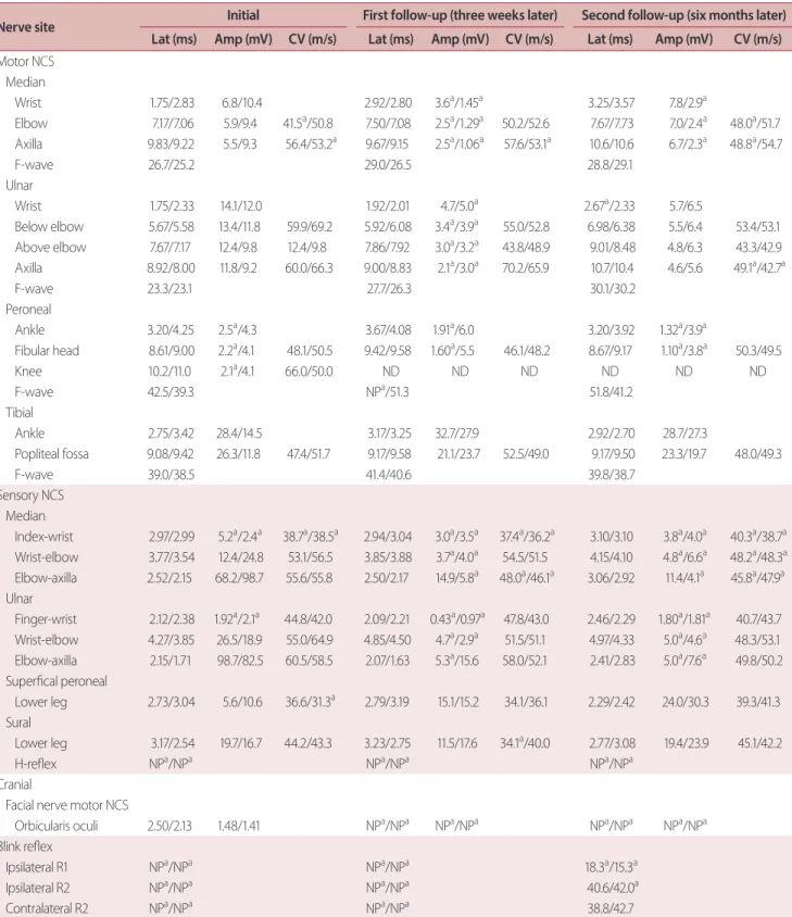
Nerve conduction studies (NCS) performed on the third day after admission revealed acute motor sensory axonal neuropathy (AMSAN) (Table 1). There were slowed median conduction velocities in both forearms and decreased com-pound motor action potentials (CMAPs) in the right peroneal nerves. Sensory NCS revealed slight decreases in the sensory nerve action potentials (SNAPs) and conduction velocities in both upper extremities and the left peroneal nerve. The H-reflex was absent in both lower limbs, while F-waves were present in all extremities. There was no response in the bilat-eral blink reflex pathways.
We started the patient on intravenous immunoglobulin (IVIg) based on the presence of PCB and MFS. During the IVIg treatment, the limb muscle weakness worsened bilaterally to MRC grade 2/3 in the upper extremities and MRC grade 4+ in the lower extremities after 3 days. Her dysarthria, dys-phagia, and dyspnea also worsened, and it became difficult for her to lie down. The amount of sputum increased and respiratory failure occurred. Intubation and L-tube insertion were performed and she was admitted to the intensive care unit. There was some improvement in leg strength from day 2 after initiating IVIg treatment, but the upper extremity muscle strength and bulbar dysfunction did not improve significantly. On the 12th day after tracheostomy she was put on a mechanical ventilator.
The findings of follow-up NCS performed at 3 weeks af-ter admission were compatible with AMSAN, with mildly decreased SNAP and CMAP amplitudes in both upper ex-tremities and the right leg. There was still no response in the bilateral blink reflex pathways.
An enzyme-linked immunosorbent assay (ELISA) was performed to detect various antiganglioside antibodies, in-cluding immunoglobulin G (IgG) and IgM antibodies against the gangliosides GM1, GM2, GD1a, GD1b, GD3, GT1a, GT1b, GQ1b, and GB1b, as described previously;1 all of the results
were negative. Additional evaluations for anti-GSC antibod-ies was performed for various subset of GSCs such as GM1/ GQ1b, GM1/phosphatidic acid, and GQ1b/sulfatide based on the patient’s main neurologic problem. Each ganglioside was used at half the usual amount in a conventional single ELISA for a complex study.7 The results showed strong
(semi-Table 1. Nerve conduction studies
Nerve site Initial First follow-up (three weeks later) Second follow-up (six months later) Lat (ms) Amp (mV) CV (m/s) Lat (ms) Amp (mV) CV (m/s) Lat (ms) Amp (mV) CV (m/s)
Motor NCS Median Wrist 1.75/2.83 6.8/10.4 2.92/2.80 3.6a/1.45a 3.25/3.57 7.8/2.9a Elbow 7.17/7.06 5.9/9.4 41.5a/50.8 7.50/7.08 2.5a/1.29a 50.2/52.6 7.67/7.73 7.0/2.4a 48.0a/51.7 Axilla 9.83/9.22 5.5/9.3 56.4/53.2a 9.67/9.15 2.5a/1.06a 57.6/53.1a 10.6/10.6 6.7/2.3a 48.8a/54.7 F-wave 26.7/25.2 29.0/26.5 28.8/29.1 Ulnar Wrist 1.75/2.33 14.1/12.0 1.92/2.01 4.7/5.0a 2.67a/2.33 5.7/6.5 Below elbow 5.67/5.58 13.4/11.8 59.9/69.2 5.92/6.08 3.4a/3.9a 55.0/52.8 6.98/6.38 5.5/6.4 53.4/53.1 Above elbow 7.67/7.17 12.4/9.8 12.4/9.8 7.86/7.92 3.0a/3.2a 43.8/48.9 9.01/8.48 4.8/6.3 43.3/42.9 Axilla 8.92/8.00 11.8/9.2 60.0/66.3 9.00/8.83 2.1a/3.0a 70.2/65.9 10.7/10.4 4.6/5.6 49.1a/42.7a F-wave 23.3/23.1 27.7/26.3 30.1/30.2 Peroneal Ankle 3.20/4.25 2.5a/4.3 3.67/4.08 1.91a/6.0 3.20/3.92 1.32a/3.9a Fibular head 8.61/9.00 2.2a/4.1 48.1/50.5 9.42/9.58 1.60a/5.5 46.1/48.2 8.67/9.17 1.10a/3.8a 50.3/49.5 Knee 10.2/11.0 2.1a/4.1 66.0/50.0 ND ND ND ND ND ND F-wave 42.5/39.3 NPa/51.3 51.8/41.2 Tibial Ankle 2.75/3.42 28.4/14.5 3.17/3.25 32.7/27.9 2.92/2.70 28.7/27.3 Popliteal fossa 9.08/9.42 26.3/11.8 47.4/51.7 9.17/9.58 21.1/23.7 52.5/49.0 9.17/9.50 23.3/19.7 48.0/49.3 F-wave 39.0/38.5 41.4/40.6 39.8/38.7 Sensory NCS Median Index-wrist 2.97/2.99 5.2a/2.4a 38.7a/38.5a 2.94/3.04 3.0a/3.5a 37.4a/36.2a 3.10/3.10 3.8a/4.0a 40.3a/38.7a Wrist-elbow 3.77/3.54 12.4/24.8 53.1/56.5 3.85/3.88 3.7a/4.0a 54.5/51.5 4.15/4.10 4.8a/6.6a 48.2a/48.3a Elbow-axilla 2.52/2.15 68.2/98.7 55.6/55.8 2.50/2.17 14.9/5.8a 48.0a/46.1a 3.06/2.92 11.4/4.1a 45.8a/47.9a Ulnar Finger-wrist 2.12/2.38 1.92a/2.1a 44.8/42.0 2.09/2.21 0.43a/0.97a 47.8/43.0 2.46/2.29 1.80a/1.81a 40.7/43.7 Wrist-elbow 4.27/3.85 26.5/18.9 55.0/64.9 4.85/4.50 4.7a/2.9a 51.5/51.1 4.97/4.33 5.0a/4.6a 48.3/53.1 Elbow-axilla 2.15/1.71 98.7/82.5 60.5/58.5 2.07/1.63 5.3a/15.6 58.0/52.1 2.41/2.83 5.0a/7.6a 49.8/50.2 Superfical peroneal Lower leg 2.73/3.04 5.6/10.6 36.6/31.3a 2.79/3.19 15.1/15.2 34.1/36.1 2.29/2.42 24.0/30.3 39.3/41.3 Sural Lower leg 3.17/2.54 19.7/16.7 44.2/43.3 3.23/2.75 11.5/17.6 34.1a/40.0 2.77/3.08 19.4/23.9 45.1/42.2 H-reflex NPa/NPa NPa/NPa NPa/NPa Cranial
Facial nerve motor NCS
Orbicularis oculi 2.50/2.13 1.48/1.41 NPa/NPa NPa/NPa NPa/NPa NPa/NPa
Blink reflex
Ipsilateral R1 NPa/NPa NPa/NPa 18.3a/15.3a
Ipsilateral R2 NPa/NPa NPa/NPa 40.6/42.0a
Contralateral R2 NPa/NPa NPa/NPa 38.8/42.7
Data are values on the right/left sides.
Lat, latency; Amp, amplitude; CV, conduction velocity; NCS, nerve conduction study; ND, not done; NP, no potential.
quantitative titers using the subtracted optical density [OD] decided as follows: 1+, OD = 0.10-0.29; 2+, OD = 0.30-0.49; 3+, OD = 0.50-0.99; and 4+, OD ≥1.0).
The complex ganglioside antibody test was performed again after 6 months, which gave a negative result. Severe ptosis and bilateral facial palsy persisted for 1 month after symptom onset, and gradually began to recover, and the tracheostomy tube was removed after 6 months.
DISCUSSION
The present case suggests that the manifestation of overlap between MFS/PCB variants and GBS is caused by the an-ti-GSC antibodies. GBS is an acute autoimmune polyneurop-athy that can be classified into various regional variants.2
Al-though GBS, MFS, and PCB variants have distinct symptoms and antibody test results, overlapping cases of GBS, MFS, and PCB variants are not uncommon in the clinical setting; Sekiguchi et al.5 found overlaps in 50% of MFS patients,
with 23% PCB-GBS and 15% conventional GBS. Previous results4,5 suggest that MFS, PCB, and GBS are likely to form a
continuous spectrum. The regional progression of the MFS spectrum can be either downward or upward,5 and it was
downward in the present case. Clarifying the classification of MFS/PCB, MFS/PCB-GBS, and MFS/GBS is difficult due to the continuity of the disease,5 and so judgment based on
the specific clinical situation in individual patients remains important.
The electrophysiologic results of our patient (Table 1) led to a classification as AMSAN. Previous studies have indicated that the electrophysiologic results for MFS and MFS/GBS were similar for acute motor axonal neuropathy (AMAN).5
The mechanisms underlying AMSAN and AMAN are similar, and AMSAN has also been recognized as a severe form of AMAN. Therefore, both previous cases and the present case indicate that MFS, MFS/GBS, and MFS/PCB-GBS may also occur as a result of AMAN or AMSAN depending on the se-verity of the disease.
The overlap syndrome has also been explained based on results obtained in studies of ganglioside antibodies. This may due to the cross reaction of gangliosides GQ1b and GT1a, although the IgG GT1a antibody did not cross react with the ganglioside GQ1b in pure PCB.8 Anti-GQ1b
anti-bodies are found in most cases of MFS,2 and IgG GT1a
anti-bodies are also found in acute bulbar palsy.3
A new test method was recently used to identify anti-GSC antibodies in patients in whom antibodies were not found in the single ganglioside antibody test.6 In addition to
isolat-ed ophthalmoplegia and isolatisolat-ed bulbar palsy, overlapping symptom involving neck, arm, and leg weakness can also present with complex antibodies.6,9 While IgG GM1/GQ1b
and GQ1b/sulfatide were found to be associated with bulbar palsy and ophthalmoplegia, respectively,7,9 the
character-istic manifestations of individual complex antibodies could be predicted only partially. The antibodies detected in the presence of such overlapping symptoms would facilitate the understanding of the extent of the regional manifestations of GBS and MFS or predicting long-term outcomes.
The results obtained in the present patient did not include those for single antibodies, but the test methods used for GSC testing did reveal various forms of complex antibodies, including anti-GQ1b antibodies. Compared to the regional spectrum found in the previous study,2,4,5 the GSC found in
this study represents an example of the manifestations of ophthalmoplegia, facial palsy, bulbar palsy, neck, and arm weakness.
As a subtype of GBS, anti-GQ1b antibodies and anti-GT1a antibodies were also found in a case of polyneuritis cranialis, which can be caused by multiple cranial neuropathies with-out limb weakness.10 However, similar to the present case, if
the initial expression is in the form of idiopathic cranial neu-ropathy or polycranial neuritis, and it appears as an overlap syndrome as it progresses to other parts of the body, a test for GSC should be considered.
Some clinicians have only recommended checking gan-glioside antibodies to GM1, GD1a, and GQ1b. However, the confirmation of an unusual complex antibody combination in the present case of overlap syndrome of MFS/PCB-GBS suggests the utility of testing a panel of simple or complex ganglioside antibodies for further diagnostic clarification. Further large-scale studies are needed to reveal the clinical significance of GSC testing. In conclusion, the presence of complex anti-ganglioside antibodies may aid the differential diagnosis in cases in which the clinical diagnosis based on the results of other conventional supportive tests is ambigu-ous or overlapping.
Acknowledgements
This study was supported by National Research Foundation of Korea (NRF) grants funded by the Korea government (MIST) (No. 2020R1G1A1008446 and 2016R1A5A2007009). Conflicts of Interest
The authors declare that they have no competing financial interests.
REFERENCES
1. Kim JK, Bae JS, Kim DS, Kusunoki S, Kim JE, Kim JS, et al. Preva-lence of anti-ganglioside antibodies and their clinical correlates with guillain-barre syndrome in Korea: a nationwide multicenter study. J Clin Neurol 2014;10:94-100.
2. Wakerley BR, Uncini A, Yuki N, the GBS Classification Group. Guil-lain-Barré and Miller Fisher syndromes--new diagnostic classifi-cation. Nat Rev Neurol 2014;10:537-544.
3. Kim JK, Kim BJ, Shin HY, Shin KJ, Nam TS, Oh J, et al. Acute bul-bar palsy as a variant of Guillain-Barré syndrome. Neurology 2016;86:742-747.
4. Nagashima T, Koga M, Odaka M, Hirata K, Yuki N. Continuous spectrum of pharyngeal-cervical-brachial variant of Guillain-Bar-ré syndrome. Arch Neurol 2007;64:1519-1523.
5. Sekiguchi Y, Mori M, Misawa S, Sawai S, Yuki N, Beppu M, et al. How often and when Fisher syndrome is overlapped by Guil-lain-Barré syndrome or Bickerstaff brainstem encephalitis? Eur J Neurol 2016;23:1058-1063.
6. Kaida K, Morita D, Kanzaki M, Kamakura K, Motoyoshi K, Hirakawa M, et al. Ganglioside complexes as new target antigens in Guil-lain-Barré syndrome. Ann Neurol 2004;56:567-571.
7. Huh SY, Lee SY, Lee JH, Lee WG, Kim JK, Yoon BA, et al. A variant Guillain-Barré syndrome with ganglioside complex anti-body. J Neurocrit Care 2018;11:134-136.
8. Koga M, Yoshino H, Morimatsu M, Yuki N. Anti-GT1a IgG in Guil-lain-Barré syndrome. J Neurol Neurosurg Psychiatry 2002;72:767-771.
9. Morikawa M, Kuwahara M, Ueno R, Samukawa M, Hamada Y, Kusunoki S. Serological study using glycoarray for detecting anti-bodies to glycolipids and glycolipid complexes in immune-me-diated neuropathies. J Neuroimmunol 2016;301:35-40.
10. Wakerley BR, Yuki N. Polyneuritis cranialis: oculopharyngeal sub-type of Guillain-Barré syndrome. J Neurol 2015;262:2001-2012.