Mutations in FAM50A suggest that Arm
field XLID
syndrome is a spliceosomopathy
Yu-Ri Lee
1,20
, Kamal Khan
2,3,4,5,20
, Kim Arm
field-Uhas
6,20
, Sujata Srikanth
7,20
, Nicola A. Thompson
8
,
Mercedes Pardo
9
, Lu Yu
9
, Joy W. Norris
7
, Yunhui Peng
10
, Karen W. Gripp
11
, Kirk A. Aleck
12
,
Chumei Li
13
, Ed Spence
14
, Tae-Ik Choi
1
, Soo Jeong Kwon
15
, Hee-Moon Park
15
, Daseuli Yu
16
,
Won Do Heo
16
, Marie R. Mooney
2,3
, Shahid M. Baig
4
, Ingrid M. Wentzensen
17
, Aida Telegra
fi
17
,
Kirsty McWalter
17
, Trevor Moreland
7
, Chelsea Roadhouse
13
, Keri Ramsey
18
, Michael J. Lyons
7
,
Cindy Skinner
7
, Emil Alexov
10
, Nicholas Katsanis
2,3,19
, Roger E. Stevenson
7
, Jyoti S. Choudhary
9
,
David J. Adams
8
, Cheol-Hee Kim
1,21
✉
, Erica E. Davis
2,3,19,21
✉
& Charles E. Schwartz
7,21
✉
Intellectual disability (ID) is a heterogeneous clinical entity and includes an excess of males
who harbor variants on the X-chromosome (XLID). We report rare FAM50A missense
var-iants in the original Arm
field XLID syndrome family localized in Xq28 and four additional
unrelated males with overlapping features. Our fam50a knockout (KO) zebra
fish model
exhibits abnormal neurogenesis and craniofacial patterning, and in vivo complementation
assays indicate that the patient-derived variants are hypomorphic. RNA sequencing analysis
from fam50a KO zebra
fish show dysregulation of the transcriptome, with augmented
spli-ceosome mRNAs and depletion of transcripts involved in neurodevelopment. Zebrafish
RNA-seq datasets show a preponderance of 3′ alternative splicing events in fam50a KO, suggesting
a role in the spliceosome C complex. These data are supported with transcriptomic
sig-natures from cell lines derived from affected individuals and FAM50A protein-protein
interaction data. In sum, Arm
field XLID syndrome is a spliceosomopathy associated with
aberrant mRNA processing during development.
https://doi.org/10.1038/s41467-020-17452-6OPEN
1Department of Biology, Chungnam National University, Daejeon, Korea.2Center for Human Disease Modeling, Duke University Medical Center, Durham,
NC, USA.3Advanced Center for Translational and Genetic Medicine (ACT-GeM), Stanley Manne Children’s Research Institute, Ann & Robert H. Lurie Children’s Hospital of Chicago, Chicago, IL, USA.4Human Molecular Genetics Laboratory, Health Biotechnology Division, National Institute for Biotechnology and Genetic Engineering (NIBGE), Faisalabad, Pakistan.5Pakistan Institute of Engineering and Applied Sciences (PIEAS), Islamabad, Pakistan.6Children’s Healthcare of Atlanta, Atlanta, GA, USA.7Greenwood Genetic Center, Greenwood, SC, USA.8Wellcome Sanger Institute, Hinxton, Cambridge, UK.
9Chester Beatty Laboratories, Institute of Cancer Research, London, UK.10Department of Physics, Clemson University, Clemson, SC, USA.11Division of
Medical Genetics, A. I. duPont Hospital for Children, Wilmington, DE, USA.12Genetics and Metabolism, Phoenix Children’s Medical Group, Phoenix, AZ, USA.13Clinical Genetics Program, McMaster University Medical Center, Hamilton, ON, Canada.14Division of Pediatric Genetics and Metabolism, University of North Carolina School of Medicine, Chapel Hill, NC, USA.15Department of Microbiology and Molecular Biology, Chungnam National University, Daejeon, Korea.16Department of Biological Sciences, Korea Advanced Institute of Science and Technology, Daejeon, Korea.17GeneDx Inc, Gaithersburg,
MD, USA.18Center for Rare Childhood Disorders, TGen, Phoenix, AZ, USA.19Department of Pediatrics, Feinberg School of Medicine, Northwestern
University, Chicago, IL, USA.20These authors contributed equally: Yu-Ri Lee, Kamal Khan, Kim Arm
field-Uhas, Sujata Srikanth.21These authors jointly supervised: Cheol-Hee Kim, Erica E. Davis, Charles E. Schwartz. ✉email:zebrakim@cnu.ac.kr;eridavis@luriechildrens.org;ceschwartz@ggc.org
123456789
I
ntellectual disability (ID) affects 1–3% of the general
popula-tion
1. Males exceed females in the ID population by 20–30%,
likely due to an enrichment of genes on the X-chromosome
that are required for neurodevelopment. X-linked ID (XLID)
disorders, resulting from hemizygous variants, contribute
sig-nificantly to the male ID population
2. Efforts by research groups
worldwide have identified 145 XLID genes contributing to 114
XLID syndromes and 63 non-syndromic XLID entities
3. Exome
sequencing has accelerated mutational analysis of the coding
regions of the X-chromosome and identified 28 of the 145 XLID
genes in the past decade
3. Despite these accomplishments, more
than 56 XLID syndromes and 33 non-syndromic XLID entities
remain without a molecular diagnosis.
In 1999, we characterized Armfield XLID syndrome and
localized the causal locus to an 8 Mb region on Xq28 using
linkage analysis
4. Affected individuals display a distinctive
phe-notype involving multiple systems: postnatal growth retardation;
variable head circumference with a prominent forehead and
dysmorphic facial features; ocular abnormalities and seizures
4.
Here, we report the causal variant that segregates with the
Armfield syndrome phenotype. As part of a screen of XLID genes
localized to Xq28, we identify an ultra-rare missense variant in
FAM50A (family with sequence similarity 50 member A; known
as XAP5 or HXC26) in affected males and unaffected carrier
females. We use GeneMatcher
5, to identify four unrelated males
who have undergone whole-exome sequencing (WES), and who
each bear a rare missense variant in FAM50A. These males
dis-play phenotypes similar to Armfield XLID syndrome.
To investigate FAM50A function, establish relevance to the
Armfield XLID clinical spectrum, and test variant pathogenicity,
we utilize zebrafish (Danio rerio). A zebrafish fam50a knockout
(KO) recapitulates the human phenotype with abnormal
devel-opment of cephalic structures. In addition, we use in vivo
com-plementation studies to show that the missense FAM50A changes
identified confer a partial loss of function. Transcriptomic studies
of fam50a KO zebrafish heads enable correlation with the human
phenotype and validate previous reports suggesting FAM50A to
be associated with the spliceosome complex
6,7. Transcriptomic
data from lymphocyte cell lines (LCL) derived from affected
males and FAM50A protein–protein interaction data further
support the previous
findings. We propose that aberrant
spli-ceosome C-complex function is the molecular mechanism
underpinning Armfield XLID, defining it as a spliceosomopathy.
Results
Clinical and genetic studies implicate
FAM50A in XLID. We
report updated clinical information for affected siblings in family
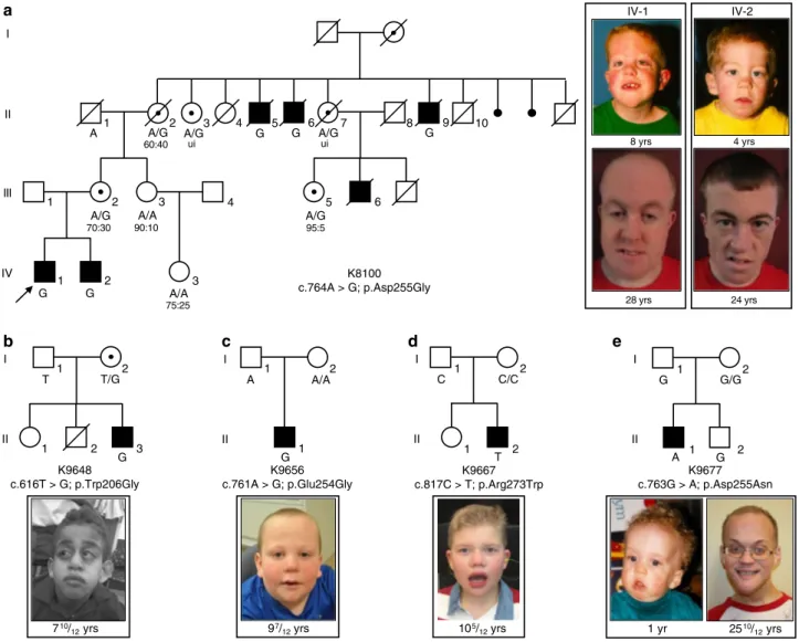
K8100 (IV-1 and IV-2; Fig.
1
a and Table
1
; Supplementary
Note 1). The causal locus was localized to Xq28
4, and within this
chromosome band, a hitherto uncharacterized gene, FAM50A/
XAP5, was reported in which the 5′ untranslated region contained
a run of GGC repeats
8. Analysis of an affected male from K8100
along with males from other XLID families localized to
Xq28 showed no expansions beyond the normal range. We
per-formed bidirectional Sanger sequencing of the coding regions and
exon–intron boundaries of five candidate genes located in Xq28
(GDI1, MECP2, L1CAM, AFF2/FMR2, FAM50A/XAP5) in
affec-ted males. These analyses revealed a missense change, c.764A>G,
p.Asp255Gly, in FAM50A (GenBank [
https://www.ncbi.nlm.nih.
gov/nuccore/NM_004699.4
]), which segregated with disease in
the family (Fig.
1
a). To exclude the possibility of a causal variant
elsewhere in Xq28, we included an affected male from K8100 in a
larger sequencing project of 718 genes located on the
X-chromosome
9. The same alteration in FAM50A was the only
likely causal change identified. This same variant was again
identified as the sole candidate in K8100 as part of an X-exome
next-generation sequencing project conducted later
10.
The p.Asp255Gly change was not present in 400
X-chromo-somes from ethnically matched controls from our in-house data
set, and is absent from 182,557 alleles in gnomAD (accessed April
2019;
https://gnomad.broadinstitute.org/
). Prediction algorithms
suggested that 255Gly was likely pathogenic (Supplementary
Table 1). Asp255 is embedded within a highly conserved string of
amino acids (KEDLI) present in vertebrates, D. melanogaster, C.
elegans, and S. pombe (Supplementary Fig. 1a–c). Secondary
structure prediction programs proposed that Asp255Gly is
located in a beta-turn of a random coil domain (Supplementary
Fig. 2a). In silico protein modeling indicated that p.Asp255Gly is
located in a short loop in the low confidence structural region of
the model (Supplementary Fig. 2). In the wild-type (WT)
structure, a hydrogen (H)-bond is formed between the
Asp255 side chain and Arg180, and the variant is expected to
affect the H-bonding network and alter the net charge. However,
binding free energy change predictions made by
five servers are
inconsistent, likely due to
fidelity of the modeled structure
(Supplementary Table 2). In sum, genetics data coupled to variant
prediction and protein modeling suggested that p.Asp255Gly is
deleterious. We then sought additional individuals through
GeneMatcher
5, and identified four unrelated males with variants
in FAM50A.
The family K9648 proband (II-3) was 10 years old at the last
clinical examination and he displayed global developmental delay,
prominent forehead, glaucoma, and small hands and feet (Fig.
1
b
and Table
1
; Supplementary Note 1). We performed
parent-proband trio WES. Subsequent to bioinformatic
filtering for rare
functional variants under autosomal recessive, dominant de novo
or X-linked paradigms, we identified a maternally inherited
FAM50A c.616T>G; p.Trp206Gly variant (Fig.
1
b; Supplementary
Fig. 1a–c). All prediction algorithms indicated that the p.
Trp206Gly variant was likely pathogenic (Supplementary Table 1).
In silico protein modeling analysis indicated that p.Trp206Gly is
located in a short loop of the high-confidence structural region,
partially buried and surrounded by hydrophobic residues
(Supplementary Fig. 2a, b). However, protein folding free energy
changes in the context of Trp206Gly are contradictory
(Supple-mentary Table 2).
The family K9656 proband (II-1) presented with global delay,
strabismus, short stature, and dysmorphic facial features (Fig.
1
c
and Table
1
; Supplementary Note 1). WES of the proband and
his healthy parents identified a hemizygous, de novo, variant in
FAM50A (c.761A>G, p.Glu254Gly; Fig.
1
c; Supplementary
Fig. 1a–c). The variant was predicted to be deleterious
(Supplementary Table 1), and occurs in a short loop in the
low confidence structural region of the protein (Supplementary
Fig. 2a, b). Glu254 makes an H-bond with nearby residue
Asn177 and the substitution with Gly will eliminate the
H-bond. The variant is also predicted to destabilize the structure
and to alter the net charge at position 254 (Supplementary
Table 2).
The clinical presentation of the family K9667 affected male
(II-2) consisted of global delay, exotropia, and myopia, although he
did not have short stature (Fig.
1
d; Table
1
; Supplementary
Note 1). WES of the proband and his unaffected parents
identified a de novo FAM50A variant (c.817C>T, p.Arg273Trp;
Fig.
1
d; Supplementary Fig. 1a–c). All bioinformatic analyses
indicated that the variant was likely pathogenic (Supplementary
Table 1). Protein modeling suggests that p.Arg273Trp is located
in a helix in the high-confidence structure (Supplementary
Fig. 2a, b); the side chain of Arg273 is buried and forms H-bonds
with Glu200 and the backbone of Ile199. This amino acid change
potentially affects protein stability, and predictions from different
servers also indicate that the Arg273Trp variant might destabilize
the protein (Supplementary Table 2).
The clinical features of the family K9677 male proband (II-1)
overlapped with affected males in K8100 (Fig.
1
e and Table
1
;
Supplementary Note 1); he exhibited global developmental delay,
dysmorphic facial features and exotropia. Trio-based WES and
bioinformatic
filtering identified a rare de novo variant in
FAM50A (c.763G>A, p.Asp255Asn; Fig.
1
e; Supplementary
Fig. 1a, b), that was predicted to be likely pathogenic
(Supplementary Table 1). Protein modeling suggests that this
change introduces a polar residue, Asn, and thus does not form
an H-bond with residue Arg180 as does the WT amino acid
(Supplementary Fig. 2). Protein stability predictions are
incon-sistent (Supplementary Table 2). However, the variant alters the
charge at residue 255, potentially affecting FAM50A function.
Of the
five families with FAM50A variants, the variants in
K8100 and K9648 were inherited. All available females in K8100
were tested for X-inactivation (XI) at the AR locus
11. We
observed no correlation between the presence of the FAM50A
alteration and the degree of skewed XI (Fig.
1
a). Both a
non-carrier female (III-3) and a non-carrier female (III-5) had significant
skewing of XI. The three remaining informative females (carriers
II-2, III-2; non-carrier IV-3) had random-to-moderate skewed XI.
Together, we identified a cohort of nine males from five
unrelated families who carry rare FAM50A variants. Affected
individuals share syndromic ID and comorbid phenotypes
impacting growth, facial gestalt, and ocular development (Fig.
1
and Table
1
). These nonsynonymous changes segregate with
disease status in pedigrees, are absent from gnomAD, and reside
within highly conserved regions of the XAP domain in the
C-terminal portion of FAM50A (Fig.
1
; Supplementary Fig. 1a, b
and Supplementary Table 1).
FAM50A has ubiquitous expression and nuclear localization.
The FAM50A cDNA was characterized through efforts to catalog
the genes on Xq28
8,12. Monitoring of FAM50A in adult and fetal
human tissue panels reported ubiquitous expression
8,13. We
1 2 3 4 5 6 7 8 9 10
A A/G A/G G G A/G G
1 2 3 4 A/G A/A 5 6 A/G 1 2 3 A/A G G 1 2 1 2 3 T T/G G 1 2 1 A/A G 1 2 2 1 C/C T 1 2 2 1 G G/G A I II III IV I II I II I II I II K8100 c.764A > G; p.Asp255Gly K9648 c.616T > G; p.Trp206Gly K9656 c.761A > G; p.Glu254Gly K9667 c.817C > T; p.Arg273Trp K9677 c.763G > A; p.Asp255Asn 28 yrs 8 yrs IV-1 IV-2 4 yrs 24 yrs 2510/ 12yrs 1 yr 105/ 12yrs 97/ 12yrs 710/ 12yrs
a
b
c
d
e
A C G 60:40 ui ui 95:5 70:30 90:10 75:25Fig. 1 Missense variants inFAM50A cause XLID in five unrelated families. a–e Pedigrees of the five families reported in this study are shown, with FAM50A genotype given for each available individual. Photographs of available affected males are provided for each pedigree. For family K8100, photographs are provided for the two affected males in generation IV at ages 8 and 4 years, when the family was originally published4; new photos from the last clinical assessment (December 2017) are shown (28 and 24 years). Ratios under females II-2, III-2, III-3, III-5, and IV-3 represent X-inactivation data. Females II-3 and II-7 were uninformative (ui) at the AR locus. Circles, females; squares, males; unfilled shapes, unaffected; black filled shapes, affected; unshaded circle with black dot, carrier female as determined by FAM50A analysis or by pedigree structure; diagonal line, deceased. Male K8100-III-6 had macrocephaly, seizure disorder, bilateral ventricular enlargement, and atrophy of the left hemisphere on a pneumoencephalogram; he was unavailable for FAM50A genotyping.
Table
1
Clinical
fi
ndings
in
fi
ve
families
with
mutations
in
FAM50A
.
Individual K8100 IV-1 K8100 IV-2 K9648 II-3 K9656 II-1 K9667 II-2 K9677 II-1 FAM50A variant c.764A>G; p.Asp255Gly c.764A>G; p.Asp255Gly c.616T>G; p.Trp206Gly c.761A>G; p.Glu254Gly (de novo ) c.817C>T; p. Arg273Trp (de novo ) c.763G>A; p. Asp255Asn (de novo ) Ethnicity Caucasian Caucasian Mixed (African-American, Middle Eastern, Mixed European) Caucasian Caucasian Caucasian Clinical characteristics Growth Birth (gestational weeks ) 4 0 4 0 3 4 3 5 38.5 ND Length, cm (%) 47 (5) 45.7 (<3) 43.2 (15) 47 (50) 48.3 (25) 53.3 (95) Weight, kg (%) 3.2 (30) 2.9 (15) 2.4 (60) 2.5 (40) 2.8 (20) 4.4 (97) HC, cm (%) 36.2 (55) 36 (50) NA NA 34.3 (20) 39 (>97) Postnatal (years –months) 28 24 7– 10 9– 78 –32 5– 10 Height, cm (%) 154.9 (<3) 154.9 (<3) 106.2 (<3) 120.5 (<3) 122 (9) 160.5 (<3) Weight, kg (%) 70 (15) 43.2 (<3) 19.4 (<3) 38.4 (85) 27.9 (63) 61.9 (<3) HC, cm (%) 58.6 (85) 58.5 (85) 50.2 (5) 54 (75) 52.1 (30) 60.25 (>97) De velopment Delay Global, special education, ambulatory, speaks in short phrases Global, special education, ambulatory (walked at 3 yrs ) single words Global, not ambulatory, no speech Global, regular classes with support Global, no speech, special education, ambulatory for short distances Global IQ 66 ND ND ND <50 63 Somatic fi ndings Craniofacial Macrocephaly, epicanthal folds, depressed nasal bridge, downslanted palpebral fi ssures, cleft palate, bow-shaped mouth, microretrognathia Broad forehead, epicanthal folds, depressed nasal bridge, downslanted palpebral fi ssures, low-set ears, micrognathia Prominent forehead, bitemporal narrowing, proptosis, hypotelorism, tubular nose, single median incisor, hypodontia, low-set ears, large left ear, prominent lips Bilateral epicanthal folds, infraorbital creases, wide nasal root, short and lightly upturned nose with underdeveloped nares, slightly posteriorly rotated ears, faint hemangiomas between brows and at back of neck Bulbous nose, excessively folded helices Prominent tallforehead, overfolded helices, micrognathia
Ocular Strabismus — Axenfeld –Rieger with glaucoma, nystagmus Strabismus Exotropia
Exotropia, keratoconus, nystagmus
Cardiac ASD, PDA — Tetralogy of Fallot, right ventricle dilation ASD —— Skeletal Small feet, pes cavus, hammertoes Small feet, pes cavus, hammertoes Small hands/feet, short limbs, crease across dorsum of feet, foot e version/ inversion, coxa valga, mild scoliosis Joint hypermobility Small hands/feet Stiff joints, small hands and feet, club foot Gastrointestinal —— G-tube Inguinal hernia Hiatal hernia, dysphagia, constipation Umbilical hernia, imperforate anus Genitourinary —— Horseshoe kidney, micropenis, undescended testis Unilateral renal agenesis, micropenis, small scrotum Cryptorchidism — Skin Capillary hemangiomas of nasal bridge and e yelids Facial capillary hemangiomas Sacral dimple, lipoma ND ND Hemangiomas Neurologic Seizures Seizures Hypotonia Hypotonia Hypotonia/ hypertonia, jerky movements, tethered cord Seizures, tremor, hypotonia, incontinent Other Obstructive sleep apnea Aggressive, quick tempered Sleep disturbance 2 vessel umbilical cord, obesity, gynecomastia Sleep disturbance, incontinent Impulsive, mood
disorder, hypothyroidism, hypodontia
MRI Enlarged 3rd ventricle, extra-axial fl uid Asymmetric ventricles Decreased white matter, small corpus callosum, small brain stem —— — ASD, atrial septal defect; G-tube, gastrostomy tube; HC, head circumfe rence; IQ, intelligence quotient; MRI, magnetic resona nce imaging; ND , n o d a ta; PDA, patent ductus arteriosus; yrs, years; (— ) not present.
performed semi-quantitative RT–PCR using a multiple human
fetal tissue cDNA panel (Clontech). FAM50A was expressed in all
eight fetal tissues assessed, including brain (Supplementary
Fig. 3a). Next, we evaluated FAM50A expression in fetal brain
using a Rapid Scan Human Brain Panel (Origene). FAM50A was
detectable in the fetal cerebellum and hypothalamus
(Supple-mentary Fig. 3b). However, we observed low expression of
FAM50A in the temporal lobe and were unable to detect
expression in the hippocampus using this method.
Mazzarella and colleagues
8reported features suggestive of
nuclear localization in the FAM50A amino acid sequence. cNLS
mapper
(
http://nls-mapper.iab.keio.ac.jp/cgi-bin/NLS_Mapper_
form.cgi
) indicated that FAM50A (GenBank ID:
NP_004690.1
)
contained a nuclear localization signal (NLS), ITTKKRKLG
(positions 149-157, score of 7.5), predicting partial localization to
the nucleus
14. cNLStradamus (
http://www.moseslab.csb.utoronto.
ca/NLStradamus
), predicted a NLS domain within FAM50A
amino
acids
88–116
(LAKKEQSKELQMKLEKLREKERK-KEAKRK)
15. To examine FAM50A cellular localization, we
performed endogenous protein immunostaining of NIH/3T3
cells. We observed dispersed nuclear localization throughout the
cell cycle (Supplementary Fig. 4a). After demonstrating that
FAM50A protein levels are not significantly different in LCLs
derived from affected males vs matched controls (Supplementary
Fig. 5), we tested whether the XLID-associated variants affected
FAM50A localization. We generated C-terminally tagged V5
plasmids containing the WT or mutant FAM50A open reading
frame (ORF) and visualized FAM50A in transfected COS-7 cells
(Supplementary Table 3). All mutant proteins tested (p.
Trp206Gly, p.Glu254Gly, and p.Asp255Gly) localized to the
nucleus and were indistinguishable from WT (Supplementary
Fig. 4b), an unsurprising result since none of the variants
impacted a predicted NLS. However, the nuclear staining in the
NIH/3T3 cells appeared more diffuse than the transfected COS-7
cells, possibly reflecting the different technologies and cell types
utilized.
fam50a KO zebrafish display patient-relevant phenotypes. The
zebrafish genome harbors a single reciprocal ortholog encoding a
protein with the same length as the human protein (339 amino
acids), which is highly conserved (86% identity; 93% similarity;
Supplementary Fig. 1c). To characterize the spatiotemporal
expression of D. rerio fam50a, we probed RNA in situ on
whole-mount embryos at eight different stages. We noted ubiquitous
expression prior to and throughout gastrulation, and in
mid-somitic embryos. From 24 h post-fertilization (hpf) onward, and
up to the latest time point assessed (72 hpf) fam50a mRNA
regionalized to all visible anterior structures including the brain,
eye and mandible (Supplementary Fig. 6a). These observations
were consistent with publicly available zebrafish RNA-seq data
(
https://www.ebi.ac.uk/gxa/experiments/E-ERAD-475/Results
).
To determine the consequences of FAM50A depletion, we
performed genome editing and identified loss of function mutant
alleles. We disrupted the highly conserved XAP domain by
injecting Cas9 mRNA and guide RNA targeting either fam50a
exon 6 or exon 7, screened for mosaic F0 founders, and isolated
two mutant alleles in the F1 generation (compound 11
+ 5 bp
deletion [KO1] or 4 bp deletion [KO2]; Supplementary Fig. 6b–e).
We used KO1 for all subsequent phenotyping (hereafter referred
to as KO). Whole-mount in situ hybridization (WISH),
immunofluorescent antibody staining, or immunoblotting of
embryo-derived protein lysate could not detect fam50a mRNA or
FAM50A protein as early as 24 hpf (Supplementary Fig. 6f, g, h).
fam50a KO was present in the expected Mendelian ratios and
displayed similar gross morphology to WT until ~3 days
post-fertilization (dpf). However, at 5 dpf KO larvae were severely
affected by abnormal anterior development impacting the brain,
eyes and cartilage (Fig.
2
a), resulting in lethality by ~6 dpf.
We evaluated cell-type specific and cellular response assays in
fam50a KO and WT larvae (Supplementary Table 3). Using a
transgenic reporter of pan-neuronal cells, tg(huc:egfp), we found a
reduction of differentiated neurons in fam50a KO brain (Fig.
2
b),
consistent with her4.1 WISH indicating diminished
neurogen-esis
16at 3 dpf (Fig.
2
c; Supplementary Fig. 7). Both markers
showed indistinguishable neuronal integrity at 2 dpf (Fig.
2
b, c).
We tested whether altered proliferation and cellular stress
responses could account for the onset of neuronal phenotypes.
We observed marked depletion of proliferation markers pcna and
ccnd1 at 3 dpf with concomitant augmentation of p53 pathway
effectors tp53, mdm2, and cdkn1a at an earlier stage, especially in
the midbrain (2 dpf; Fig.
2
d; Supplementary Fig. 8). Blood vessel
development was normal up to 2.5 dpf in tg(kdrl:egfp);fam50a KO
zebrafish (Supplementary Fig. 9a), and was confirmed by in situ
analysis with endothelial molecular markers etv2, cdh5, and
cldn5b (Supplementary Fig. 9b). These data recapitulate specific
early neurogenesis defects that are present in Armfield XLID
syndrome.
Affected males with mutations in FAM50A show dysmorphic
facial features (Fig.
1
and Table
1
). We stained zebrafish larvae
with Alcian blue at different time points (2.5, 3, and 4.5 dpf) to
study orthologous structures. At 2.5 dpf, we observed no major
differences between KO and WT siblings; cartilage structures
such as the ceratohyal, palatoquadrate, and ethmoid plate were
present. However, by 3 dpf, we observed anterior-posterior
shortening of the pharyngeal skeleton with delayed branchial arch
patterning. This persisted up to 4.5 dpf (Fig.
2
e). To quantify
these defects, we generated
−1.4col1a1:egfp;fam50a larvae and
performed live ventral imaging of
fluorescent signal at 3 dpf and
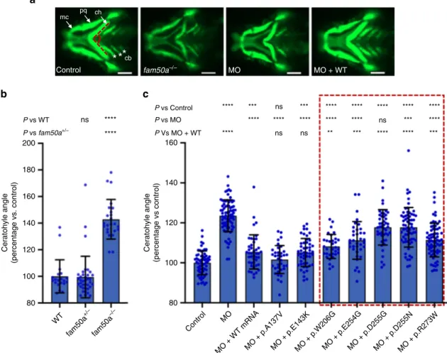
measured the ceratohyal angle as a proxy for mandibular
development. We observed a significantly wider ceratohyal angle
in fam50a KO compared to heterozygotes or WT (p
= 5.3E−13:
unpaired Student’s t-test, two-sided; repeated; Fig.
3
a, b;
Supplementary Fig. 10a and Supplementary Table 4).
Zebrafish studies show that FAM50A variants are
hypo-morphic. Cross-species in vivo complementation testing is a
sensitive and specific method to establish missense variant
pathogenicity
17. We targeted the splice donor site of D. rerio
fam50a exon 4 with a morpholino (MO), and RT–PCR showed
deletion of exons 3 and 4, and a subsequent frameshift and
premature transcript termination (Supplementary Fig. 11a–c).
We injected increasing doses of fam50a splice-blocking MO (3, 6,
and 9 ng) into
−1.4col1a1:egfp embryos and assessed ventral
cartilage at 3 dpf using the live automated imaging paradigm. We
observed a dose dependent exacerbation of craniofacial features
(p
= 0.34, 1.5E−38, and 4.3E−31 for 3, 6, and 9 ng vs control,
respectively; unpaired Student’s t-test, two-sided, repeated;
Sup-plementary Fig. 11d and SupSup-plementary Table 4), and
cartilage-patterning defects matched the fam50a KO (Figs.
2
e and
3
a;
Supplementary Fig. 10a). We rescued this phenotype by
co-injecting either 150 pg of human FAM50A WT mRNA or
mRNAs carrying common variants (gnomAD) as negative
con-trols (p.Ala137Val and p.Glu143Lys; p
= 2.4E−12, 9.9E−13, and
5.7E−11 for WT or variant rescue vs MO, respectively; unpaired
Student’s t-test, two-sided, repeated; Figs.
3
a, c; Supplementary
Fig. 10b, Supplementary Tables 3 and 4).
Next, we tested the patient-specific allelic series (p.Trp206Gly,
p.Glu254Gly, p.Asp255Gly, p.Asp255Asn, and p.Arg273Trp)
using equivalent doses of MO and mRNA across experiments
(Supplementary Tables 3 and 4). Co-injection of MO with all
patient variants resulted in a mean ceratohyal angle significantly
broader than FAM50A WT (p
= 0.0025, 0.0003, 3.3E−13,1.06E
−09, and 0.0004, respectively, unpaired Student’s t-test,
two-sided, repeated; Fig.
3
c; Supplementary Fig. 10b–e). The failure of
mutant mRNA to rescue morphant phenotype was unlikely due
to dominant toxic effects; expression of FAM50A mRNAs alone
did not result in significant defects (replicated; Supplementary
Fig. 12; Supplementary Table 4). We performed similar rescue
experiments using fam50a KO zebrafish and the formation of the
swim bladder as a qualitative criterion for morphology
18. KO
phenotypes were rescued by WT FAM50A, but were only partially
improved by a subset of three FAM50A patient variants
(Supplementary Fig. 13). In vivo complementation data,
gener-ated in either transient or stable fam50a models, supported a
partial loss of FAM50A function in Armfield XLID syndrome.
fam50a KO zebrafish have altered expression profiles. We
hypothesized that impaired FAM50A function might affect
tran-scriptional regulation or mRNA processing. These predictions are
supported by affinity-purified complexes that suggested FAM50A as
a potential spliceosome protein
7; and an in vitro study that classified
FAM50A as a candidate mRNA binding protein
6. We performed
RNA-seq analyses on total RNA obtained from WT and fam50a
KO larvae harvested at 2 dpf, prior to the onset of major
mor-phological defects (Fig.
2
; Supplementary Fig. 8). We obtained
embryos from
five pairs of fam50a
+/−(heterozygous) adults;
decapitated larvae for RNA extraction (heads) and genotyping
(tails); and combined 20 genotype-matched heads per pool (n
= 5
biological replicates of sibling-matched WT and KO; Fig.
4
a). We
generated ~37 million 50 bp single-read sequences per library and
assessed global transcriptomic profiles in KO vs WT.
WT KO 3 d.p.f. 5 d.p.f. WT KO 2 d.p.f. 3 d.p.f. Tg(huc:egfp) her4.1 2 d.p.f. 3 d.p.f. WT KO WT KO cdkn1a tp53 mdm2 WT KO 2.5 d.p.f. 3 d.p.f. 4.5 d.p.f. WT KO ***** mcpq ch cb
a
b
c
d
RNA in situ hybridization (2 d.p.f.)e
Alcian blue staining (cartilage)Fig. 2fam50a KO zebrafish display central nervous system and craniofacial patterning defects. a Representative bright field lateral images of WT and fam50a KO are shown at 3 and 5 days post-fertilization (dpf). Morphology of fam50a KO was relatively normal at 3 dpf; repeated four times. However, at 5 dpf, fam50a KO showed craniofacial abnormalities; repeatedfive times. Number of larvae assessed with similar results: 3 dpf, n = 56; 5 dpf, n = 44. b Fluorescent lateral images of WT and fam50a KO larvae on a Tg(huc:egfp) neuronal reporter. No difference between WT and fam50a KO was detected in the anterior structures at 2 dpf. However, at 3 dpf, a prominent reduction of GFP-positive neurons was observed in KO larvae. Number of larvae assessed with similar results: 2 dpf, n= 20; 3 dpf, n = 20. c Whole-mount in situ hybridization (WISH) of her4.1, a molecular marker of neurogenesis, indicated a depletion of neurons in fam50a KO compared to WT at 3 dpf; repeated. Number of larvae assessed with similar results: 2 dpf, n= 24; 3 dpf, n = 21. d Apoptosis markers are elevated in fam50a KO larvae at 2 dpf. Note the induction of tp53 and tp53 target genes, mdm2 and cdkn1a (p21) in the cell proliferative zone in the midbrain region fam50a KO larvae at 2 dpf; repeated. Number of larvae assessed with similar results: tp53, n= 41; mdm2, n = 52; cdkn1a, n= 39. e Representative ventral images of Alcian blue staining of cartilage structures shows severe defects in cartilage development that become apparent at 3 dpf. Meckel’s cartilage, mc; palatoquadrate, pq; ceratohyal arch, ch; and ceratobranchial arches, cb. Number of larvae assessed with similar results: 2.5 dpf, n= 28; 3 dpf, n = 21; 4.5 dpf, n = 14. For all images: anterior, left; posterior, right. Scale bars; 200 μm.
As expected, fam50a was the most significantly reduced coding
mRNA between the two genotypic groups. Clustering analysis
suggested a marked effect of genotype on global transcription
(Supplementary Fig. 14). This observation was supported by
gene-level expression analysis: ~12% of genes had significantly altered
levels in fam50a KO compared to WT (n
= 2804 genes, p < 0.05,
Wald
test,
FDR-corrected
using
the
Benjamini–
Hochberg method), of which ~48% were downregulated (n
=
1359 genes) and ~52% were upregulated (n
= 1445 genes; Fig.
4
b;
Supplementary Fig. 15). To test whether dysregulated genes in
fam50a KO have been implicated in human pathologies
over-lapping Armfield XLID, we overlaid our RNA-seq data with
Human Phenotype Ontology (HPO,
https://hpo.jax.org/
) and
Online Mendelian Inheritance in Man (OMIM,
https://omim.
org/
) annotations. Among genes with significantly altered gene
expression in fam50a KO, some cause clinically similar genetic
disorders. Examples include downregulated genes gss (logFC
−1.4;
p < 4E−26; Glutathione synthetase deficiency) and aaas (logFC
−1.2; p < 1.8E−12; Achalasia-addisonianism-alacrimia syndrome)
and upregulated genes ggt1b (logFC 2.9; p < 3.7E−06; ID); and
eftud2 (logFC 1.5; p < 1E−142; Mandibulofacial dysostosis; Wald
test, FDR-corrected using the Benjamini–Hochberg method;
Table
2
). Although a substantial fraction of the transcriptome is
differentially expressed in the context of FAM50A loss, we could
not identify a single altered gene driver of Armfield XLID
syndrome.
Next, we performed gene set enrichment analysis (GSEA) on
WT and fam50a KO RNA-seq data sets. The top ten pathways
with significant downregulation (familywise-error rate [FWER]
p < 0.03, Kolmogorov–Smirnov test) can be mapped to patient
phenotypes, especially neurodevelopment, brain function, and
cartilage patterning (Fig.
4
c). However, for the top ten ranking
gene sets with significant upregulation (FWER p < 0.008,
Kolmogorov–Smirnov test), we observed that nine out of ten
are involved in mRNA processing or splicing (Fig.
4
c). These data
suggest that FAM50A impairment leads to cellular compensation
P vs WT ns 200 180 160 140 Ceratohyle angle (percentage vs. control) Ceratohyle angle (percentage vs control) 120 100 80 WT Control MO
MO + WT mRNAMO + p.A137VMO + p.E143KMO + p.W206GMO + p.E254GMO + p.D255GMO + p.D255NMO + p.R273W fam50a +/– fam50a –/– 160 140 120 100 80 **** P vs fam50a+/– ****
b
Control MO MO + WTa
mc pq ch P vs Control P vs MO P Vs MO + WTc
*
*
*
cb fam50a–/– **** *** ns *** **** **** **** **** **** **** **** **** **** **** ns *** **** **** ns ns ** *** **** **** ***Fig. 3 In vivo assays indicate thatFAM50A missense variants confer a partial loss of function. a Representative ventral views of craniofacial structures imaged live using the VAST BioImager in -1.4col1a1:egfp zebrafish larvae at 3 days post-fertilization (dpf). mc, Meckel’s cartilage; pq, palatoquadrate; ch, ceratohyal arch; cb, ceratobranchial arches. Red dashed lines on control image indicate the ch angle measured to quantify altered cartilage patterning in wild-type (WT) control, KO (homozygous mutants) or morphants. 5 ng morpholino (MO) and/or 150 pg of human FAM50A mRNA were injected for all assays. Scale bars, 100μm. b Quantification of ch angle in fam50a KO, fam50a+/−(heterozygous mutant) and WT. Heterozygous and WT animals are indistinguishable; ch angle was significantly increased in fam50a KO. **** indicates p < 0.0001. ns, not significant (unpaired Student’s t-test, two-sided). See Supplementary Table 4 for exact p-values. Left to right: n= 20, 37, and 24 larvae per condition, respectively. c In vivo complementation studies indicate that FAM50A variants in XLID males are pathogenic. Quantification of ch angle as indicated by measurements of ventral images (a), and statistical comparison of variant mRNA vs WT mRNA rescue of MO effect indicates that patient-associated variants are hypomorphic (partial loss of function; red dashed box). p.Ala137Val (A136V; rs149558328) and p.Glu143Lys (E143K; rs782017549) are present in hemizygous males in gnomAD and were scored as benign; *, **, ***, **** indicate p < 0.05; 0.01; 0.001; and 0.0001, respectively. ns, not significant (unpaired Student’s t-test, two-sided). Left to right: n = 61, 78, 43, 36, 49, 32, 36, 46, 66, 69 larvae per condition, respectively; replicated. Data are presented as mean values ± standard deviation. See Supplementary Table 4 for exact p-values.
of mRNA processing effectors, arguing that neuronal and
cartilage-related biological processes are particularly susceptible
to FAM50A loss of function.
To validate a subset of RNA-seq results, we probed the
transcript levels of 25 genes using WISH at 2 and 3 dpf (Fig.
5
and Table
2
; Supplementary Fig. 16 and Supplementary Table 3).
We prioritized the top eight significantly upregulated genes
(snapc4, ice1, tp53, mdm2, mettl16, prpf3, prpf31, and eftud2), the
majority of which comprise the splicing machinery. We validated
additional mRNA splicing effectors (n
= 7: prpf8, prpf4, prpf6,
snrnp200, snrpe, sf3b4, and eif4a3)
19–25. Many of these genes
involved in spliceosomal function are related to human disorders,
2 d.p.f. fam50a+/– X fam50a+/– RNAseq n = 5 replicates (20 heads/pool)
–3
–2
–1
0
1
2
3
Normalized enrichment score (mutant vs control)
a
c
Genotyping Step 1 Step 2 Head RNA extraction DNA extraction Genotype matched Tail Step 3 Differential expression Gene set enrichment analysis HPO and OMIM disease annotation Splicing analysis 88% 12% 48% 52% Normal Dysregulated Downregulated Upregulatedb
Alternative splicing events GO terms (alternative splicing)
Alternative 3′ Alternative 5′ Mutually exclusive exon Retained intron Skipped exon
d
e
49% 4% 6% 10% 30% 70% 20% 7% 4% (0) Collagen trimer (0) Extracellular matrix structural constituent (0.002) Neuron–neuron synaptic transmission (0.003) Glutamate receptor signaling pathway (0.008) Biological adhesion and cell adhesion (0.009) Protein DNA complex (0.019) Glutamate receptor activity (0.025) Synaptic membrane (0.027) Negative regulation of cell motility and cell migration (0.027) Ion channel complexmRNA metabolic process (0)
mRNA splicing via transesterification reactions, spliceosome (0) mRNA processing (0)
RNA processing (0) RNA splicing (0) Spliceosomal complex (0)
Small nuclear ribonucleoprotein complex (0) Ribonucleoprotein complex (0)
DNA templated transcription initiation (0.001) Positive regulation of peptidase activity(0.008)
N N
especially those with craniofacial or ocular phenotypes
26,27. We
also validated
five transcripts that are significantly depleted in the
fam50a KO (gss, aaas, vwa7, and eda); and one transcript
involved in XLID (huwe1)
28but not significantly altered in
fam50a KO. For all 18 genes, the RNA ISH data confirmed our
transcriptomic profiling (Table
2
and Fig.
5
; Supplementary
Fig. 16). In addition, we queried the RNA-seq data for genes
assessed via WISH during the initial characterization of the
fam50a KO. Accordingly, p53 pathway markers tp53, cdkn1a and
mdm2 showed significantly augmented expression (p < 9.8E−103,
Wald test, FDR-corrected using the Benjamini–Hochberg
method;
≥2-fold increase; Fig.
2
d). Further, her4.1, pcna, and
ccnd1 WISH showed no significant differences, consistent with
the spatiotemporal expression studies at 48 hpf (Fig.
2
c and Table
2
; Supplementary Figs. 7 and 8).
fam50a KO are enriched for mRNA mis-splicing events. To
probe the involvement of FAM50A in mRNA splicing, we applied
replicate multivariate analysis of transcript splicing (rMATS), a
statistical method that can detect: alternative 5′ splice sites;
alternative 3′ splice sites; retained introns; mutually exclusive
exons; or skipped exons from multiple replicate samples
29. We
queried splice sites and found representation of significantly
augmented or depleted splicing events in each of the
five
cate-gories. However, we noticed an uneven distribution of aberrant
splicing events by splicing category that withstood an
FDR-corrected p < 0.05 threshold (likelihood-ratio test; Fig.
4
d).
Aberrant 3′ splicing was predominantly affected (n = 235, 49%),
followed by exon skipping (n
= 145, 30%), retained intron (n =
50, 10%), mutually exclusive exons (n
= 29, 6%), and alternative
5′ splicing (n = 18, 4%).
We applied the DAVID functional annotation clustering tool
(
https://david.ncifcrf.gov/
) to the rMATS output to identify
enrichment of transcript modules impacted significantly by
aberrant splicing. The majority of significant gene category hits
(uncorrected p < 0.05, Fisher’s exact test) also impacted
alter-native 3′ splice sites (n = 32, 70%; Fig.
4
e; Supplementary
Table 5). There were only six Bonferroni-corrected (p < 0.05,
Fisher’s exact test) gene ontology (GO,
http://geneontology.org/
)
terms cumulatively spanning all splicing categories. These GO
groups were either impacted in both alternative 3′ splicing and
exon skipping: RNA binding (GO:0003723); or were exclusive to
alternative 3′ splicing events: nucleus (GO:0005634); DNA
binding (GO:0003677); regulation of transcription,
DNA-templated (GO:0006355); nucleic acid binding (GO:0003676);
and transcription factor complex (GO:0005667). In sum, mRNA
splicing analysis uncovered aberrant events biased toward 3′
alternative splicing or exon skipping. These alterations occur late
in mRNA splicing, consistent with phenomena expected to be
downstream of a C-complex impairment
30.
Transcriptomic profiling of a zebrafish eftud2 mutant model
demonstrated global RNA splicing deficiency with concomitant
p53-dependent apoptosis
31, concordant with our RNA ISH and
RNA-seq data. tp53 was significantly upregulated in fam50a KO,
similar to its downstream target mdm2, compared to WT
(Table
2
). To test whether p53 pathway activation is correlated
with apoptosis, we examined fam50a morphants via TUNEL
staining (Supplementary Table 4). TUNEL positive cells were
significantly augmented in an anterior region of interest (ROI)
(p
= 4.8E−06, unpaired Student’s t-test, two-sided). This defect
was rescued by co-injection of human WT FAM50A mRNA (p
=
1.5E−06, unpaired Student’s t-test, two-sided; Supplementary
Fig. 17a, b). Cell-cycle progression was also increased in fam50a
morphants as determined by significantly increased
phospho-histone H3 immunostaining, a marker of G2/M transition (p
=
1.5E−07; unpaired Student’s t-test, two-sided; Supplementary
Fig. 17c, d; Supplementary Table 4). To examine whether the p53
pathway is a cause or effect of the fam50a KO phenotype, we
crossed fam50a
+/−and tp53
−/−(homozygous) mutant lines.
fam50a KO and fam50a/tp53 double KO displayed similar
phenotypes at 5 dpf (Supplementary Fig. 18). These data reinforce
the involvement of p53 independent apoptosis in the fam50a KO
phenotype that is likely correlated, directly or indirectly, with
aberrant mRNA splicing.
Patient LCLs have mRNA expression and splicing defects. To
test for transcriptional dysregulation and spliceosome disruption
in LCLs derived from patients with Armfield XLID, we performed
RNA-seq (K9648, p.Trp206Gly and K9656, p.Glu254Gly: Fig.
1
b,
c and Table
1
). Differential expression analysis of triplicates
showed significant dysregulation in a substantial fraction of the
transcriptome in affected individuals compared to control (~38%;
FDR-corrected p < 0.05, Wald test, FDR-corrected using the
Benjamini–Hochberg method). Mapping of dysregulated
tran-scripts in human to zebrafish RNA-seq data showed that 42% of
upregulated and 39% of downregulated human transcripts,
respectively, were also significantly altered in the fam50a KO.
GSEA identified the spliceosome as the fourth most significantly
enriched functional group (FWER p < 0.0001, Kolmogorov–
Smirnov test; Supplementary Fig. 19). Similar to fam50a KO, we
noticed an uneven distribution of aberrant splicing events by
category that withstood an FDR-corrected p < 0.05 threshold
(likelihood-ratio test). Exon skipping was predominantly affected
(n
= 443, 53%), followed by retained intron (n = 172, 20%),
alternative 3′ splicing (n = 136, 16%), alternative 5′ splicing (n =
75, 9%), and mutually exclusive exons (n
= 15, 2%). We
per-formed DAVID analysis using rMATS data, however none of the
Bonferroni-corrected functional groups identified in LCLs could
be mapped back to the gene sets identified in the zebrafish mutant
rMATS analysis (Fisher’s exact test, Supplementary Table 6).
These data offer consistency in upregulation of the spliceosome,
with modest differences in alternative splicing events possibly
because lymphocytes are not known to be a site of pathology in
Armfield XLID syndrome.
FAM50A interacts with spliceosome U5 and C-complex
pro-teins. To validate a potential affiliation for FAM50A in the
Fig. 4 RNA-seq analysis revealed mRNA splicing defects infam50a KO zebrafish. a Schematic describing the RNA-seq experiment from sample preparation to data analysis. Steps 1 and 2 describe sample collection and preparation, whereas step 3 indicates the RNA data analysis and interpretation. Each replicate pool (n= 5 per genotype) contained total RNA from 20 genotype-matched larval heads at 2 days post-fertilization (dpf). fam50a+/− (heterozygous mutant).b Pie charts representing differential expression analysis results for KO vs WT. Transcripts with FDR-corrected p < 0.05 were included (Wald test, FDR-corrected with the Benjamini–Hochberg method). c Gene set enrichment analysis was performed using normalized enrichment score for KO vs WT. The top ten significantly disrupted pathways (depleted or augmented) are plotted along the x axis. Downregulated gene sets, orange; upregulated gene sets, blue. p-values (FDR-corrected) are indicated in parentheses (Kolmogorov–Smirnov test). d Pie chart representing percentage of alternative splicing events (by category) that are enriched in fam50a KO. p < 0.05, likelihood-ratio test with FDR correction.e Pie chart representing the distribution of GO terms impacted by discrete alternative splicing events (by category) in fam50a KO. p < 0.05, Fisher’s exact test.Table
2
Validation
of
RNA-seq
data
using
whole-mount
in
situ
hybridization
in
zebra
fi
sh.
Gene name Descr iption Human pheno type RNA-s eq log 2 (FC) RNA-seq p -value In situ hybridiz ation fam50a Family with seque nce similar ity 50 , me mber A ND − 3 3.47E-233 Downregulat ed gss Glu tathione syn thetase Glu tathione syn thetase de fi cienc y − 1.39 4.08E-26 Downregulat ed aaas Ac halasia, adrenocortical insuf fi cienc y, alacrim ia Achala sia-a ddisonianism-a lacrimia syn drome − 1.21 1.83E-12 Downregulat ed huwe1 HEC T, UBA and WW E domain cont aining 1 Menta l retar dation, X-li nked syn dromic, Turner type − 0.14 0.71 Downregulat ed vwa7 von Willebrand fact or A domain cont aining 7 ND − 0.99 1.07E-49 Downregulat ed eda Ect odysplasin A Ectode rmal dysplasia, X-linked recess ive; tooth agene sis − 0.60 0.02 Downregulat ed pcna Pr oliferating cell nucl ear antige n Atax ia-telangie ctasia-like disorder 2 − 0.08 0.72 Downregulat ed her4. 1 Hair y -rel ated 4, tand em duplicate 1 ND − 0.07 0.9 Downregulat ed ccnd1 Cycli n D 1 N D − 0.01 0.97 Downregulat ed snapc4 Sma ll nucl ear RN A act ivating comp lex, poly peptide 4 ND 2.79 9.16E-28 1 Upregulated cdkn1a Cycli n-de pendent kina se inhibitor 1A ND 2.40 9.87E-103 Upregulated ice1 KIA A0947-like (H. sapien s) N D 2.29 9.81E-22 0 Upregulated mettl1 6 Me thyltransferase-like 16 ND 2.23 4.93E-156 Upregulated tp53 Tumo r protein p53 ND 2.16 3.14E-21 7 Upregulated mdm 2 MDM2 oncogene, E3 ubiquitin protein liga se ND 1.98 3.88E-163 Upregulated prpf31 PRP31 pre-m RNA processing factor 31 homolog (y east) Retinit is pigme ntosa 1.75 1.25E-147 Upregulated prpf3 PRP3 pre-mRNA proc essing factor 3 homolog (y east) Retinit is pigme ntosa 1.7 5.23E-1 49 Upregulated eftud 2 Elong ation factor Tu GT P binding domain cont aining 2 Mand ibulofacial dysostosis 1.45 1.01E-142 Upregulated prpf4 PRP4 pre-mR NA proc essi ng factor 4 homolog (y east) Retinit is pigme ntosa 1.38 3.95E-74 Upregulated snrpe Sma ll nucl ear ribon ucleoprotein poly peptide E Hypot richosis 1.30 1.16E-15 Upregulated snrnp2 00 Sma ll nucl ear ribon ucleoprotein 20 0 (U5) NA 1.09 5.49E-63 Upregulated prpf8 Pr e-mRNA proc essi ng factor 8 Retinit is pigme ntosa 0.87 2.46E-33 Upregulated prpf6 PRP6 pre-mRN A proc essi ng factor 6 homolog (S. cere visiae ) Retinit is pigme ntosa 0.72 6.35E-49 Upregulated eif4a3 Euka ryotic translation init iation fact or 4A3 Robin seque nce with cleft mandible and limb anomalies 0.55 9.95E-2 1 Upregulated p -values determined with a Wald test. FC, fold change; ND, none described.spliceosome C-complex
7, we performed a FAM50A pulldown
assay followed by mass spectrometry (nanoscale liquid
chroma-tography with tandem MS). We transfected HEK-293T cells with
a C-terminally tagged dual streptavidin and
flag epitopes on a
FAM50A PiggyBac transposon system and expanded cells under
puromycin selection. Strepavidin capture identified a combined
repertoire of 99 FAM50A interactors with a SAINT probability
cutoff value >0.7 (Supplementary Data 1) compared to controls in
two independent replicates. The top 20 functional groups involve
RNA processing, with U5 snRNA binding (GO:0030623) ranked
with
the
most
significant fold-enrichment (p = 1.3E−02;
Kolmogorov–Smirnov test, Supplementary Table 7).
To show direct interaction of FAM50A with bona
fide
spliceosome effectors, we performed co-immunoprecipitation
(co-IP) assays. We transiently transfected U-87 glioblastoma
cells with V5-tagged EFTUD2 and DDX41 plasmids, which are
part of the spliceosome U5 and C-complex, respectively
7(Fig.
6
a,
b). Immunoblotting using anti-V5 and anti-GAPDH antibodies
detected the overexpressed tagged proteins in input lysates
(Fig.
6
c, d). Next, we immunoprecipitated proteins with
anti-FAM50A antibody in transfected and negative control samples.
Western blot against FAM50A in co-IP lysates discovered the
pulled down FAM50A protein in all samples. Immunoblot with
anti-V5 in IP lysates detected both Co-IP partners in their
respective transfected samples but not in negative controls,
indicating a specific physical interaction of FAM50A with
spliceosome binding partners that are active during the
two-step splicing reaction (Fig.
6
c, d).
Discussion
We report partial loss-of-function missense variants in FAM50A
as the genetic basis of Armfield XLID syndrome. Our work
epi-tomizes the challenges of understanding rare disease
pathogen-esis. Using candidate gene sequencing, we identified the causal
FAM50A variant in the original Armfield XLID syndrome family
in 2001. Next-generation sequencing technology and data sharing
platforms were required to identify four additional cases with
FAM50A variants several years later. Even with bolstered support
for genetic causality, this work required a vertebrate model to
gain insight into variant pathogenicity and cellular mechanism.
Our experience is not unique. Of the estimated 9000 Mendelian
phenotypes that have been described
32, a substantial proportion
of gene-phenotype pairs identified in the last 5 years required
partnering of a rare human
finding with a model organism or
relevant in vitro functional assay
33.
XLID conditions range from isolated ID to multi-organ
dis-orders. Males with FAM50A variants overlap phenotypically with
other XLID syndromes presenting with seizures and dysmorphic
facial features (Table
1
). We noted variable head circumference
with an inconsistent facial phenotype. However, Armfield XLID
syndrome also includes postnatal growth retardation and ocular
findings, which are less common among XLID syndromes
3,34.
Affected males with FAM50A variants had normal intrauterine
growth but statural growth slowed postnatally, eventually falling
more than 2 SD below the mean for height. Small hands and feet
are also signature features of this cohort. Ocular anomalies were
pervasive with all patients having strabismus, keratoconus, and
anterior chamber anomalies. In sum, this constellation of
cog-nitive, craniofacial, growth, and ocular features define Armfield
XLID syndrome as a distinct clinical entity.
To investigate FAM50A function, we generated zebrafish KO
models, however, fam50a KO zebrafish larvae are not viable long
term. Homozygous mutants for two independent loss-of-function
alleles exhibited defects impacting neuronal proliferation and
craniofacial patterning, which mirror the phenotypes in FAM50A
mutation-bearing males (Figs.
1
and
2
; Table
1
). However, there is
a key difference between the KO and Armfield XLID syndrome
cases: strength of allele effect. Although we were limited to testing
variant pathogenicity using either a quantitative comorbid feature
(craniofacial patterning) or a clearly defined qualitative
mor-phological defect (swim bladder), data generated through in vivo
complementation assays suggested that males with FAM50A
WT KO prpf8 prpf3 prpf4 prpf6 snrnp200 prpf31 eftud2
sf3b4 snrpe eif4a3 snapc4 ice1
WT
KO
Fig. 5 Major spliceosome effectors are upregulated infam50a KO zebrafish. Representative lateral images of whole-mount in situ hybridization performed on 3 day post-fertilization larvae validate RNA-seq data (Table2) from fam50a KO (homozygous mutant) vs wild-type (WT). Number of embryos assessed with similar results: prpf3, n= 11; prpf4, n = 10; prpf6, n = 11; prpf8, n = 10; prpf31, n = 11; eftud2, n = 10; snrnp200, n = 11; sf3b4, n = 11; snrpe, n= 11; eif4a3, n = 9; snapc4, n = 11; ice1, n = 10. Scale bars; 200 μm.
variants have some residual protein function (Fig.
3
;
Supple-mentary Fig. 13). Even without variant testing data from a direct
neurodevelopmental phenotype, we would still expect humans
with null FAM50A variants to be embryonic lethal, as supported
by a low observed/expected (o/e) constraint metric in >125,000
exomes in gnomAD (o/e
= 0.06).
mRNA splicing is a dynamic process that involves intron
branching and exon ligation
30(Fig.
6
a). Electrophoretic
separa-tion of the spliceosome initially identified complexes associated
with each splicing step
35,36. Proteomics and cryo-electron
microscopy have refined further this process enabling the
con-tents and relative ratios of spliceosome complexes to be
140 260 kDa 40 50 100 140 90 kDa 40 50 100 140 90 kDa 35 40 140 260 35 40 kDa IP: Anti-FAM50A IP: Anti-FAM50A Input (50 μ g Lysate) Input (50 μ g Lysate) Anti-V5 (EFTUD2) Anti-GAPDH Anti-V5 (DDX41) Anti-GAPDH Anti-V5 (EFTUD2) Anti-FAM50A Anti-V5 (DDX41) Anti-FAM50A pcDNA 3.1/nV5-EFTUD2 +–
pcDNA 3.1/nV5-DDX41+
–
pcDNA 3.1/nV5-DDX41+
–
pcDNA 3.1/nV5-EFTUD2+
–
+
–
80% 20% 100%c
d
a
pcDNA 3.1/nV5_EFTUD2 OR pcDNA 3.1/nV5_DDX41 60 h.p.t. U87 cells 50%–70% confluency TransfectionImmunoblot using anti-V5 and anti-FAM50A antibodies IP with anti-FAM50A antibody
b
ILS mRNA Complex C FAM50A DDX41 BΔU1 Pre-spliceosome U4/U6.U5tri-snRNP 3′SS BP A 5′SS Pre-mRNAExon Intron Exon
Complex B′ Complex B
Lariat
Two-step splicing reaction
U2 U5 U5 U5 EFTUD2 EFTUD2 EFTUD2 U2 U2 U2 U2 U6 U6 U6 Step 1 Step 2 U6 U5 EFTUD2 U1 U1 U1 U1 U4 U4Fig. 6 FAM50A interacts with U5 and C-complex proteins. a Schematic representation of the two-step splicing reaction (adapted from ref.70). EFTUD2 (U5); FAM50A and DDX41 (C-complex).b Graphical representation of semi-native co-immunoprecipitation assay. Candidate interactors were overexpressed in U-87 cells, harvested in immunoprecipitation (IP) lysis buffer at 60 h post-transfection and immunoprecipitated with anti-FAM50A antibody. The interaction partners were detected in IP lysate using anti-V5 and anti-FAM50A antibodies, respectively.c, d Western blot of proteins after co-immunoprecipitation. Top: total protein lysate input (50μg/lane) was migrated on 4–15% polyacrylamide gels to detect EFTUD2 (c) or DDX41 (d) using anti-V5 mouse monoclonal antibody. GAPDH was used as loading control. Bottom: Anti-FAM50A antibody was used to immunoprecipitate native FAM50A protein in total input lysate of 2.3 mg/condition (c) or 3.5 mg/condition (d). The IP lysate was separated in two parts (20% and 80%) and migrated independently on 4–15% polyacrylamide gels. The proteins of interest with predicated band sizes are indicated with black arrows. Plus and minus signs indicate presence and absence of relevant plasmids, respectively. EFTUD2 and DDX41 were detected in independent experiments using protein lysates derived from replicate batches of U-87 cells.