A THESIS
FOR THE DEGREE OF DOCTOR OF PHILOSOPHY
REPRODUCTION OF THE ANTARCTIC CLAM,
LATERNULA ELLIPTICA COLLECTED FROM
MARIAN COVE IN KING GEORGE ISLAND,
ANTARCTICA
DO-HYUNG KANG
DEPARTMENT OF MARINE BIOLOGY GRADUATE SCHOOL
CHEJU NATIONAL UNIVERSITY
REPRODUCTION OF THE ANTARCTIC CLAM,
LATERNULA ELLIPTICA COLLECTED FROM
MARIAN COVE IN KING GEORGE ISLAND,
ANTARCTICA
DO-HYUNG KANG
(Advised by Professor Kwang-Sik Choi)
A thesis submitted in partial fulfillment of the requirement for the degree of
DOCTOR OF PHILOSOPHY
August 2006
This thesis has been examined and approved by
DEPARTMENT OF MARINE BIOLOGY GRADUATE SCHOOL
CONTENTS ạⱬ 㟈㚱……….. 1 LIST OF FIGURES……….. 3 LIST OF TABLES………..……….. 8 SUMMARY……….……….. 9 INTRODUCTION..……… 12
Part I. Quantitative assessment of reproductive condition of the Antarctic clam, Laternula elliptica (King & Broderip), using image analysis……… 18
Abstract…………...……… 18
Materials and Methods…..……… 19
Sampling efforts………... 19
Histological preparation and analysis of gonadal tissues ……... 19
Results...………...………... 21
Quantitative assessment of L. elliptica reproductive condition . 21 Comparison of the quantitative indices in the clams from the two austral summers ………... 22
Discussion..………...………... 23
Computer-based image analysis of the reproductive condition of L. elliptica ………... 23
Reproductive traits of L. elliptica at Marian Cove of ………... 25
Oocyte size ……….………. 25
Size and putative age at first reproduction ……….……. 26
Variation in reproductive output with body size ………. 27
References..………...……….. 37 (Continued)
Part II. Annual gametogenesis of the Antarctic clam, Laternula
elliptica from Marian Cove ………... 42
Abstract…………...……… 42
Materials and Methods…..……… 43
Sample collection ………….……….. 43
Histological preparation ……….….... 44
Analysis of annual gametogenesis ………. 44
Results...………...………... 45
Seawater temperature and total chlorophyll ……….. 46
Microscopic observation of gonad ………..….. 47
Annual variations of MI ………..….. 49
Annual variations of oocyte diameters and growth .………….. 50
Resorption process of the relict ova ……….. 53
Discussion..………...……….. 54
Large lecithotrophic eggs of L. elliptica ……… 54
Food availability and the clam reproduction ……….. 55
Resorption process for regulating nutritional energy source …. 60 References..………...……….. 63
ACKNOWLEDGEMENT..………...……… 87
DEDICATIONG
G G G G G G GThis dissertation is dedicated to
my wife, Ae-Jung, and daughters, Seo-Hyun and Seo-Young,
my mother and father- and mother-in-law, and my family
GạⱬG 㟈㚱G G
1. 䀨䘜䉤䞈ㇵ㷌⫠ὤⷉ㡸 㢨㟝䚐 ⇜Ἥ䆤⢔㦤ᵐ, Laternula elliptica (King & Broderip)㢌 ㈑㐑㋀ⵐ␠ 㥉⣽㡸 㠸䚐 㜤ẠG
1.1.㢨 㜤Ạ⏈ ⇜Ἥ 䞌ᷱⷴ䞈㜄 ♤⪬ 䚨㛅㤴㉐㈑Ɒ ⷴ┍㜄 䚐 㣙ὤḴ㽕 㜤Ạ㢌G 㢰䞌㡰⦐㉐SG 䚐ⴰạG ㉬㦹ὤ㫴ᴴG 㠸㾌䚐G 䇭㦤㫴㉠㢌 ⫼⫠㙼 ㋀⬀ 㦤䚌㜄 ㉐㐑䚌⏈ ⇜Ἥ䆤⢔㦤ᵐ(Laternula elliptica)⏈ SCUBA⪰ 㢨㟝䚌㜠 1996≸ 12㠈Ḱ 1998≸ 1㠈㜄 㵸㬅䚌㜴ḔSG 㦤㫵䚍㤵G ⵝⷉḰG 䀨䘜䉤䞈ㇵ㷌⫠ὤⷉ㜄G 㢌䚨G ㈑㐑㋀G ⵐ␠Ḱ㥉G ⺸㉑㡸G 㥉⣽䞈G 䚌㜴␘UG G 1.2.⇜Ἥ㦤ᵐ ⇐㋀㋀㜱 ⇨ ⇐⯜㉬䔠㢌 ⮨㤵⽸ 㫴㍌(the percentage egg area
within a follicle, FI)㝴 䓽ἔ ⇐⯜㉬䔠 㫵ᷱ 㫴㍌(mean oocyte diameter, MOD)⏈ ㉐⦐ ␘⪬ 㵸㬅 㐐ὤ㢬 1996≸ 12㠈Ḱ 1998≸ 1㠈㢌 ㈑㐑㋀ⵐ ␠ 㵜㢨⪰ 㡔㢌䚌᷀ ⇌䇴⇼㫴⬀, 㢰ⵌ㤵㢬 ㉥㍍⓸ 㫴㍌(maturity index, MI)㝴 㤸㷨㡕㫼⺴ ⮨㤵㜄 䚐 ㈑㐑㋀ ⮨㤵⽸ 㫴㍌(percentage gonad area, PGA)⏈ ▄ 㐐ὤ㢌 ⇜Ἥ㦤ᵐ㜄 Ḵ㵤䚐 ㈑㐑㋀ ⵐ␠㢌 㵜㢨⪰ ⇌䇴 ⇨㫴 ⯯䚌㜴␘. 㢨⤠䚐 ᷤḰ⏈ ⇜Ἥ㦤ᵐ ㈑㐑㋀ ⵐ␠㡸 䑄㥉䚌⏈ 㫴㍌ ⦐㉐ FI㝴 MOD⏈ 㐔⧤㉥Ḱ ᵄ⓸⮨㜄㉐ ␘⪬ ▄ 㫴㍌⸨␘ ⋆㚌␘. 1.3.㈑㐑㋀ 㢨㫴⪰ 㢨㟝䚐 ㈑㐑㉬䔠㢌 ⮨㤵㽕㥉ⵝⷉ(planimetric technique) 㡴 ㇠㟝㢨 㟝㢨䚔 ⬀ 㙸⏼⢰ 㵸㬅 ᵐ㷨㍌ᴴ ⬂㡸 ᷱ㟤㜄⓸ ⒤㢨䉤 㷌⫠ ㋁⓸ᴴ ⾔⪨Ḕ, 㢰ⵌ㤵㢬 㫴㍌☘⸨␘ 㥉⣽㤵㢬 㥉⸨⪰ 㥐ḩ䚌⦐ ⇜Ἥ䆤⢔㦤ᵐ L. elliptica ⶼ㐑 㜤Ạ㜄 䟜㡜㤵㡰⦐ 㢨㟝╔ ㍌ 㢼␘. 1.4.䏭䢼 76-85 mm 䆠ὤ㢌 ⇜Ἥ䆤⢔㦤ᵐ ᵐ㷨☘㡴 ␘⪬ 䆠ὤ㜄 ⽸䚨 ⋆㡴 ⶼ㐑⣽㡸 ⇌䇴⇨⦐ ⇜Ἥ䆤⢔㦤ᵐ ⶼ㐑䑜䉨 ⷴ┍㢌 㣙ὤḴ㽕㡸 㠸䚨 ㉐⏈ 㢨 䆠ὤ㢌 ᵐ㷨☘㡸 㨰⦐ ㇠㟝䚌⏈ ᶷ㢨 ⸨␘ 䟜㡜㤵㢰 ᶷ㡰⦐ 䑄␜═␘. 2. ⇜Ἥ 䇭㦤㫴㉠㢌 ⫼⫠㙼 ㋀⬀ 㦤䚌㜄 ㉐㐑䚌⏈ ⇜Ἥ䆤⢔㦤ᵐ, Laternula elliptica㢌 㜤㩅 ⶼ㐑 䏭㉥ 2.1. ⇜Ἥ䆤⢔㦤ᵐ㢌 㜤㩅 ⵤ㟤㣄䝉㉥Ḱ㥉Ḱ ᷸㤼ⷸ ⶼ㐑䏭㉥㡸 㜤Ạ䚌ὤ 㠸䚨 1998≸ 2㠈⺴䉤 2000≸ 1㠈ᾀ㫴 2≸ ┍㙼 SCUBA⪰ 㢨㟝䚌㜠 ᵐ 㷨☘㡸 㵸㬅䚌㜴␘. ⇜Ἥ䆤⢔㦤ᵐ ㈑㐑㋀ⵐ␠, ㉥㍍ ⵃ ⢴ ☥㡴 㠸 Part 1㜄㉐ ᵐⵐ䚐 ⵝⷉḰ 㢰ⵌ㤵㢬 ⵝⷉ㡸 㢨㟝䚌㜠 㥉㉥㤵, 㥉⣽㤵㡰 ⦐ 㽕㥉䚌㜴␘. G X
2.2. ㈑㐑㋀ⵐ␠ 㫴㍌㢬 FI㝴 MI⏈ 1998≸ 10㠈⺴䉤 11㠈ᾀ㫴 㡔㢌䚌᷀ 㫑 ᴴ䚌㜠 12㠈㜄 㺐Ḕ㾌⪰ ⇌䇴⇼Ḕ, 1999≸ 1㠈Ḱ 2㠈㜄 Ἵᷝ䚌᷀ ᵄ㋀䚼 ␘. 䝉 ㉥㍍⇐㢌 䆠ὤ(100-190µm)⪰ Ḕ⥘䚼㡸 ⚀, ⇜Ἥ䆤⢔㦤ᵐ⏈ ⇐ 䞝㜵㛅䝉(lecithotrophic) 㡔㈑Ḱ㥉㡸 ᶤ㾌⏈ ᶷ㡰⦐ 䑄␜═␘. ⢴ 䟸 ⇐ ㋀㜄 ⇜㙸 㢼⏈ 㣈㦨⇐☘㡴 䜼Ạ㉬䔠㜄 㢌䚐 㐑㉬䔠㣅㟝㜄 㢌䚨 㜤㩅 㣠䢕㍌╌㛼Ḕ, 2㠈Ḱ 3㠈㜄 㬅㩅㤵㡰⦐ ⇌䇴⇠␘. 㢨⤠䚐 㣠䢕㍌ Ḱ㥉㡴 ⢴ 䟸㜄 㢰㛨⇌⏈ 㷨⇨ ␜ⵥ㫼㠄 㣠㇠㟝 ὤ㤸㢌 㢰ⵌ㤵㢬 䜸ㇵ㡰⦐㉐, 㜤㩅 ⭭㢨ḩἽ㢨 㜠⪸㷔㜄 Ἥ䢼 㥐䚐═ ⇜Ἥ 㜤㙼㜄 ㉐㐑䚌⏈ ⇜Ἥ䆤⢔ 㦤ᵐ㢌 䟜㡜㤵㢬 㷨⇨ 㜄∼㫴 Ḵ⫠⪰ 㠸䚐 㤵㢅ὤ㤸㡰⦐ 䑄␜═␘. 2.3. ⇜Ἥ䆤⢔㦤ᵐ㢌 ㈑㐑㋀ ㉥㍍㡴 1998≸Ḱ 1999≸ ⯜▄ ⫼⫠㙼 ㋀⬀ ⇨ 䙀⣅䆠䋘 ⶼ㐑 㐐ὤ㝴 ⬒Ɒ⫤ 10㠈⺴䉤 12㠈ᾀ㫴 㬅㩅㤵㡰⦐ 㢨⨜㛨 㦀␘. 䚐䓬 ㍌㩅㡰⦐⺴䉤㢌 ⭭㢨ḩἽ㢨 ㇵ㤵㡰⦐ ⋆㚌⒌ 1998≸ 10㠈 㢌 ㈑㐑㋀ ㉥㍍㢨 1999≸ 10㠈⸨␘ ⾔⪨␘⏈ ᶷ㡸 Ḕ⥘䚼㡸 ⚀, ㍌㩅㢌 ⭭㢨㠄㡴 ⇜Ἥ䆤⢔㦤ᵐ㢌 ㈑㐑㋀ ㉥㍍Ḱ ⢴㜄 ᷤ㥉㤵㢬 㜡䚔㡸 䚌⏈ ᶷ㡰⦐ 䑄␜═␘. 2.4. ⇜Ἥ䆤⢔㦤ᵐ, L. elliptica㢌 ㈑㐑㋀ ⺸㉑Ḱ ⵤ㟤㣄䝉㉥Ḱ㥉㡸 2≸ᴸ Ḵ 㵤䚐 ᷤḰ, ㈑㐑㋀㢌 ㉥㍍Ḱ ⢴㡴 㜤㩅 㢰㛨⇌㫴 㙾Ḕ ⇜Ἥ 㜠⪸㷔㢌 㬛㡴 ὤᴸ㜄 㢨⨸㫴⏈ ᶷ㡰⦐ 䑄␜═␘. 㢨⤠䚐 ⶼ㐑䑜䉨㢌 ⟐⥫䚐 ᷸ 㤼㉥㡴 ⵤ㟤㣄䝉㉥Ḱ㥉㜄㉐⓸ Ḵ㵤╄␘: 1) 䢕㍌ὤ㝴 䡨㫴ὤ㢌 㨰㟈 㐐 ὤ⏈ 2㠈⺴䉤 3㠈, 2) ㈑㐑㋀ⵐ␠Ḱ ㉥㍍㢌 㨰㟈 㐐ὤ⏈ 10㠈⺴䉤 12㠈, 3) ⢴㡴 12㠈⺴䉤 2㠈ᾀ㫴 㨰⦐ Ḵ㵤╄␘. G G Y
LIST OF FIGURES
Fig. 1. The Antarctic clam, Laternula elliptica. …..……….17 Fig. 2. Sampling location of L. elliptica at Marian Cove. ………..….28 Fig. 3. Cross-section of L. elliptica. Scale bar = 1 cm.…..…..……….29 Fig. 4. Photomicrographs of ovaries from L. elliptica collected in
December 1995 (scale bar = 200 µm). (A) Developing stage:
ovary contains a small oogonium (OG), previtellogenic oocytes
(PVO), and vitellogenic oocytes (VO) along the follicle wall
(FW). Follicles expand into the vesicular connective tissue
(VC) and few gaps are found between follicles. (B) Fully
developed stage: large eggs are attached to germinal epithelium
and the follicle is completely filled with ova (O). (C) Spawning
stage: partially spawned ovaries contain loosely packed relict
ova (RO) and spaces in the lumen (LM). The follicle wall is
noticeably thinner. (D) Spent stage: follicle wall is withered
and some relict ova still remain within follicles. Early
vitellogenic oocytes (EVO) are attached to follicle wall. (E)
Absorbing stage: ovary degenerates and relict ova are
reabsorbed (AO) into the thin follicle epithelium by phagocytic
activities.………...…..……….30
Fig. 5. Photomicrographs of testes (scale bar = 200 µm). (A)
Developing stage: columns of spermatozoa (SZ) move to center
G
[
of a growing testis with a thick follicle wall (FW). Most of the
lumen is filled with dense radiating bands of spermatozoa (CT,
connective tissue). (B) Fully developed stage: mature testes are
filled with spermatocytes (SC) and spermatozoa, and are
largely devoid of nutritive tissue. Dense bands of spermatozoa
fill the majority of the lumen and often spiral into its center. (C)
Spawning stage: partially spawned testes contain spaces
vacated by spawned spermatozoa and nutritive phagocytes (RS,
relict sperm). (D) Spent stage: spent spermaries (SS) are
partially empty, although relict spermatozoa may be present.
Follicle wall is thin and testis volume is small (CFT, connective
fibromuscular tissue; DG: digestive gland)..…..…..…………32
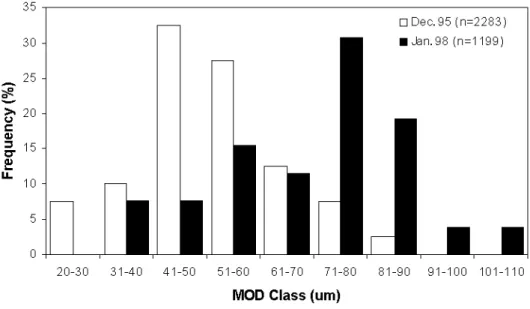
Fig. 6. Frequency distribution of MOD in December 1995 and January
1998 (n: number of oocytes measured).…..…..………...33
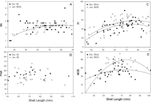
Fig. 7. Comparisons of MI, PGA, FI and MOD between L. elliptica
collected in December 1995 and January 1998. There was no
significant difference in the MI and PGA between the two years
(A and B), while the FI and MOD values were significantly
higher in January 1998 than in December 1995 (C and D).
Regression lines: (A) MI (Jan. 98) = -0.000864SL2+ 0.142SL
- 2.40, r2 = 0.41, n=42, P < 0.001; No significant size relation
was found for both years. (C) FI (Dec. 95) = -0.0133SL2+ 2.26SL - 43.35, r2 = 0.36, n=49, P < 0.001; FI (Jan. 98) =-0.0158SL2+ 2.58SL – 46.88, r = 0.19, n=42, P < 0.05. (D) 2 MOD (Dec. 95) = -0.0132SL2+ 2.024SL – 21.82, r2 = 0.15, n=38, P < 0.05; MOD (Jan. 98) = -0.0405SL2+ 6.40SL - 172.2, r2 = 0.39, n=26, P < 0.01.…..…..……….34
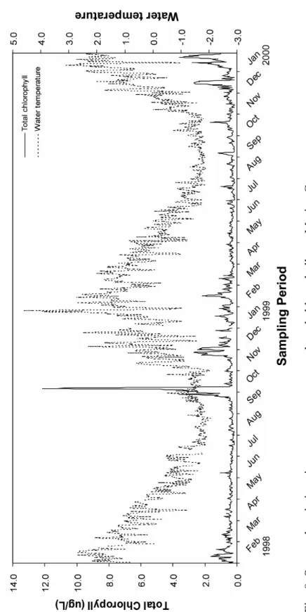
Fig. 8. Seasonal variations in water temperature and total chlorophyll a
at Marian Cove..…..…..………..…….72
Fig. 9. Photomicrographs of ovaries. (A) Resting stage: gonadal tissues
are little or no visible along the thin follicle wall. (B)
Developing stage: growing ovary with primary oocytes (PO),
previtellogenic oocytes (PVO) and vitellogenic oocytes (VO)
along the follicle wall (FW). (C) Fully developed stage: mature
ovaries packed with ova (O) and a thin layer along follicle wall.
In germinal epithelium, very small PO is present. (D)
Spawning stage: partly spawned ovaries with loosely packed
ova and the spaces vacated in the lumen (LM). (E) Spent stage:
ovaries largely devoid of ova and shrinking ovary with VO and
relict ova (RO). (F) Absorbing stage: relict oocytes will be
reabsorbed (AO) and a thin follicle epithelium. Scale bar: 100
µm..…..…..……….…….73
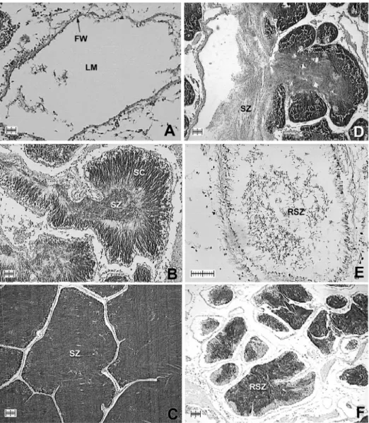
Fig. 10. Photomicrographs of testes. (A) Resting stage: resting ovaries
are vacated with lumen (LM) and thin follicle wall (FW). (B)
Developing stage: columns of spermatocytes (SC),
spermatozoa (SZ) move to central part in growing testes. (C)
Fully developed stage: mature testes filled with SZ and largely
devoid of nutritive tissue. (D) Spawning stage: partly spawned
testes with spaces vacated by spawned SZ. (E) Spent stage:
partly devoid of content, although relict spermatozoa (RSZ)
may be present. (F) Absorbing stage: shrinking testes with thin
follicle wall. Scale bar: 100 µm.…..…..……….…….75
Fig. 11. Temporal changes in proportional occurrence of various oocyte
with size-frequency distributions in MOD.…..…..………….77
Fig. 12. Temporal changes in respective percentages in each oocyte
classes characterizing the developmental size of oocytes
distinguished in this study.…..…..………..…….78
Fig. 13. Photomicrographs of different developmental stages of oocytes
between vitellogenic oocytes (VO) and mature oocytes (MO).
A: histological feature of the oocytes in gonad; B: extracted
oocytes from gonad. Thick arrows indicate the gelatinous
layers in each developmental stages of oocyte. Scale: 100
µm.…..…..………..….79
Fig. 14. Monthly changes in FI of L. elliptica. …..…..………..….….80 Fig. 15. Photomicrographs show resorption process in degenerative
ovaries of L. elliptica. A-B: invaded hemocytes (H) in the
follicular lumen, and the process of phagocytosis began to
relict ova (RO) with nucleus (N), yolk granules (YG) and
wound gelatinous layers (GL); C: transported eosinophil
granules (EG) by H with fluttered GL; D: RO and
phagocytosed ova (PHO) remaining in follicle and transported
phagocytosed materials (PM) to follicular epithelium (FE); E:
magnified image of transported PM by H; F: magnified image
of deposited PM on follicle. Scale: 20 µm. ….……….….….81
LIST OF TABLES
Table 1. Classification of Laternula elliptica gonad developmental
stages (I–VI) with numerical scores (1–6).……...……….35
Table 2. Mean follicle index (FI) and mean percentage gonad area
(PGA) and at various stages of gonadal development in L.
elliptica. Data from December 1995 and January 1998 are
pooled.–,data are not available.…..………...….36
Table 3. Developmental stage of L. elliptica gonad. ……….…..83 Table 4. Shell length (SL), tissue wet weight (TWW) and percentage of
gonadal maturation with maturity index of L. elliptica used in
this study . ……….…..….84
Table 5. Partial correlation coefficients between total chlorophyll,
water temperature and reproductive indices of Laternula
elliptica used for analysis. MI Maturity index, FI Follicle
index, MOD Mean oocyte diameter, GI Gonad index.
Significance: ** 0.001 < P < 0.01, * 0.01 < P < 0.05 ..…...85
Table 6. Reproductive characteristics of L. elliptica reported from
various studies. ……….…...…..…....86
SUMMARY
1. Quantitative assessment of reproductive condition of the Antarctic clam, Laternula elliptica (King & Broderip), using image analysis
1.1. The reproductive state of Antarctic clam, L. elliptica, collected
from a small cove on King George Island in the spawning season
was assessed quantitatively using computer-based image analysis.
1.2. The percentage egg area within a follicle (FI) and mean oocyte
diameter (MOD) values differed significantly between December
and January but conventional maturity index (MI) and percentage
gonad area (PGA) values did not, suggesting that FI and MOD are
the more reliable and sensitive indicators for differentiating
reproductive condition.
1.3. This study also showed that a planimetric technique using
computer-based image analysis is fast, convenient to use, and better
than the conventional MI at providing reliable quantitative
information about L. elliptica reproduction.
1.4. These indices varied with body size. The FI and MOD values
peaked in clams of 76 to 85 mm shell length, indicating that clams
of this size have the highest reproductive output at spawning time.
Therefore, animals of a standard size (76 to 85 mm) should be
monitored seasonally or over a longer term.
2. Annual gametogenesis of the Antarctic clam, Laternula elliptica from Marian Cove, King George Island
2.1. The annual gametogenesis of the Antarctic soft-shelled clam,
Laternula elliptica inhabited in Marian Cove, King George Island
was studied over a two-year period from February 1998 to January
2000. Annual changes in gonad maturation were investigated using
histology-based techniques as previous mentioned.
2.2. In 1998, monthly mean FI and MI increased significantly from
October to November and reached a peak in December. The FI and
MI values dropped rapidly from December to January. In 1999,
degenerated eggs were observed from the spent follicles in
February and March. Degeneration and absorption of the residual
eggs by phagocytosis occurred mostly in February and March in
1998 and 1999, although the resorption process was observed all
year round. The gonad maturation during October to December in
both years was corresponded with the annual chlorophyll maximum
in the Cove, suggesting that food supply is crucial to gonad
maturation and subsequent spawning.
2.3. Gametogenesis of L. elliptica at Marine Cove investigated for two
years confirmed that the clam spawns annually, and the gonad
maturation takes only a few months during the Austral summer. A
distinct seasonality was also observed in the annual gametogenesis
as 1) absorbing and resting during February and March, 2)
developing and maturation during October and December and 3)
spawning during December and February.
INTRODUCTION
G
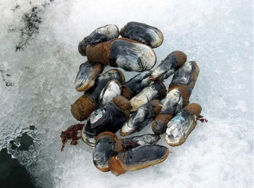
The Antarctic soft-shelled clam, Laternula elliptica (Fig. 1) is
one of the most common marine organisms inhabiting the shallow
subtidal area around Antarctic coastal waters (Ahn 1994a; Ansell and
Harvey 1997; Sahade et al. 1998; Urban and Mercuri 1998). The clam
is endemic to the Antarctic and is widely distributed in shallow waters
around the continent and adjacent islands (Ahn 1994; Mercuri et al.
1998; Cattaneo-Vietti et al. 2000; Brockington 2001). This
deep-burrowing, large (§110 mm in shell length, Ahn 1994) filter-feeder
occurs in dense patches (a few hundred/m2) and appears to play a key
role in the Antarctic nearshore marine ecosystem (Zamorano et al.
1986; Ahn 1993; Arntz et al. 1994; Ahn et al. 2001). Especially, the
clam occurs in dense patches at a depth between 20 and 30 m at King
George Island, South Shetland Islands and is considered to be one of
the well adapted benthic animals in the harsh shallow Antarctic water
(Zamorano et al. 1986; Ahn 1994b; Sahade et al. 1998). Despite its
wide distribution and abundance, few studies have concerned the
reproductive biology and ecology of L. elliptica (Pearse et al. 1991;
Urban and Mercuri 1998; Bigatti et al. 2001; Ahn et al. 2000).
Laternula elliptica is a simultaneous hermaphrodite which
produces unusually large encapsulated eggs (§200 µm in diameter), which develop as encapsulated lecithotrophic larvae (Pearse et al. 1991;
Ansell and Harvey 1997). Spawning probably occurs during summer
months along a latitudinal cline: mid-December at King George Island
(Ahn et al. 2000) and from March to mid May at McMurdo Sound,
King George Island (Bosch and Pearse 1988). The reproductive
patterns and related characteristics of L. elliptica, however, need to be
further elucidated with seasonal or long-term studies. Like other
benthic animals in Antarctica, the clam has slow growth and slow
gonad maturation (Pearse et al. 1991; Ansell and Harvey 1997; Urban
and Mercuri 1998; Bigatti et al. 2001). Slow gonad maturation and
pelagic non-feeding larval development of L. elliptica is considered to
be a reproductive adaptation to the harsh Antarctic environment (Bosch
and Pearse 1988; Pearse et al. 1991; Urban and Mercuri 1998). Like
other benthic organisms inhabiting in the Antarctic, limited information
is available on growth and reproduction of L. elliptica due to the
technical difficulties involved in direct observation and sampling.
Recently studies have been conducted to investigate
reproduction and growth of L. elliptica. These studies have reported
that the clam spawns during austral summer although the spawning has
a latitudinal cline; mid-December to February at Marian Cove (Ahn et
al. 2000, 2003), and from March to mid May at McMurdo Sound, King
George Island (Bosch and Pearse 1988; Pearse et al. 1991). Urban
and Mercuri (1998) investigated annual gametogenesis of L. elliptica
collected from Potter Cove, King George Island over a one year period
using histology. They reported that L. elliptica at Potter Cove spawn
during February and March. Interestingly, they were unable to
observe any early developing gametes in the gonad while they found
ripe oocytes in the follicle all year round. Based upon histological
observation, they concluded that the oocyte development cycle last
longer than one year (Urban and Mercuri 1998). Bigatti et al. (2001)
also analyzed the same clams sampled from Potter Cover by Urban and
Mercuri (1998) by measuring the size of the oocytes. Contrary to Urban
and Mercuri (1998), they concluded a complete oocyte growth cycle
takes less then a year and the clam may be ready for spawning at any
time during the whole year, although intensive spawning is believed to
occur in February.
Numerous techniques have been used to assess the reproductive
condition of marine molluscs. Histological methods have been the most
widely and frequently used, since histology provides visual information
about gonadal tissues. Reproductive condition is usually classified as
one of several numerical codes on the basis of gonadal tissue
characteristics viewed under a microscope (see review of
Lango-Reynoso et al. 2000). The process of gonadal development is also
monitored by measuring increases in oocyte diameter or the degree to
which a microscopic field is occupied by gonadal cells (see a review by
Heffernan and Walker 1989). With the aid of a computer and video
camera, the microscopic image of the gonad is digitized into a
computer graphic file and software is used to calculate objectively the
area of eggs or gonadal areas (Heffernan et al. 1989; Lango-Reynoso et
al. 2000).
In general gametogenesis of bivalves is categorized into 1)
formation of germ cells, 2) growth and enrichment of gametes known
as vitellogenesis 3) spawning and discharging of the gametes, and 4)
resorption of residual gamete (Loosanoff 1942; Kennedy and Battle
1964; Braley 1982; Olive 1984; Shafee 1989). Such cyclic changes in
gametogenesis are in part governed by changes in environmental
parameters such as water temperature, salinity and food (Bayne and
Newell 1983; Soniat and Ray 1985; Heffernan et al. 1989; Hofmann et
al. 1992; Kang et al. 2000). The amount of food in the water column for
filter feeders often shows a strong seasonality in temperate region with
water temperature playing a key role in governing the seasonality
(Mann 1979; Clarke 1987; Heffernan et al. 1989; Hooker and Creese
1995; Saucedo et al. 2001). Numerous studies have reported that gonad
development and subsequent spawning of marine bivalves is
synchronized with seasonal change of available food in the water
column (Hofmann et al. 1992; Grant and Creese 1995; Kennedy et al.
1996; Kang et al. 2000). Along with water temperature, the quantity of
food in the water column determines the amount of gametes produced
(i.e. reproductive output) and the timing of spawning (Kang et al. 2000;
Llodra 2002; Park and Choi 2004). Primary production of coastal
Antarctic is extremely seasonal and the phytoplankton blooming occurs
during a short period of austral summer (Chang et al. 1990, Rivkin
1990; Kang et al. 1997; Ahn et al. 2003). Therefore, available food is
an important factor involving reproduction of Antarctic marine
invertebrates (Urban and Mercuri 1998; Peck et al. 2000; Ahn et al.
2003; Clarke et al. 2004). Laternula elliptica are possibly adapted on
reproductive strategy as reproductive output, spawning time and mode
in relatively poor food condition.
The objectives of the present study were to 1) establish standard
methods for use in a seasonal or longer-term study of L. elliptica
reproduction; 2) investigate the seasonal variations of oocytes growth
and the gametogenic cycle, resorption process to the relict eggs and the
correlation between available food and reproduction of L. elliptica
collected from Marian Cove, King George Island in Antarctica.
Fig. 1. The Antarctic clam, Laternula elliptica.
Part I
Quantitative assessment of reproductive condition of the Antarctic clam,Laternula elliptica (King & Broderip), using image analysis
1. Abstract.
The reproductive state of Antarctic clam, L. elliptica, collected
from a small cove on King George Island was assessed quantitatively in
the spawning season. Using computer-based image analysis, percentage
gonad area (PGA), percentage egg area within a follicle (FI), and mean
oocyte diameter (MOD) were determined from gonadal cross-sections
prepared for histological studies. These indices were then compared on
the basis of scores from 1 to 6 on a conventional maturity index (MI),
which is scored using subjective criteria determined by microscopic
examination. Clams collected during two different spawning periods
were analyzed. The FI and MOD values differed significantly between
the two groups but MI and PGA values did not, suggesting that FI and
MOD are the more reliable and sensitive indicators for differentiating
reproductive condition. This study also showed that a planimetric
technique using computer-based image analysis is fast, convenient to
use, and better than the conventional MI at providing reliable
quantitative information about L. elliptica reproduction. I also
investigated whether these indices varied with body size. The FI and
MOD values peaked in clams of 76 to 85 mm shell length, indicating
that clams of this size have the highest reproductive output at spawning
time. Therefore, animals of a standard size (76 to 85 mm) should be
monitored seasonally or over a longer term.
2. Materials and Methods
2.1. Sampling efforts
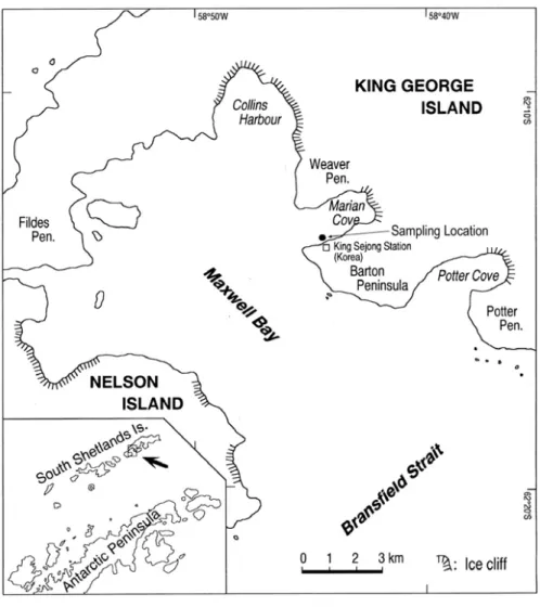
Marian Cove is a small embayment located in northeast
Maxwell Bay (Ahn et al. 2000) (Fig. 2). Every summer for the past
several years, L. elliptica has been collected from depths between 20
and 30 m at Marian Cove (62°13'S, 58°47'W) by SCUBA. For our
study, clams collected during two austral summers (Dec 12th -22th, 1995
and Jan 12th 1998) were selected, as clams of a wide size range were
available from the specimen sets collected. Shell length (SL, the
longest dimension of the shell) was measured to 0.01 mm. Each clam’s
soft body was then removed and the soft tissues were fixed in 10%
neutral formalin for further analysis.
2.2. Histological preparation and analysis of gonadal tissues
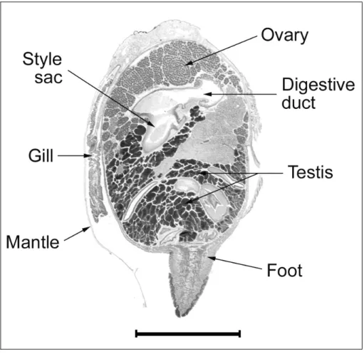
A 1-cm-thick longitudinal cross-section was removed from the
center of the soft body mass of each clam for histological analysis (Fig.
3). Each cross-section contained gonads, visceral mass, a foot, gills,
and a mantle, all which are recommended for complete histological
sectioning of marine bivalves (Howard and Smith 1983). The tissues
were dehydrated in an alcohol, cleared in xylene, embedded in paraffin,
sliced to 6 µm and stained with Harris’ hematoxylin and eosin Y. The
sections were examined under a light microscope equipped with a
video camera connected to a personal computer.
L. elliptica is a hermaphrodite, gonadal development of testis
and ovary were scored from 1 to 6 according to Table 1. Three
microscopic fields on average were examined for each clam and the
mean score was used as the maturity index (MI). Gonad development
of ovary was also analyzed quantitatively using the following
planimetric technique. A microscopic image of gonad was captured by
a video camera and sent to a personal computer. Surface areas of ovary
and testis in each cross-section were then measured from the digitized
images using Image Pro® image analysis software. The ratio of
gonadal/total cross-sectional area was also determined and expressed as
percentage gonad area (PGA, µm2) (Morales-Alamo and Mann 1989).
The percentage area occupied by oocytes in a follicle, or follicle index
(FI, %), was determined using three to six follicles that were randomly
selected from the histological preparation of each clam. Mean oocyte
diameter (MOD, µm) was determined using 40 to 50 oocytes in three to
six follicles measured from the computerized microscopic image.
I investigated variation in these indices associated with body
size and age using specimens over a wide size range. In addition, some
reproductive traits of L. elliptica are discussed.
3. Results
3.1. Quantitative assessment of L. elliptica reproductive condition
In mid December 1995, the reproductive condition of 51 clams
was examined. Of these, 3.9% were sexually undifferentiated (stage I),
11.8% were developing (stage II, Fig. 4A), 51.0% were fully developed
and sexually mature (stage III, Fig. 4B), 29.4% were spawning (stage
IV, Fig. 4C); the remaining 4.0% were either spent (stage V, Fig. 4D) or
absorbing (stage VI, Fig. 4E). In mid January 1998, 47 clams were
examined. Of these, 23.4% were developing, 53.2% were fully
developed, and 23.4% were spawning. No clams from this year had
gonads that were in the early developing, spent, or absorbing stages. In
both years, most clams were ready to spawn or already spawning.
Oocyte diameter L. elliptica from Marian Cove ranged from
20.0 to 192.1 ȝm. The diameter of fully developed eggs averaged 156.6 ȝm. Each fully developed egg was enveloped in a 9.1-ȝm membrane that stained strongly with Harris’ hematoxylin. There were oocytes of
various sizes within each follicle, which lowered the MOD of each
clam. The mean MOD of oocytes was 57.79 µm in developing ovaries,
62.85 ȝm in fully developed ovaries, 49.30 µm in spawning ovaries, and 22.75 ȝm in spent ovaries. Mean values of PGA and FI were highest in fully developed ovaries, followed by spawning ovaries
(Table 2). The area occupied by the testis was always larger than that of
the ovary regardless of body size or reproductive stage.
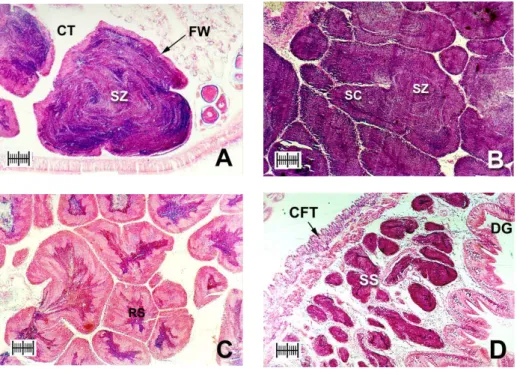
In the developing stage of the testes, follicles expanded toward
connective tissues and spermatogenic cells began to proliferate around
follicle walls (Fig. 5A). Fully developed, spawning, and partially
spawned testes were observed simultaneously in both years (Figs. 5B,
C). Spent testes often exhibited relict spermatozoa (Fig. 5D).
Gametogenesis of the testis progresses synchronously with that of the
ovary.
3.2. Comparison of the quantitative indices in the clams from the two austral summers
Mean SL of the clams collected in December 1995 and in
January 1998 was 65.6 mm and 64.9 mm respectively, i.e. no
significant difference in SL was found between the two populations.
Small oocytes of >60 µm diameter were dominant in the clams
collected in December 1995, while relatively bigger oocytes of <71 µm
diameter were dominant in January 1998 (Fig. 6). Since clams of a
wide size range were used in this study, direct comparisons for MI,
PGA, FI and MOD values in the two austral summers could not be
made from the mean values. The MI, PGA, FI, and MOD values of
clams collected in each year, therefore, were plotted against clam SL
(Fig. 7). Each of the four indices was compared using regression
analysis (ANCOVA). There was no significant difference in the MI and
PGA between the two years (Figs. 7A, B). However, FI and MOD did
differ significantly between sampling years: FI and MOD were
significantly higher in January 1998 than in December 1995 (P < 0.05
for FI, P < 0.001 for MOD) (Figs. 7C, D).
Distinct correlations of FI and MOD with body size (SL) were
observed in clams collected in both years (Figs. 7C, D). In the case of
MI, however, a significant size relation was found only for January
1998, and for PGA, none of the years showed a significant relation
(Figs. 7A, B). The relations of these indices with body size were best
fitted by quadratic regression curves that peaked between 76 and 85
mm SL.
4. Discussion G
4.1. Computer-based image analysis of the reproductive condition ofL. elliptica
Microscopic examination of histological sections of gonads has
been used to study marine bivalve reproduction over many decades.
Reproductive status is often categorized according to a numerical scale
that describes various stages of gonadal development (Loosanoff 1942;
Kennedy and Krantz 1982; Shafee 1989; Barber 1996). While direct
microscopic examination is relatively simple, scoring or grading
gonadal condition using the numerical scale is rather subjective (see
reviews in Lucas 1982 and Heffernan and Walker 1989). Reproductive
condition is also often evaluated by using objective measurements such
as gonad thickness or width, which are not applicable in most cases
(Loosanoff 1965; Gauthier and Soniat 1989). Oocyte size, percentage
area occupied by eggs in a follicle, and cross-sectional area occupied by
gonads determined by stereology and planimetry have also been used
widely to evaluate the status of gonadal development in bivalves
(Morvan and Ansell 1988; Morales-Alamo and Mann 1989).
In our study, MI and PGA did not differ significantly between
clams from the two years of collection, but FI and MOD did (Fig. 7).
This finding suggests that planimetric indices, particularly FI and MOD,
are more reliable and sensitive than the conventional MI in quantitative
assessment of L. elliptica reproductive status. The planimetric
technique used in this study employed computer-based image analysis
and was fast and convenient to use; this technique would also be useful
for processing large numbers of samples for seasonal or longer-term
studies.
It is interesting to note that both FI and MOD values were
higher in January 1998 than in December 1995. The higher MOD
values in January 1998 indicate that oocytes were more mature (Fig. 6),
probably at their developmental peak and ready to spawn, in January.
The percentage of developing and mature oocytes (> 100 ȝm) in January 1998 (§32%) was about double that in December 1995 (§16%).
4.2. Reproductive traits of L. elliptica at Marian Cove
4.2.1. Oocyte size
As shown in Figure 2, ripe eggs were enclosed in a thick
(9.1-µm) gelatinous layer. Mature eggs, measured using image analysis,
averaged 156.6 µm in diameter, with a maximum of 192.1 µm in
diameter, excluding the gelatinous layer. At Potter Cove, a few
kilometers away from the study area, Bigatti et al. (2001) reported a
mean mature oocyte size of 195.2 µm in diameter, with a maximum of
220.7 µm in diameter, including the gelatinous layer. The difference in
oocyte diameter measured in this study and that by Bigatti et al. (2001)
can be explained in part by variation in thickness of the gelatinous layer.
I did not include the gelatinous layer in oocyte size measurements,
since its thickness varies with developmental stage: on average, 34.8
µm in developing oocytes versus 9.1 µm in ripe eggs. Ansell and
Harvey (1997) reported that developing eggs (from non-preserved
oocytes) were 162.3 ± 5.4 µm in diameter. This oocyte size is similar to
our measurements when shrinkage of oocytes during fixation and
dehydration for histology is taken into account.
4.2.2. Size and putative age at first reproduction
I estimated putative age of the clam using the L. elliptica
growth curve reported by Ralph and Maxwell (1977); clams ranged
from 3 years (34.4 mm SL) to 44 years old (106.0 mm SL). The
3-year-old clam collected in December 1995 contained fully mature eggs
covered by a gelatinous layer and which were considered to be in the
spawning stage. These data suggest that L. elliptica may achieve sexual
maturity as young as three years old at Marian Cove. Bigatti et al.
(2001) described an individual L. elliptica near Potter Cove (27 mm
SL), estimated to be less than two years old, that had ovarian follicles
and testicles. Their result pushes back the age at first reproduction to as
early as two years. Due to the limited number of clams collected in this
study, no clams with age of two or younger were included in our data
set. It is very likely that L. elliptica at Marian Cove also achieve sexual
maturity at the age of two as suggested by Bigatti et al. (2001) since our
study area is geographically close Potter Cove.
4.2.3. Variation in reproductive output with body size
As shown in Figure 5, MI, FI and MOD were best fitted to a
quadratic regression curve. These indices were significantly described
by the curve with a peak between 76 and 85 mm SL. These data
indicate that L. elliptica in this size range have the highest reproductive
output. Several studies of reproduction in marine bivalves, including
oysters, mussels, and hard clams, have shown that gamete production
tends to increase with size and age and then plateau (Morvan and
Ansell 1988; Levitan 1991; Choi et al. 1993; Franz 1996; Kang et al.
2003). Urban and Mercuri (1998) reported that gonad development in L.
elliptica rapidly increased with size in individuals of 65 to 90 mm SL.
The reproductive effort of older and larger clams (> 90 mm SL) may
decrease after they have reached an optimal size for gamete production.
G
G G
Fig. 2. Sampling location of L. elliptica at Marian Cove.
Fig. 3. Cross-section of L. elliptica. Scale bar = 1 cm.G
G
hG
G G
Fig. 4. Photomicrographs of ovaries from L. elliptica collected in December 1995 (scale bar = 200 µm). (A) Developing stage: ovary contains a small oogonium (OG), previtellogenic oocytes (PVO), and vitellogenic oocytes (VO) along the follicle wall (FW). Follicles expand into the vesicular connective tissue (VC) and few gaps are found between follicles. (B) Fully developed stage: large eggs are attached to germinal epithelium and the follicle is completely filled with ova (O). (C) Spawning stage: partially spawned ovaries contain loosely packed relict ova (RO) and spaces in the lumen (LM). The follicle wall is noticeably thinner. (D) Spent stage: follicle wall is withered and some relict ova still remain within follicles. Early vitellogenic oocytes (EVO) are attached to follicle wall. (E) Absorbing
stage: ovary degenerates and relict ova are reabsorbed (AO) into the thin follicle epithelium by phagocytic activities.
G
G
hG
G
Fig. 5. Photomicrographs of testes (scale bar = 200 µm). (A) Developing stage: columns of spermatozoa (SZ) move to center of a growing testis with a thick follicle wall (FW). Most of the lumen is filled with dense radiating bands of spermatozoa (CT, connective tissue). (B) Fully developed stage: mature testes are filled with spermatocytes (SC) and spermatozoa, and are largely devoid of nutritive tissue. Dense bands of spermatozoa fill the majority of the lumen and often spiral into its center. (C) Spawning stage: partially spawned testes contain spaces vacated by spawned spermatozoa and nutritive phagocytes (RS, relict sperm). (D) Spent stage: spent spermaries (SS) are partially empty, although relict spermatozoa may be present. Follicle wall is thin and testis volume is small (CFT, connective fibromuscular tissue; DG: digestive gland).
Fig. 6. Frequency distribution of MOD in December 1995 and January 1998 (n: number of oocytes measured).
G Z[
Fig. 7. Comparisons of MI, PGA, FI and MOD between L. elliptica collected in December 1995 and January 1998. There was no significant difference in the MI and PGA between the two years (A and B), while the FI and MOD values were significantly higher in January 1998 than in December 1995 (C and D).
Regression lines: (A) MI (Jan. 98) = -0.000864SL2+ 0.142SL
- 2.40, r2 = 0.41, n=42, P < 0.001; No significant size relation
was found for Dec. 95. (B) PGA; No significant size relation
was found for both years. (C) FI (Dec. 95) = -0.0133SL2 +
2.26SL - 43.35, r2 = 0.36, n=49, P < 0.001; FI (Jan. 98)
=-0.0158SL2+ 2.58SL – 46.88, r2 = 0.19, n=42, P < 0.05. (D)
MOD (Dec. 95) = -0.0132SL2+ 2.024SL – 21.82, r2 = 0.15,
n=38, P < 0.05; MOD (Jan. 98) = -0.0405SL2+ 6.40SL - 172.2,
T
able 1. Classification of
Laternula elliptica
gonad developm
ental stages (I–VI) with num
erical scores (1–6). Num erical score Developm ental stages Histological description Sexually un dif ferentiated (I) 1
Little or no gonadal tissue visib
le.
Follicle exp
anded; grow
ing ovary with vitellogenic oocytes and prem
ature oocytes. Som e mature gam ete present. Developing (II) 2
Fully developed (III)
3
Most gam
etes m
ature; little connective tissue rem
aining.
Fully m
ature eggs detached from
the follicle. S p awning ovaries h ave thin follicle wall. Spawning (IV) 4 Reduced num ber of gam etes; som e m ature gam
etes still rem
aining;
evidence of renewed reproductive activity
.
Spent (V)
5
Apparent phagocytic activities around gonadal tissues;
ragged eggs or sperm ary . Absorbing (VI) 6 G Z\
G
Z]
T
able 2
. Mean follicle index (FI) and m
ean pe
rcentage go
nad area (P
GA) and at various stag
es of gonadal development in
L.
elliptica.
Data from
Decem
ber 1995 and January 1998 are po
oled.
–,
data are not available
. PGA Mean FI (%) T estis Ovary T o tal I0 0 0 0 II – – – – III 52.75 23.43 16.65 40.08 IV 37.70 16.04 12.05 28.09 V 8.17 5.46 0.07 5.53 Developm ental stages VI – 18.17 7.22 25.39
5. References
Ahn I-Y (1993) Enhanced particle flux through the biodeposition by
the Antarctic suspension feeding bivalve Laternula elliptica in
Marian Cove, King George Island. J Exp Mar Biol Ecol 171:75-90
Ahn I-Y (1994) Ecology of the Antarctic bivalve Laternula elliptica
(King and Broderip) in Collins Harbor, King George Island:
Benthic environment and an adaptive strategy. Mem Nat Instit
Polar Res Special Issue 50:1-10
Ahn I-Y, Cho KW, Choi K-S, Seo Y, Shin J (2000) Lipid content and
composition of the Antarctic lamellibranch, Laternula elliptica
(King&Broderip) (Anomalodesmata: Laternulidae), in King
George Island during an austral summer. Polar Biol 23:24-33
Ahn I-Y, Chung H, Choi K-S (2001) Some ecological and
physiological features of the Antarctic clam, Laternula elliptica
(King and Broderiop) in a nearshore habitat on King George
Island. Ocean Polar Res 23:419-424
Ansell AD, Harvey R (1997) Protected larval development in the
Antarctic bivalve Laternula elliptica (King and Broderip)
(Anomalodesmata: Laternulidae). J Mollus Stud 63:285-286
Arntz WE, Brey T, Gallardo VA (1994) Antarctic zoobenthos.
Oceanogr Mar Biol Ann Rev 32:241-304
Barber BJ (1996) Gametogenesis of Eastern oyster, Crassostrea
virginica (Gmelin, 1791), and Pacific oyster, Crassostrea gigas
(Thunberg, 1793) in disease-endemic lower Chesapeake Bay. J
Shellfish Res 15:285-290
Bigatti G, Penchaszadeh PE, Mercuri G (2001) Aspects of the gonadal
cycle in the Antarctic bivalve Laternula elliptica. J Shellfish Res
20:283-287
Bosch I, Pearse JS (1988) Seasonal pelagic development and juvenile
recruitment of the bivalve Laternula elliptica in McMurdo Sound,
Antarctica. Am Zool 28:89A
Brockington S (2001) The seasonal energetics of the Antarctic bivalve
Laternula elliptica (King and Broderip) at Rothera Point, Adelaide
Island. Polar Biol 24:523-530
Cattaneo-Vietti R, Chiantore M, Schiaparelli S, Albertelli G (2000)
Shallow- and deep-water mollusc distribution at Terra Nova Bay
(Ross Sea, Antarctica). Polar Biol 23:173-182
Choi KS, Lewis DH, Powell EN, Ray SM (1993) Quantitative
measurement of reproductive output in the American oyster,
Crassostrea virginica (Gmelin), using an enzyme-linked
immunosorbent assay (ELISA). Aquacult Fisher Manag
24:375-398
Franz DR (1996) Size and age at first reproduction on the ribbed
mussel Geukensia demissa (Dillwyn) in relation to shore level in a
New York salt marsh. J Exp Mar Biol Ecol 205:1-13
Gauthier JD, Soniat TM (1989) Changes in the gonadal state of
Louisiana oysters during their autumn spawning season. J
Shellfish Res 8:83-86
Heffernan PB, Walker RL (1989) Quantitative image analysis methods
for use in histological studies of bivalve reproduction. J Mollus
Stud 55:135-137
Heffernan PB, Walker RL, Carr JL (1989) Gametogenic cycle of three
marine bivalves in Wassaw Sound, Georgia II Crassostrea
virginica. J Shellfish Res 8:61-70
Howard DW, Smith CS (1983) Histological techniques for marine
bivalve mollusks. NOAA Technical Memorandum
NMFS-F/NEC-25
Kang S-G, Choi K-S, Bulgakov A-A, Kim Y, Kim S-Y (2003)
Enzyme-linked immunosorbent assay (ELISA) used in
quantification of reproductive output in the pacific oyster,
Crassostrea gigas, in Korea. J Exp Mar Biol Ecol 282:1-21
Kennedy VS, Krantz LB (1982) Comparative gametogenic and
spawning patterns of the oyster Crassostrea virginica (Gmelin) in
central Chesapeake Bay. J Shellfish Res 2:133-140
Lango-Reynoso F, Chávez-Villalba J, Cochard JC, Pennec ML (2000)
Oocyte size, a means to evaluate the gametogenic development of
the Pacific oyster, Crassostrea gigas (Thunberg). Aquaculture
190:183-199
Levitan DR (1991) Influence of body size and population density on
fertilization success and reproductive output in a free-spawning
invertebrate. Biol Bull 181:261-268
Loosanoff VL (1942) Seasonal gonadal changes in the adult oysters,
Ostrea virginica, of Long Island Sound. Biol Bull 82:195-206
Loosanoff VL (1965) Gonad development and discharge of spawn in
oysters of Long Island Sound. Biol Bull 129:546-561
Lucas A (1982) Evaluation of reproductive effort in bivalve mollusks.
Malacologia 22:183-187
Mercuri G, Iken K, Ledesma B, Dubois RF (1998) The Potter cove
coastal ecosystem, Antarctica. In: Wiencke C, Ferreyra G, Arntz W,
Rinaldi C (eds) On the distribution patterns and density of the
Antarctic infaunal bivalve Laternula elliptica in Potter Cove, King
George Island. Ber Polarforsch 299:137-143
Morales-Alamo R, Mann R (1989) Anatomical features in histological
sections of Crassostrea virginica (Gmelin, 1791) as an aid in
measurements of gonad area for reproductive assessment. J
Shellfish Res 8:71-82
Morvan C, Ansell AD (1988) Stereological methods applied to
reproductive cycle of Tapes rhomboids. Mar Biol 97:355-364
Pearse JS, McClintock JB, Bosch I (1991) Reproduction of Antarctic
benthic marine invertebrates: tempos, modes, and timing. Am Zool
31:65-80
Ralph R, Maxwell JGH (1977) Growth of two Antarctic
lamellabranchs: Adamussium colbecki and Laternula elliptica.
Mar Biol 42:171-175
Shafee MS (1989) Reproduction of Perna picta (Mollusca: Bivalvia)
from the Atlantic coast of Morocco. Mar Ecol Progr Ser
53:235-245
Urban H-J, Mercuri G (1998) Population dynamics of the bivalve
Laternula elliptica from Potter Cove, King George Island, South
Shetland Islands. Antarct Sci 10:153-160
Zamorano JH, Durate WE, Moreno CA (1986) Predation upon
Laternula elliptica (Bivalvia, Anatinidae): a field manipulation in
South Bay, Antarctic. Polar Biol 6:139-143
Part II
Annual gametogenesis of the Antarctic clam, Laternula elliptica from Marian Cove, King George Island
1. Abstract
The annual reproductive cycle of the Antarctic soft-shelled
clam, Laternula elliptica, in Marian Cove, King George Island, was
studied over a 2-year period from February 1998 to January 2000.
Annual changes in gonad maturation were investigated using
histological techniques, including numerical scoring of the
microscopic appearance of the gonad (maturity index, MI), measuring
the percentage of area occupied by oocytes in a follicle (follicle index,
FI), and assessing the oocyte size. In 1998, the monthly mean FI and
MI increased significantly from October to November, peaked in
December, and dropped rapidly from December to January. In
February and March 1999, degenerated eggs were observed in the
spent follicles. Degeneration and absorption of residual eggs by
phagocytosis occurred mostly in February and March in 1998 and
1999, although the resorption process was observed year-round.
Gonad maturation and spawning of L. elliptica have significant year to
year differences; the gonad maturation and spawning occurred rapidly
when food levels in the water column become sufficient, suggesting
that food supply is critical to gonad maturation and subsequent
spawning. L. elliptica spawns annually and gonad maturation takes
only a few months during the austral summer. There was a distinct
seasonality to the annual gametogenesis, with absorption and resting
in February and March, development and maturation in October and
December, and spawning from December to February.
2. Materials and Methods
2.1. Sample collection
Clams were collected by SCUBA divers from depths of 20 to
30 m near King Sejong Station (62°13' S, 58°47' W) in Marian Cove,
King George Island (Fig. 1). Twenty to thirty clams were collected on
a monthly basis between February 1998 and January 2000, except in
April, July, and September 1998, when we were unable to collect
clams because of inclement weather. Collected clams were preserved
in 10% buffered formalin in situ for histological analyses. Daily total
chlorophyll concentration and water temperature data were obtained
from a long-term monitoring program conducted at the station
(KORDI 1999). Total Chlorophyll (Chlorophyll a + phaeopigments)
concentration was determined in vivo using a fluorometer (Turner
10-AU-005). Water temperature was measured using a conductivity meter
(YSI 610-D).
2.2. Histological Preparation
Upon arrival to the laboratory, shell length and height was
measured to 0.01 mm. The soft body of the clams was then removed
and wet weight of the tissue recorded to 0.01 g. Clam tissues were
preserved in 75% ethanol. A longitudinal cross section of 1 cm
thickness was made along the central part of the soft body. After
dehydration, clearing and embedding in paraffin, the block was sliced
to 5 µm and stained with Harris' hematoxylene and eosin Y.
Degeneration of relict ova by hemocytes (i.e., phagocytosis)
was examined using the hemocyte-specific Giemsa staining for
paraffin sections. Briefly, the dewaxed section was rinsed in ethanol
and distilled water. The section was stained in a glass jar in a mixture
of Giemsa stock and distilled water in a water bath at 56ºC for 40 min.
After a rinse in distilled water, staining was contrasted using acetic
acid. After blotting dry, clearing and mounting, phagocytosis was
observed by activities of hemocyte stained blue color and
phagocytosed granules stained eosinophilic red.
2.3. Analysis of annual gametogenesis
The reproductive condition of L. elliptica was examined under
a light microscope equipped with a video camera connected to a
personal computer. Since L. elliptica is a hermaphrodite, gonadal
development of testis and ovary were scored according to Table 3.
Four microscopic fields were randomly selected from each slide and
graded from 1 to 6 according to the criteria in Table 3. The qualitative
reproductive condition of each clam was determined with an average
score of the four microscopic fields, and the average score was used as
the maturity index (MI).
Gonad and oocytes development of the ovary was also
analyzed using a planimetric technique (Kang et al. 2003). From the
digitized images of gonadal tissue, oocyte diameter and area in a
follicle were measured using an image analyzing software. An oocyte
diameter was measured from a microscopic image of the ovary, and
mean oocyte diameter (MOD, µm) was calculated using 40 to 50
oocytes selected in follicles. Then, the frequency distribution of the
oocytes was analyzed to identify the monthly variation of oocyte size
and growth. The normality of the frequency distribution of oocytes
was tested the symmetric and/or asymmetric distributions using the
Kolmogorov–Smirnov tests (Sokal and Rohlf 1981). One-way
ANOVA test and Pearson’s correlation were conducted to compare
similarity and/or difference of the frequency distributions in the
sampling period. Various sizes of oocytes were classified according to
the different oogenic stages. The percentage area occupied by
oocytes in a follicle, or follicle index (FI, %), was determined using
three to six follicles selected from the histological slide of each clam.
The percentage between follicle area and total area of oocytes was
then calculated. The average FI per month and standard deviation of
each clam were calculated to analyze the temporal fecundity and
spawning time of the clam. One-way ANOVA was used to analyze the
annual and interannual differences of the FI among sampling dates.
Two samples t-test was used for comparisons after one-way ANOVA
analysis. Regression analysis was performed to test the correlation
between monthly FI and total chlorophyll concentrations in the water
column at a significance level higher than 99 %.
3. Results
3.1. Seawater temperature and total chlorophyll
Water temperature and total chlorophyll concentrations in
surface seawater varied monthly (Fig. 8). Water temperature varied
from –1.8°C in August 1998 to +4.5°C in January 1999. Water
temperature dropped from February to August, and gradually
increased from October to January. Daily total chlorophyll
concentrations varied widely from 0.1 to 12.2 µg/L, with an irregular
seasonal pattern year-round. Algal blooms (1.07–12.2 µg/L) occurred
from late September to early October 1998 in the early austral spring,
and the algal blooms in the time was highly increased relative to the
same time in 1999. In addition, another bloom occurred from
December 1999 to January 2000 in the austral summer. Blooms were
most intense in early spring. Daily water temperature and total
chlorophyll concentrations were averaged in each sampling month.
The averaged values were then correlated with the reproductive
indices (MI, FI and MOD).
3.2. Microscopic observation of gonad
Seasonal changes of the female part of gonadal tissues are
shown in Fig. 9. Figure 9A shows resting stage, and shrunken or
vacated follicles are commonly observed in this stage. Primary
oocytes (PO) and relic ova (RO) can be infrequently observed along
the follicle walls. Figure 9B shows developing stage, with large
number of early vitellogenic oocytes (EVO), previtellogenic oocytes
(PVO) and vitellogenic oocytes (VO) are dominant along the germinal
epithelium resulting in expansion of the follicles. Developing stage
was observed from March to September, and a few ripe eggs are often
present in the follicles in this stage. A fully developed stage is
characterized by follicles which are greatly expanded and completely
filled with fully mature eggs (Fig. 9C). The proportion of mature
females increased in December when the mature gonad enlarged into
the connective tissues and occupied much of the gonad space. The
outer margin of ripe eggs was covered with a gelatinous membrane
with 12-20 µm in thickness. A few number of PO are often found
along the germinal epithelium. The spawning stage of the clams
collected in January 1999 (Fig. 9D). Partly spawned ovaries were
loosely packed with eggs in the follicles. Spent ovaries were observed
in late February to March in 1999 (Fig. 9E). Small numbers of RO
and VO can be observed in the follicles, and the follicles tend to
decrease the size. In the absorbing stage, ROs are reabsorbed by
phagocytic activities in the follicles (Fig. 9F). Some degenerating ova
are surrounded by the hemocytes, indicating that the eggs were
phagocytosed by hemocytes. Reabsorbing eggs in the ovaries were
observed throughout the year (2-28%).
Figure 10 shows seasonal changes of male part of gonadal
tissues. In resting stage, most of the follicle walls were thin and
lumens were empty (Fig. 10A). In the developing stage, spermatocytes
(SC) began to proliferate to the center of the lumen, and maturation of
spermatozoa (SZ) rapidly progressed in the center of follicle. Thick
differentiated bands between SC and SZ were observed in the follicles
(Fig. 10B). In mature spermaries, the mass of mature SZ increased in
volume (Fig. 10C). Most of the SZ was observed in the center of the
lumen. In spawning spermaries, follicles have numerous gaps with
spawning SZ and relict spermatogenic cells still occupy the lumen
(Fig. 10D). Figure 10E shows spent testes, and the follicle walls
were constricted while lumens are empty except for relict SZ. In the
absorbing stage, shrunken follicles were observed with relict or
degenerating spermatogenic cells (Fig. 10F).
3.3. Annual variations of MI
Of the 637 clams examined, the smallest clam analyzed was
34.5 mm, estimated to be three years old (Ralph and Maxwell 1977),
and exhibited a mature gonad. Microscopic observations of the gonad
revealed that gonads of L. elliptica showed cyclic changes year round
(Table 4). Mean MI of the clams varied from 1.7 in March 1998 to 4.5
in February 1999, and reached a peak when water temperature was
near 1.3ºC. The mean MI from May to August was similar in both
years. However, we found significant interannual variation in the MI
value during the austral summer periods. The mean MI was
significantly higher in October and November 1998 than the MI at the
both months in 1999 (P < 0.01). The seasonal variation of MI was
significantly correlated to the variation of total chlorophyll
concentration (P < 0.001) (Table 5).
Most of the clams displayed in early or mid-developing stages
in February 1998 (72.6%). The proportion of fully matured and
spawning stages increased from October (60.0%, 25.3% respectively)
while the spawning stage steadily increased from November (32.0%)
to December (38.0%). In 1999, an extended spawning stage was
observed in January (47.4%), and the ovaries also displayed a spent
stage (30.3%). Spawning gradually decreased from February (12.9%).
Occurrence of the absorbing stage was observed in year round while
the stage was highest in February (27.9%). In March, the gonad
displayed a developing stage (88.6%) and the stage increased up to
October (88.5%). Sexually ripe clams were observed from November
1999 (25.0%) to January 2000 (60.8%) when water temperatures
reached on 0.9 and 1.8°C, respectively. In addition, the clams
collected from December-January were also in spawning (19.5-22.6%).
Gonadal development of the clams showed certain cyclic changes in
this study despite a protracted development time, March to October.
3.4. Annual variations of oocyte diameters and growth
Monthly frequency distributions of the oocyte size class are
shown in Fig. 11. The class cohorts showed a clear growth pattern of
small oocytes. The small oocytes (< 60 µm in diameter) were
dominant in March in both years. The oocytes size class of 61-100 µm
moved up to the size class of 81-120 µm from May to October 1998.
The high frequencies of the oocyte of 81-150 µm in diameters were
observed in October and November 1998 (P < 0.05). The size class of
121-150 µm reached a peak in December 1998 (48%). The size of
oocyte was decreased in January 1999 while the size class of 101-120
µm increased about 20% in February 1999. In March 1999, the female
gonad resumed oogenesis with the small oocytes with 21-60 µm in
diameter (65%). The size class of 61-100 µm began to increase from
April-July 1999. The size class of 81-120 µm moved up to the size
class of 121-150 µm from November 1999 to January 2000. The size
class of 120-150 µm reached a peak (35%) in January 2000. The
asymmetric distributions (i.e. bimodal distributions) of oocyte size
were observed from February to June 1998 and from March to May
1999 when the small oocytes were mainly developed in these months.
The symmetric or less asymmetric distributions (i.e. unimodal
distributions) were showed in December 1998, January to February
1999 and June 1999 to January 2000.
Seasonal variations of the different size classes of oocytes are
shown in Fig. 12. The small oocytes, such as the PO and PVO with
21-40 µm in diameter, were about 60% of the oocytes in March 1999
(Fig. 12a). The proportion of oocytes of 41-60 µm (i.e. EVO) was
about 35% in April-May 1999 (Fig. 12b). The oocyte size with 61-80
µm (i.e. VO) reached a peak at 34% in October 1999 while the lowest
rate (1.3%) was observed in March 1999 (Fig. 12c). Figure 12d shows
the oocytes with 81-100 µm in diameter (i.e. premature oocytes)
reached a peak at 25% in February 1999 and the lowest frequency was
about 1% in March 1999. The thick gelatinous membrane was
associated with the 81-100 µm size class (Fig. 13), indicating that the
membrane-covered oocytes were close to maximum growth and ready
to spawn. The oocytes of size class with 101-120 µm reached a peak
at 34.5% in February 1999 (Fig. 12e). The oocytes decreased rapidly
from February-March. The number of large oocytes (> 121 µm)
increased from October-December while the oocytes decreased
suddenly from December to January (Fig. 12f), indicating that the
clam spawned ripe eggs in the time.
Figure 14 shows seasonal variations of the monthly FI from
February 1998 to January 2000. The highest FI was recorded in
December 1998 (70%), and was similar with the FI (68%) in
December 1999. The minimum FI was estimated in March of both
years (19%) when the clams were in spent and absorbing stages. The
FI were not differed from May to August 1998 and from May to
September 1999. The FI values significantly increased from October
(38%) 1998 (P < 0.01). The maximum FI was observed in December
1998 (70%) when the clams were sexually mature or ready to spawn.
The FI value significantly decreased to 35% in January 1999, and also
decreased about 20% from February to March 1999 (P < 0.01). The
main spawning of clams commenced from December 1998 to January
1999 and continued through February and March 1999, and 90% of
the ripe eggs were released for spawning in two spawning peaks. In
addition, the variation of FI was strongly correlated to the variation of
total chlorophyll concentration in the water column (P < 0.01).
3.5. Resorption process of the relict ova
The absorbing stage was observed year round, while
degenerating and absorbing the residual eggs was mainly observed in
February-March (Table 4). Figure 15 shows typical degenerating
ovaries of L. elliptica collected in February 1999. Invaded hemocytes
began phagocytic activity to the relict ova (RO) with wounding
gelatinous layers (Fig. 15A). The resorption process was started with
the degeneration of the gelatinous membranes. The membranes and
germinal vesicle of degenerative ova were phagocytosed by
hemocytes, although some RO remain in the follicular lumen.
Invading of hemocytes into the follicular lumen, the hemocytes began
the phagocytosis to the remaining yolk granules (Fig. 15B).
Hemocytes were round in shape with biased basophilic oval nucleus
(Fig. 15C). After phagocytosis, resorption was completed in the inner
membrane. Hemocytes were also observed around the follicular
epithelium (Fig. 15D). At the end of the process, relics of
phagocytosed materials were observed at follicular epithelium as
rounded opaque and eosinophilic corps (Figs. 15E, F).
4. Discussion
4.1. Large lecithotrophic eggs of L. elliptica
Laternula elliptica eggs, excluding the gelatinous layer, varied
from 120 to 150 µm in size, and were much larger than eggs of the
Antarctic scallop, Adamussium colbeki, which exhibits planktotrophic
larval development (approximately 55 µm in diameter; Berkman et al.
1991). The relatively large egg size of L. elliptica suggests that these
clams produce lecithotrophic eggs (Bosch and Pearse 1988; Berkman
et al. 1991; Arntz et al. 1994; Ansell and Harvey 1997). Pearse et al.
(1991) suggested that pelagic lecithotrophy may be an adaptation to
poor food availability in Antarctic waters. Ansell and Harvey (1997)
reported that trochophore larvae of the clam were observed 5 days
after fertilization, and the larvae had developed to shelled larvae of
about 200 µm in size, 18 days after fertilization. Larval settlement of
L. elliptica has been mainly observed in late April and May (Bosch
and Pearse 1988). The size of shelled larval L. elliptica as previous
study is somewhat larger than the size of mature eggs (120–150 µm in
diameter) in the present study, indicating that fertilized eggs rapidly
developed to pelagic larvae just after the spawning season (January–