저작자표시-비영리-변경금지 2.0 대한민국 이용자는 아래의 조건을 따르는 경우에 한하여 자유롭게
l 이 저작물을 복제, 배포, 전송, 전시, 공연 및 방송할 수 있습니다. 다음과 같은 조건을 따라야 합니다:
l 귀하는, 이 저작물의 재이용이나 배포의 경우, 이 저작물에 적용된 이용허락조건 을 명확하게 나타내어야 합니다.
l 저작권자로부터 별도의 허가를 받으면 이러한 조건들은 적용되지 않습니다.
저작권법에 따른 이용자의 권리는 위의 내용에 의하여 영향을 받지 않습니다. 이것은 이용허락규약(Legal Code)을 이해하기 쉽게 요약한 것입니다.
Disclaimer
저작자표시. 귀하는 원저작자를 표시하여야 합니다.
비영리. 귀하는 이 저작물을 영리 목적으로 이용할 수 없습니다.
변경금지. 귀하는 이 저작물을 개작, 변형 또는 가공할 수 없습니다.
의학석사 학위논문
눈부속기 변연부 B 세포 림프종에서 임상병리학적 특징과 그
임상적 중요성
Clinicopathologic characteristics and their clinical significance in ocular adnexal extranodal
marginal zone B cell lymphoma
울 산 대 학 교 대 학 원 의 학 과
최 소 연
눈부속기 변연부 B 세포 림프종에서 임상병리학적 특징과 그
임상적 중요성
지 도 교 수 최 혜 정
이 논문을 의학석사학위 논문으로 제출함
2 0 2 2 년 2 월
울 산 대 학 교 대 학 원 의 학 과
최 소 연
최소연의 의학석사학위 논문을 인준함
심사위원 차 희 정 인 심사위원 최 혜 정 인 심사위원 이 주 향 인
울 산 대 학 교 대 학 원
2 0 2 2 년 2 월
국문요약
배경 : 점막 관련 림프조직 변연부 림프종은 눈부속기에 발생하는 가장 흔한 림프종 아형이 다. 이 종양의 좋은 예후에도 불구하고 일부 환자에서는 완전관해를 보이지 못하였다. 우리는 치 료에 반응하지 않았거나, 반응하였으나 완전관해를 보이지 못한 환자들에서 보이는 임상병리의 특징 차이를 찾고자 하였다.
방법 : 이 연구는 2002년 3월부터 2018년 8월까지 울산대학교병원에서 눈부속기 변연부 B세 포 림프종을 진단 받은 47명의 환자 자료를 후향적 분석하였다. 6개월 내로 관찰된 환자들은 제외 하였다. 환자들의 임상병리학적 특징들을 분석하였고, 평균 관찰기간 42.6달 (range 7-109달) 동안
47명 환자 중 33명은 완전관해를 보였으며, 14명의 환자들은 완전관해를 보이지 못하였다. 단변량
분석에서 혈청 LDH 값이 완전관해를 보이지 못한 군에서 더 높은 것을 발견하였다. 다변량 분석 에서는 방사선 치료와 항암 치료를 포함하는 치료 여부가 완전관해와 관련이 있음을 발견하였다 (odds ratio 6.347, 95% confidence interval 1.040–38.724, p = 0.045). 침범하는 기관에 따라 결막 침범군과 결막외 침범군을 나눈 하위집단 분석에서 두 그룹 간에 차이를 보이는 변수는 없었다. 6명 (12.8%) 의 환자들이 재발하였으며, 원발 부위 외에 다른 곳에서 재발한 환자들의 Ki-67 index가 유의하게 높은 값을 보였다.
결론 : 이 연구는 눈부속기 변연부 B세포 림프종의 여러 병리학적 특징을 제시하였고, 침범 기관에 따른 분석을 제시하였다. 이 결과는, 초기 병기라도 완전관해를 위해서는 적극적인 치료를 하여야 하며, 혈청 LDH 값이 높거나, Ki-67 index가 높을 때는 특히 주의를 요하여야 하겠다.
Contents
List of Tables and Figures...iii
Abbreviations...iv
Introduction...1
Materials and Methods ...1
Treatment and follow-up ...2
Clinical features ...2
Histological features...3
IgH gene rearrangement study ...6
Subgroup analysis ...6
Statistical analyses ...6
Results ...7
Clinicopathologic characteristics of patients...7
Risk factors for non-complete remission ...10
Clinicopathologic characteristics and risk factors for recurrence in the conjunctiva and non-conjunctiva groups...17
Clinicopathologic characteristics for recurred patients...29
Discussion...30
Conclusion ...37
References ...38
List of Figures and Tables
Figure 1. Images of pathological characteristics ...4 Figure 2. Images of Ki-67 index in case5 and diffuse large B cell lymphoma transformed case ...35 Table 1. Clinical characteristics of ocular adnexal MALT lymphoma...8 Table 2. Univariable analysis of clinicopathologic characteristics of the complete remission group versus partial
response, stable disease, and progression group in ocular adnexal MALT lymphoma... 11 Table 3. Multivariable analysis of clinicopathologic factors regarding the non-complete response outcome ....16 Table 4. Univariable analysis of clinicopathologic characteristics of conjunctival and non-conjunctival MALT
lymphoma ...18 Table 5. Univariable analysis of clinicopathologic characteristics of conjunctival and non-conjunctival MALT
lymphoma ...23 Table 6. Clinical characteristics of recurred cases ...30
Abbreviation
MALT lymphoma: Extranodal marginal zone lymphoma of mucosa-associated lymphoid tissue; PR: Partial Response; PD: Progressive Disease; CR: Complete Remission; IHC: Immunohistochemistry; CT: Computed Tomography; MRI: Magnetic Resonance Image; [F18]FDG PET: [18F]Fluorodeoxyglucose Positron Emission Tomography; RT: Radiotherapy; SD: Stable Disease; SUVmax: Maximum Standardized Uptake Value; LDH:
Lactate Dehydrogenase; 2βMG: 2β-Microglobulin; CRP: C-Reactive Protein; IgG: Immunoglobulin G; Hp:
Helicobacter pylori; H&E: Hematoxylin and Eosin; CK: Cytokeratin; Gy: Gray; Chlamydia psittasi: Cp; RLH:
Reactive Lymphoid Hyperplasia; ALH: Atypical Lymphoid Hyperplasia
Introduction
Lymphoma is the most common malignancy that develops in the ocular adnexa and 3rd most common malignancy in the conjunctiva [1]. Extranodal marginal zone lymphoma of mucosa-associated lymphoid tissue (MALT lymphoma) is the most common subtype accounting for approximately 60% followed by follicular lymphoma or diffuse large B-cell lymphoma or mantle cell lymphoma [2]. The incidences of MALT lymphoma in Asian countries are higher and account for 79.5% to 91%, than in, Western countries, where the incidences account for 35% to 70% [3,4]. The prognosis of lymphoma in the ocular adnexa is dependent primarily on the histologic subtype [3,5,6]. MALT lymphoma and follicular lymphoma have a better prognosis than diffuse large B-cell lymphoma and mantle cell lymphoma [3,6,7]. Despite the excellent prognosis of MALT lymphoma, some patients show partial remission (PR) or progressive disease (PD) [8] .
The overall remission rate of doxycycline is 48% [9]. Radiation therapy is generally used for early stages with a high local control rate of 85% to 100% [10-12]. Chemotherapy, immunotherapy, or clinical trial enrollment is performed in higher stages. However, the treatment guideline for MALT lymphoma in sites other than the stomach is not established.
To the best of our knowledge, there are few studies on the clinicopathologic prognostic factors associated with ocular adnexal MALT lymphoma. Moreover, since the 8thedition of the American Joint Committee on Cancer tumor-node-metastasis (AJCC/TNM) cancer staging system has simplified the T staging system compared with the 7thedition, studies proposing prognostic factors are sparse. We aimed to identify not only the clinical factors but also pathological factors associated with the outcome in patients with ocular adnexal MALT lymphoma. To identify prognostic factors, we compared the complete remission (CR) group to the non-CR group.
Materials and Methods
The data obtained from 47 patients who were diagnosed with ocular adnexal MALT lymphoma from March 1, 2002, to August 31, 2018, at Ulsan University Hospital were retrospectively analyzed. Ocular adnexal MALT
lymphoma was diagnosed by histologic confirmation with immunohistochemistry (IHC) studies. Staging work- up was performed by general physical examination, ophthalmologic examination, serologic test, chest, abdomen and pelvic, and orbit computed tomography (CT), orbit magnetic resonance image (MRI), [18F]fluorodeoxyglucose ([18F]FDG) positron emission tomography (PET), and bone marrow biopsy. All procedures were staged according to the 8thedition of the AJCC/TNM cancer staging system. Patients who were followed up for < 6 months were excluded. This study was approved by the Institutional review board (IRB) of Ulsan University Hospital, and the need for informed consent was waived (IRB No. 2019-06-009).
Treatment and follow-up
Treatment was performed according to the stages. Tumors confined to the conjunctiva were on observation, which included antibiotic therapy and excisional surgery considering the extent. Radiotherapy (RT) was performed when the tumor was present in the orbit and selectively in the conjunctiva considering the tumor size.
Patients were followed up by ophthalmologic examination every 3 months in the outpatient clinic, and abdomen and pelvic, chest, and orbit CT were performed every 6 months. The response evaluation was based on imaging studies and additional slit-lamp examination for lesions in the conjunctiva. The treatment response was classified into 4 groups as follows: CR, the dis2appearance of all evidence of disease; PR, regression of measurable disease and no new sites; stable disease (SD), failure to attain CR/PR or PD; relapsed disease or PD, any new lesion or increase by ≥ 50% of previously involved sites from nadir [13].
Clinical features
Patients’ clinical demographic information, including age, sex, underlying diseases, presenting symptoms at the time of diagnosis, duration of the symptoms, location, localization, TNM stage, treatment modality, radiation dose, treatment response, maximum standardized uptake value (SUVmax) of PET, and SUVmaxafter RT were collected.
Further, serologic results including lactate dehydrogenase (LDH), β2-microglobulin (β2MG), C-reactive protein
(CRP), immunoglobulin G (IgG), and IgG4 level, IgH gene rearrangement results, presence of Helicobacter pylori infection, follow-up period, and failure-free survival were collected.
Histological features
All the hematoxylin and eosin (H&E) slides and result of IHC studies were reviewed by two pathologists (H.J.C.
and S.C.). Dense and diffuse infiltration of neoplastic lymphoid cells were usually present with lymphoepithelial lesions. The patterns classified are demonstrated in Fig 1.
Figure 1. Images of pathological characteristics
(A) Neoplastic cells infiltrate in a confluent pattern (H&E, x100). (B) Neoplastic cells infiltrate in a non- confluent pattern (H&E, x200). (C, D) Tumor cells can be observed to be differentiated into sheets of plasma cells.
D is a magnified view of the inlet box in C (H&E, C; x200, D; x400). (E) Multifocal sclerosis can be observed to be intermingled with neoplastic lymphoid cells (H&E, x100). (F) Immunohistochemistry staining with cytokeratin (CK) reveals lymphoepithelial lesions (CK, x400). (G) Medium-sized lymphoid cells can be observed to be intermingled with cells transformed into large cells. Large cells are indicated with a circle (H&E, x400). (H) The high power field view shows lymphovascular invasion (H&E, x400).
Confluent or nonconfluent :
Neoplastic cells forming confluent sheets were classified as a confluent pattern, and if they were infiltrated in cords or small groups in the soft tissue or mixed pattern, they were classified as a non-confluent pattern [14].
Plasma cells :
Plasma cells scattered in the neoplastic cells were classified as patchy and sheets composed of only plasma cells were classified as a sheet.
Sclerosis :
Intermittent sclerosis was classified as mild and frequent sclerosis with thick collagen bundles was classified as moderate to severe.
Lymphoepithelial lesions :
Aggregates of ≥ 3lymphocytes exocytosis that destroyed epithelium were regarded as positive.
Large cell change :
An enlarged nucleus with an irregular nuclear membrane and resembling centroblasts or immunoblasts was regarded as positive.
Lymphovascular invasion :
Neoplastic cells invading the lymphovascular spaces were regarded as positive.
IHC :
IHC was performed on 4µm macrodissected formalin-fixed paraffin-embedded tissue sections. Polymer method was used with autostainer BOND-MAX(LEICA, Buffalo Grove, IL, US). Generally, CD20, CD3, Bcl-2, CD10, CyclinD1, and Ki-67 IHC studies were performed. The antibodies used were CD20 (DAKO, Santa Clara, CA, US, 1:500 diluted), CD3 (DAKO, Santa Clara, CA, US, 1:250 diluted), Bcl-2 (DAKO, Santa Clara, CA, US, 1:300 diluted), CD10 (LEICA, Buffalo Grove, IL, US, 1:50 diluted), Cyclin D1 (LEICA, Buffalo Grove, IL, US, 1:50 diluted), Ki-67 (DAKO, Santa Clara, CA, US, 1:300 diluted), IgG (Cell Marque, Darmstadt, Germany, 1:300 diluted), and IgG4 (Cell Marque, Darmstadt, Germany, 1:1200 diluted).
CD20 stained diffuse and strongly in the membrane of neoplastic cells, CD3 stained reactive T cells, and Bcl- 2 stained similarly to the CD20 stained pattern. CD10 and CyclinD1 were used to differentiate follicular and mantle cell lymphoma.
Ki-67 labeling index :
Ki-67 labeling index was assessed by counting the number of tumor cells with clear nuclear positivity for Ki- 67 per 5 × 100 tumor cells using the 40x objective according to the AJCC cancer staging manual. Reactive cells were not counted.
IgG4/IgG ratio :
IgG4+/IgG+ plasma cell ratio of >40% or IgG4 positive plasma cells > 50/HPF were regarded as positive in the IHC study [15].
IgH gene rearrangement study
IgH gene rearrangement study was performed with sections of formalin-fixed paraffin-embedded tissues by capillary electrophoresis with laser-induced fluorescence detection method. The 3500 Genetic Analyzer (Applied Biosystem, Waltham, MA, USA) with IdentiClone® IGH Gene Clonality Assay (Invivoscribe, San Diego, CA,USA) were used. The primer sets used were designed by the BIOMED-2 Concerted Action.
Subgroup analysis
We performed a subgroup analysis of prognostic factors associated with PD in cases including only the conjunctiva and other sites in ocular adnexa, separately.
Statistical analyses
Continuous variables were analyzed using the independent t-test or the Mann–Whitney U test, and categorical variables were analyzed using the chi-square test or Fisher’s exact test. Multivariable analysis was performed using logistic regression analysis. All statistical analyses were performed using IBM SPSS Statistics, version 24.0 (IBM Corp., Armonk, NY, USA). All the tests were two sided and a p value < 0.05 was considered significant.
Results
Clinicopathologic characteristics of patients
The clinical characteristics of patients with ocular adnexal MALT lymphoma are summarized in Table 1. The mean age of the patients was 51.8 years (range 15–75 years) and 27 (57.4%) cases were male. Two (4.2%) had a history of ocular trauma, 6 (12.8%) had Hp associated gastritis, and 2 (4.2%) were positive for hepatitis B. Salmon patch was the most common presenting symptom at the time of diagnosis followed by mass and eyelid swelling, accounting for 10 (21.3%), 9 (19.1%), and 7 (14.9%) cases, respectively. Twelve (25.5%) cases were bilateral and conjunctiva (23, 48.9%) was the most common site. Most were in T1 (22, 46.8%) or T2 (22, 46.8%) stage. Patients were followed up for a mean time of 42.6 months (range 7–109 months). Fifteen patients (31.9%) were on observation after mass excision, 30 patients (63.8%) received RT, and 2 patients (4.3) received chemotherapy leading to CR, PR, stable disease (SD), and PD in 33 (70.2%), 6 (12.8%), 2 (4.3%), and 6 (12.8%), respectively.
The mean radiation dose of the RT was 26.52Gy (range 24–36Gy). Fifteen (15/32, 46.9%) cases were positive for monoclonality in the IgH gene rearrangement study. Microscopically, 10 (21.3%) cases were found with a confluent pattern, 30 (63.8%) cases had plasma cell infiltration, 31 (65.9%) cases had sclerosis, and 31 (66.0%) cases had lymphoepithelial lesions. Large cell change transformation was observed in 4 (9.5%) cases and most of the cases had lymphovascular invasion (45 cases, 95.7%). The mean value of the Ki-67 index was 10.23% (range 1.0–40.0%). Four cases (8.5%) were positive for IgG4/IgG ratio which were consistent with the criteria as described earlier.
Table 1. Clinical characteristics of ocular adnexal MALT lymphoma.
Variable Ocular adnexal MALT lymphoma
(n = 47)
Clinical measurements
Age [years (range)] 51.8 (15.0, 15–75)
Sex
Male 27 (57.4%)
Female 20 (42.6%)
Localization
Unilateral 35 (74.5%)
Bilateral 12 (25.5%)
Mean duration of symptom (months) (n = 37) 11.4 (21.3, 0–96)
Location
Conjunctiva only 23 (48.9%)
Lacrimal gland 6 (12.8%)
Extraocular muscle 1 (2.1%)
Diffuse orbit 17 (36.2%)
T stage
T1 22 (46.8%)
T2 22 (46.8%)
T3 2 (4.3%)
T4 1 (2.1%)
Treatment
Observation 15 (31.9%)
Radiotherapy 30 (63.8%)
Chemotherapy 2 (4.3%)
Follow-up period (months) 42.6 (24.0, 7–109)
SUVmax(n = 44) 2.55 (1.42, 1.3–7.8)
SUVmax(post-RT) (n = 18) 2.27 (0.99, 0–4.4)
Laboratory measurements
LDH (U/L) (n = 45) 252.22 (83.56, 148–537)
β2MG (mg/L) (n = 43) 1.75 (0.47, 1.06–3.41)
CRP (n = 44)
Normal 19 (43.2%)
Elevated 25 (56.8%)
IgG (mg/dL) (n = 27) 1258.38 (286.87, 865.7–2025.0)
IgG4 (mg/dL) (n = 27) 242.47 (412.29, 2.7–1480.0)
SUV, standardized uptake value; RT, radiotherapy; LDH, lactate dehydrogenase; β2MG, β 2-microglobulin; CRP,
C-reactive protein.
Values are presented as n (%) or mean (standard deviation, range).
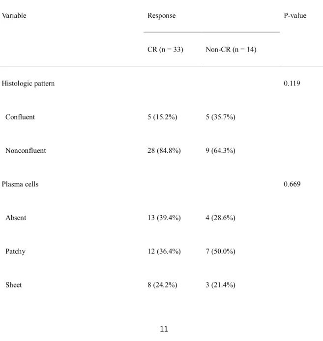
Risk factors for non-CR
Thirty-three patients were included in the CR group and 14 patients were included in the non-CR group (Table
normal cutoff value of 280U/L and the non-CR group was slightly above (237.33 U/L vs. 293.17 U/L, p = 0.046).
There were no statistically significant differences between the CR and non-CR groups for other histologic or clinical variables.
Table 2. Univariable analysis of clinicopathologic characteristics of the complete remission group versus
partial response, stable disease, and progression group in ocular adnexal MALT lymphoma.
Variable Response P-value
CR (n = 33) Non-CR (n = 14)
Histologic pattern 0.119
Confluent 5 (15.2%) 5 (35.7%)
Nonconfluent 28 (84.8%) 9 (64.3%)
Plasma cells 0.669
Absent 13 (39.4%) 4 (28.6%)
Patchy 12 (36.4%) 7 (50.0%)
Sheet 8 (24.2%) 3 (21.4%)
Sclerosis 0.283ᵇ
Absent 9 (27.3%) 7 (50.0%)
Mild 10 (30.3%) 2 (14.3%)
Moderate to severe 14 (42.4%) 5 (35.7%)
Lymphoepithelial lesions 0.199
Absent 13 (39.4%) 3 (21.4%)
Present 20 (60.6%) 11 (78.6%)
Large cell change 0.342
Absent 31 (93.9%) 12 (85.7%)
Present 2 (6.1%) 2 (14.3%)
Lymphovascular invasion 0.488
Absent 2 (6.1%) 0 (0%)
Present 31 (93.9%) 14 (100.0%)
Ki-67 9.94 (9.53) 10.93 (13.49) 0.806
IgG4/IgG ≥0.4 (n = 46) 0.251
Negative 29 (87.9%) 13 (100.0%)
Positive 4 (12.1%) 0 (0%)
IgH rearrangement (n = 35) 0.606
Monoclonal 11 (42.3%) 4 (44.4%)
Polyclonal 15 (57.7%) 5 (55.6%)
Age 52.4 (14.6) 50.5 (16.3) 0.701
Sex, n (%) 0.361
Male 20 (60.6%) 7 (50.0%)
Female 13 (39.4%) 7 (50.0%)
Laterality, n (%) 0.511
Unilateral 25 (75.8%) 10 (71.4%)
Bilateral 8 (24.2%) 4 (28.6%)
Mean duration of symptom (months) (n = 37) 7.6 (18.3) 21.7 (26.4) 0.144
Location
Conjunctiva only 16 (48.5%) 7 (50.0%)
Lacrimal gland 5 (15.2%) 1 (7.1%)
Extraocular muscle 0 (0%) 1 (7.1%)
Diffuse orbit 12 (36.4%) 5 (35.7%)
T stage 1.000ᵇ
T1 15 (45.5%) 7 (50.0%)
T2 15 (45.5%) 7 (50.0%)
T3 2 (6.1%) 0 (0%)
T4 1 (3.0%) 0 (0%)
Treatment 0.058ᵇ
Observation 7 (21.2%) 8 (57.1%)
Radiotherapy 24 (72.7%) 6 (42.9%)
Chemotherapy 2 (6.1%) 0 (0%)
RT dose (Gy) (n = 29) 24.00 (6.00) 24.60 (6.00) 0.902ᵃ
Failure-free survival 40.6 (21.8) 31.3 (21.2) 0.182
SUVmax(n = 44) 2.52 (1.30) 2.64 (1.78) 0.802
SUVmax(post-RT) (n = 18) 1.95 (0.67) 3.50 (2.07) 0.079ᵃ
LDH (U/L) (n = 45) 237.33(75.51) 293.17 (94.08) 0.046*
β2MG (mg/L) (n = 43) 1.82 (0.45) 1.58 (0.48) 0.133
CRP (n = 44) 0.191
Normal 17 (51.5%) 8 (72.7%)
Elevated 16 (48.5%) 3 (37.3%)
Serum IgG (mg/dL) (n = 27)
1268.850
(375.12)
1202.00 (442.55) 0.212ᵃ
Serum IgG4 (mg/dL) (n = 27) 63.65 (68.22) 183.00 (864.30) ᵃ0.618
Follow-up period (month) 40.6 (21.8) 47.3 (28.9) 0.387
CR, complete response; Gy, gray; SUV, standardized uptake value; RT, radiotherapy; LDH, lactate dehydrogenase;
β2MG, β2-microglobulin; CRP, C-reactive protein.
*p < 0.05,
ᵃMann–Whitney U test;
ᵇFisher’s exact test.
Values are presented as n (%) or mean (standard deviation) or median (interquartile range).
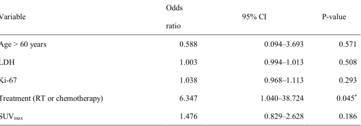
According to the result of the multivariable analysis, among the factors, namely, age over 60 years, LDH, Ki- 67, treatment including RT and chemotherapy, and SUVmaxof PET, it was found that the application of treatment was significantly associated with CR (odds ratio 6.347, 95% confidence interval 1.040–38.724, p = 0.045) (Table 3).
Table 3. Multivariable analysis of clinicopathologic factors regarding the non-complete response outcome
CI, confidence interval; LDH, lactate dehydrogenase; RT, radiotherapy; SUV, standardized uptake value.
Variable Odds
ratio 95% CI P-value
Age > 60 years 0.588 0.094–3.693 0.571
LDH 1.003 0.994–1.013 0.508
Ki-67 1.038 0.968–1.113 0.293
Treatment (RT or chemotherapy) 6.347 1.040–38.724 0.045*
SUVmax 1.476 0.829–2.628 0.186
*p < 0.05;
Nagelkerke R2= 0.247.
Clinicopathologic characteristics and risk factors for recurrence in the conjunctiva and non-conjunctiva groups
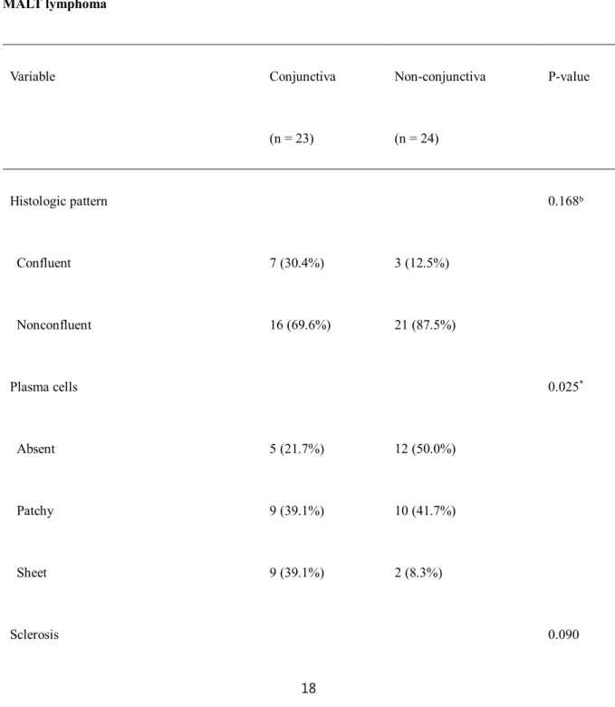
In the subgroup analysis based on the site of involvement, including only conjunctiva or other sites in ocular adnexa, some clinicopathologic characteristics differed between groups (Table 4). Histologically, the pattern of plasma cell infiltration and presence of lymphoepithelial lesions were different significantly (p = 0.025 and p = 0.000). The cases with conjunctival MALT lymphoma had more plasma cell infiltration in sheets. On the contrary, only 2 cases of non-conjunctival MALT lymphoma had plasma cell infiltration in sheets. Lymphoepithelial lesions were also observed more often in conjunctival MALT lymphoma. Sclerosis was more frequent in the non- conjunctival group but it was statistically not significant (19, 79.2% vs. 12, 52.2%, p = 0.090). Lymphovascular invasion was observed in most specimens and was seen in all cases in the non-conjunctiva group (91.3% vs. 100%, p = 0.234). A high IgG4/IgG ratio and IgH gene monoclonality were more common in the non-conjunctival groups, but were not statistically significant (4.5% vs. 12.5%, p = 0.609, 31.6% vs 56.3%, p = 0.142, respectively).
Clinically, eyelid swelling was the most common symptom in the non-conjunctiva group, and salmon patch was the most common symptom in the conjunctiva group (10, 41.8% and 8, 34.8%). Nine patients (39.1%) had bilateral conjunctival involvement and 3 patients (12.5%) in the non-conjunctiva group had bilateral involvement (p = 0.036). Most of the patients were T2 (21, 87.5%) in the non-conjunctiva group and most of the patients were T1 (22, 95.7%) in the conjunctiva group. Three cases (13.0% vs. 12.5%) had disease progression in both the conjunctiva group and the non-conjunctiva group. Treatment modality and SUVmaxdiffered significantly between the two groups (p = 0.001, and p = 0.001). Observation was more common in the conjunctiva group than in the non-conjunctiva group. The SUVmaxwas higher in the non-conjunctiva group (3.23 vs. 1.81, p = 0.001). More
patients had elevated serum CRP in the non-conjunctiva group than in the conjunctiva group but it was statistically not significant (15, 65.2% vs. 10. 47.6%, p = 0.239).
Table 4. Univariable analysis of clinicopathologic characteristics of conjunctival and non-conjunctival
MALT lymphoma
Variable Conjunctiva
(n = 23)
Non-conjunctiva
(n = 24)
P-value
Histologic pattern 0.168ᵇ
Confluent 7 (30.4%) 3 (12.5%)
Nonconfluent 16 (69.6%) 21 (87.5%)
Plasma cells 0.025*
Absent 5 (21.7%) 12 (50.0%)
Patchy 9 (39.1%) 10 (41.7%)
Sheet 9 (39.1%) 2 (8.3%)
Sclerosis 0.090
Absent 11 (47.8%) 5 (20.8%)
Mild 6 (26.1%) 6 (25.0%)
Moderate to severe 6 (26.1%) 13 (54.2%)
Lymphoepithelial lesions 0.000*
Absent 2 (8.7%) 14 (58.3%)
Present 21 (91.3%) 10 (41.7%)
Large cell change 1.000ᵇ
Absent 21 (91.3%) 22 (91.7%)
Present 2 (8.7%) 2 (8.3%)
Lymphovascular invasion 0.234ᵇ
Absent 2 (8.7%) 0 (0%)
Present 22 (91.3%) 24 (100.0%)
Ki-67 10.96 (13.08) 9.54 (8.06) 0.660
IgG4/IgG ≥ 0.4 (n = 46) 0.609ᵇ
Negative 21 (95.5%) 21 (87.5%)
Positive 1 (4.5%) 3 (12.5%)
IgH rearrangement (n = 32) 0.142
Monoclonal 6 (31.6%) 9 (56.3%)
Polyclonal 13 (68.4%) 7 (43.8%)
Age 47.9 (17.0) 55.5 (11.9) 0.084
Sex, n (%) 0.642
Male 14 (60.9%) 13 (54.3%)
Female 9 (39.1%) 11 (45.8%)
Laterality, n (%) 0.036*
Unilateral 14 (60.9%) 21 (87.5%)
Bilateral 9 (39.1%) 3 (12.5%)
Mean duration of symptom (months) (n = 37) 6.9 (15.6) 14.2 (24.1) 0.324
Location
Conjunctiva only 23 (100.0%) 0 (0%)
Lacrimal gland 0 (0%) 6 (25.0%)
Extraocular muscle 0 (0%) 1 (4.2%)
Diffuse orbit 0 (0%) 17 (70.8%)
Relapse 3 (13.0%) 3 (12.5%) 1.000ᵇ
Treatment 0.001*ᵇ
Observation 13 (56.5%) 2 (8.3%)
Radiotherapy 10 (43.5%) 20 (83.3%)
Chemotherapy 0 (0%) 2 (8.3%)
RT dose (Gy) (n = 29) 24.00 (3.00) 24.06 (6.00) 0.235ᵃ
Failure-free survival 39.1 (27.5) 36.5 (15.0) 0.689
SUVmax(n = 44) 1.81 (0.34) 3.23 (1.69) 0.001*
SUVmax(post-RT) (n = 18) 1.80 2.20 (1.50) 0.076ᵃ
LDH (U/L) (n = 45) 265.27 (95.78) 239.74 (69.83) 0.311
β2MG (mg/L) (n = 43) 1.70 (0.44) 1.80 (0.50) 0.480
CRP (n = 44) 0.239
Normal 11 (52.4%) 8 (34.8%)
Elevated 10 (47.6%) 15 (65.2%)
IgG (mg/dL) (n = 27) 1319.00 (434.250) 1218.70 (396.1) 0.781ᵃ
IgG4 (mg/dL) (n = 27) 50.60 (97.70) 77.85 (537.4) 0.561ᵃ
Follow-up period (month) 43.0 (27.8) 42.2 (20.4) 0.907
RT, radiotherapy; Gy, gray; SUV, standardized uptake value; RT, radiotherapy; LDH, lactate dehydrogenase;
β2MG, β2-microglobulin; CRP, C-reactive protein.
*p < 0.05;
ᵃMann–Whitney U test;
ᵇFisher’s exact test.
Values are presented with n (%) or mean (standard deviation) or median (interquartile range).
Three patients each had a recurrence in the group of patients with MALT lymphoma in the conjunctiva and
recurrence group, either in the conjunctiva or non-conjunctiva subgroup, but considering the 3 patients who had a recurrence in the non-conjunctiva group, Ki-67 labeling index in the recurred slide of the recurrence group were 10%, 10%, and 30%, respectively, which were higher than the mean value. In addition, serum LDH levels were higher in the recurrence group in both the conjunctiva and non-conjunctiva subgroups, although they were statistically not significant (median 225.00 U/L vs. 378.00 U/L, p = 0.408 and 217.00 U/L vs. 286.00 U/L, p = 0.309).
Table 5. Univariable analysis of clinicopathologic characteristics of conjunctival and non-conjunctival
MALT lymphoma
Conjunctival MALT lymphoma Non-conjunctival MALT lymphoma
Variable Non-
recurrence
(n = 20)
Recurrence
(n = 3)
P-value Non-
recurrence
(n = 21)
Recurrence
(n = 3)
P-
value
Histologic pattern 1.000 1.000
Confluent 6 (30.0%) 1 (33.3%) 3 (14.3%) 0 (0%)
Nonconfluent 14 (70.0%) 2 (66.7%) 18 (85.7%) 3 (100%)
Plasma cells 0.771 0.674
Absent 5 (25.0%) 0 (0%) 11 (52.4%) 1 (33.3%)
Patchy 8 (40.0%) 1 (33.3%) 8 (38.1%) 2 (66.7%)
Sheet 7 (35.0%) 2 (66.7%) 2 (9.5%) 0 (0%)
Sclerosis 0.404 0.769
Absent 8 (40.0%) 3 (100.0%) 4 (19.0%) 1 (33.3%)
Mild 6 (30.0%) 0 (0%) 6 (28.6%) 0 (0%)
Moderate to severe 6 (30.0%) 0 (0%) 11 (52.4%) 2 (66.7%)
Lymphoepithelial
lesions
1.000 0.059
Absent 2 (10.0%) 0 (0%) 14 (66.7%) 0 (0%)
Present 18 (90.0%) 3 (100.0%) 7 (33.3%) 3 (100%)
Large cell change 0.249 1.000
Absent 19 (95.0%) 2 (66.7%) 19 (90.5%) 3 (100%)
Present 1 (5.0%) 1 (33.3%) 2 (9.5%) 0 (0%)
Lymphovascular
invasion
1.000
Absent 2 (10.0%) 0 (0%) 0 (0%) 0 (0%)
Present 18 (90.0%) 3 (100.0%) 21 (100%) 3 (100%)
Ki-67 4.00 (9.00) 30.00 0.196 10.00 (7.50) 5.00 0.805
IgG4/IgG ≥ 0.4 (n =
22)
1.000 1.000
Negative 18 (94.7%) 3 (100.0%) 18 (85.7%) 3 (100%)
Positive 1 (5.3%) 0 (0%) 3 (14.3%) 0 (0%)
IgH rearrangement (n
= 19 and n = 16)
1.000 1.000
Monoclonal 5 (29.4%) 1 (50.0%) 8 (57.1%) 1 (50.0%)
Polyclonal 12 (70.6%) 1 (50.0%) 6 (42.9%) 1 (50.0%)
Age 54.5 (30.2) 35.0 0.898 56.0 (17.0) 56.0 0.805
Sex 0.538 0.082
Male 13 (65.0%) 1 (33.3%) 13 (61.9%) 0 (0%)
Female 7 (35.0%) 2 (66.7%) 8 (38.1%) 3 (100%)
Laterality 0.235 1.000
Unilateral 11 (55.0%) 3 (100.0%) 18 (85.7%) 3 (100%)
Bilateral 9 (45.0%) 0 (0%) 3 (14.3%) 0 (0%)
Mean duration of
symptom (month) (n =
14 and n = 23)
3.0 (3.7) 1.0 (0.0) 0.571 3.0 (14.3) 2.7 0.332
Location
Lacrimal gland 5 (23.8%) 1 (33.3%)
Extraocular muscle 1 (4.8%) 0 (0%)
Diffuse 15 (71.4%) 2 (66.7%)
T stage 1.000 1.000
T1 19 (95.0%) 3 (100.0%) 0 (0%) 0 (0%)
T2 1 (5.0%) 0 (0%) 18 (85.7%) 3 (100%)
T3 0 (0%) 0 (0%) 2 (9.5%) 0 (0%)
T4 0 (0%) 0 (0%) 1 (4.8%) 0 (0%)
Treatment 0.229 0.437
Observation 10 (50.0%) 3 (100.0%) 1 (4.8%) 1 (33.3%)
Radiotherapy 10 (50.0%) 0 (0%) 18 (85.7%) 2 (66.7%)
Chemotherapy 0 (0%) 0 (0%) 2 (9.5%) 0 (0%)
RT dose (Gy) (n = 9
and n= 20)
24.00 (3.00) 27.00 (6.00) 24.60 0.589
Failure-free survival
(month)
33.0 (39.5) 27.0 0.763 37.0 (20.0) 35.0 0.301
SUVmax(n = 21 and n
= 23)
1.80 (0.42) 1.80 0.814 3.00 (1.70) 2.20 0.268
SUVmax (post-RT) (n
= 3 and n = 15)
1.80 2.30 (1.50) 1.70 (0.00) 0.400
LDH (U/L) (n = 22
and n = 23)
225.00
(92.00)
378.00 0.408 217.00
(46.30)
286.00 0.309
β2MG (mg/L) (n = 20
and n= 23)
1.82 (0.75) 1.42 0.358 1.73 (0.50) 1.41 0.166
CRP (n = 21 and n =
23)
0.586 1.000
Normal 10 (55.6%) 1 (33.3%) 7 (35.0%) 1 (33.3%)
Elevated 8 (44.4%) 2 (66.7%) 13 (65.0%) 2 (66.7%)
IgG (mg/dL) (n = 9
and n = 18)
1319.00
(434.25)
1203.90
(374.80)
1419.00
(0.00)
0.444
IgG4 (mg/dL) (n = 9
and n = 18)
50.60 (97.70) 77.10
(280.60)
1480.00
(0.00)
0.111
Follow-up period
(month)
33.0 (39.5) 74.0 0.139 37.0 (25.0) 55.0 0.145
RT, radiotherapy; Gy, gray; SUV, standardized uptake value; RT, radiotherapy; LDH, lactate dehydrogenase;
β2MG, β2-microglobulin; CRP, C-reactive protein.
*P < 0.05.
Values are presented as n (%) or median (interquartile range).
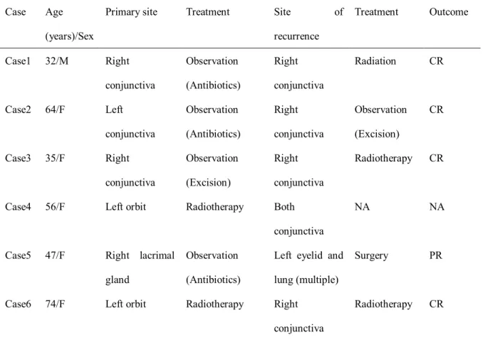
Clinicopathologic characteristics for recurred patients
The characteristics of recurred cases are shown in Table 6. Six cases (12.8%) had recurred. Three cases were primarily in the conjunctiva, and all were on observation. All recurred in the ipsilateral or contralateral conjunctiva.
Two received RT and 1 underwent surgery. All achieved CR. Of the three cases that recurred in non-conjunctiva, one case was present diffusely in the left orbit and after RT, and MALT lymphoma recurred in both conjunctiva after 35 months. Follow-up was lost since the patient moved to another hospital. One case recurred in the right lacrimal gland. She was on immunosuppression therapy for idiopathic thrombocytopenic purpura. She was on observation for MALT lymphoma with doxycycline there was recurrence in the left eyelid and lung after 35 months. Surgery was performed and she achieved a partial response. The last case was of MALT lymphoma diffusely in the left orbit including the conjunctiva. She received RT resulting in CR and had recurrence in 9 months in the right conjunctiva. RT was performed again and she achieved CR. Regarding the 4 cases with recurrence in sites other than the primary site, the Ki-67 labeling indexes were significantly higher in the primary site than in the site of recurrence (8.86% vs. 25.00%, p = 0.003).
Table 6. Clinical characteristics of recurred cases
M, male; F, female; CR, complete remission; NA, not available; PR, partial remission.
Discussions
While most ocular adnexal MALT lymphomas are reported to occur in the fifth to seventh decade of life, in the Korean population, it is reported to occur at a significantly younger age at the time of diagnosis, with a predominance in males rather than females [10]. The mean age at the time of diagnosis in our study was 51 years and a slight predominance in males was observed. The most common symptoms were salmon patch and mass,
Case Age
(years)/Sex
Primary site Treatment Site of
recurrence
Treatment Outcome
Case1 32/M Right
conjunctiva
Observation (Antibiotics)
Right conjunctiva
Radiation CR
Case2 64/F Left
conjunctiva
Observation (Antibiotics)
Right conjunctiva
Observation (Excision)
CR
Case3 35/F Right
conjunctiva
Observation (Excision)
Right conjunctiva
Radiotherapy CR
Case4 56/F Left orbit Radiotherapy Both
conjunctiva
NA NA
Case5 47/F Right lacrimal gland
Observation (Antibiotics)
Left eyelid and lung (multiple)
Surgery PR
Case6 74/F Left orbit Radiotherapy Right
conjunctiva
Radiotherapy CR
According to our study, serum LDH levels were higher in the non-CR group in the univariable analysis, although it was not significant in the multivariable analysis. It was higher in the recurrence group in both the conjunctiva and non-conjunctiva group but it was statistically not significant, partly because of a small number of recurred cases. According to the Warburg effect, altered metabolic pathway resulted in upregulation of LDH and it has been studied in many cancer types to show whether it can serve as a diagnostic, prognostic, or predictive marker [18]. LDH is an important prognostic factor for non-Hodgkin’s lymphoma described in the International Prognostic Index and is also reported in many studies [19,20]. Lu et al. showed that the 5th isotype of LDH level was associated with stage, extra-nodal site involvement, and WHO performance status in patients with non- Hodgkin’s lymphoma. Furthermore, they showed it was associated with tumor hypoxia and unfavorable prognosis, although there was no MALT lymphoma in the recruited cases [21]. In addition, SUVmax(both before and after RT) may be a potential prognostic factor although it was statistically not significant. [18F]FDG avidity has been reported to be related to the Ki-67 index, plasmacytic differentiation, tumor size, site of the disease, and morphological pattern of presentation [22-24]. The SUVmaxand post-RT SUVmaxwere higher in the non-CR group.
Considering the size and the stage, SUVmaxwas higher in the non-conjunctiva group compared to the conjunctiva group and post-RT SUVmaxwas also higher although it was statistically not significant. The histologic feature of the plasma cell infiltration pattern, which was more frequent in the conjunctiva was not consistent with other studies. Furthermore, the result of the subgroup analysis was limited by its small sample size. Whether [18F]FDG PET is useful in evaluating post-RT results and after other therapeutic modalities, remains unanswered in our study. According to multivariable analysis, treatments including chemotherapy and RT were important for CR.
The effectiveness of RT in localized disease has been proven to be associated with excellent outcomes in numerous studies [25,26]. Previously, the cutoff value of RT dose has been proposed to be approximately 30Gy, according to the outcome [11,12,27-29]. We also recommend RT rather than observation, surgery, or antibiotics. However, cataracts and dry eye syndrome were the main complications. In our study, only 2 cases received chemotherapy, and a few patients were in the advances stages, hence, we could not reach a conclusion. Two patients in stage T3 had lymphoma diffusely in the orbit and received RT which resulted in CR. One patient in stage T4 had lymphoma in the left orbit, both neck, left axilla, and right iliac lymph nodes, and was confirmed with orbital mass excision.
He had 8 cycles of chemotherapy including rituximab, cyclophosphamide, vincristine, and prednisone (R-CVP), which resulted in partial response. Generally, chemotherapy with rituximab is recommended for advanced stages [26].
Although MALT lymphoma shares many characteristics regardless of the anatomical site, there are also site- specific differences with respect to etiology, morphological features, molecular cytogenetic abnormalities, and the clinical course. Plasmacytic differentiation is observed in one-third of gastric MALT lymphoma cases and is frequent in cutaneous MALT lymphoma [30]. In our study, it was more frequent in conjunctival MALT lymphoma than in non-conjunctival MALT lymphoma (p = 0.025) but it was not related to treatment response in univariable analysis. In a study by Coupland et al., plasmacytic differentiation in ocular adnexal MALT lymphoma had a weak correlation with the development of systemic disease in univariate analyses but no statistical significance was found in multivariate analyses regarding development of local recurrence, systemic disease, and death related to lymphoma after adjusting for age and Ki-67 index [31]. However, there are conflicting results considering the relation between plasmacytic differentiation and prognosis, hence, further investigation may be helpful in the future.
CRP is an acute phase protein produced in the liver and is induced by interleukin-6 (IL-6) and other cytokines [32]. High levels of CRP in other cancer types, such as non-small cell carcinoma of the lung, breast, colon, and rectum, esophagus, head and neck, liver, ovary, prostate, pancreas, urinary system, and sarcomas are reported to be associated with disease severity or activity or prognosis [33]. Some also have reported the high serum level of CRP is related to the poor prognosis in lymphoma [32,34,35]. Twenty-five (56.8%) patients showed elevated serum CRP and the proportion of patients with elevated CRP was higher in the non-conjunctiva group compared to the conjunctiva group although it was not statistically significant. However, it was not associated with treatment response in our study.
In the result comparing conjunctiva and non-conjunctiva MALT lymphoma, bilaterality was more common in the conjunctiva group. In a study by Jung et al. concerning conjunctival lymphoproliferative lesions in South Korea, bilaterality was present in 50 patients (41.7%) in a total of 120 patients and it was found to be a much
higher proportion than reported in other studies. The study also found out that bilaterality was more common in follicular appearance than in salmon-patch appearance. Considering follicular appearance by slit-lamp examination was a predominant clinical presentation in chronic inflammation and lymphoid hyperplasia, according to their studies, the authors suggested follicular appearance reflects an extension from chronic inflammation and it was more likely to be bilateral [36]. Chronic antigenic stimulation has been postulated as etiology in MALT lymphoma and Chlamydia psittaci(Cp) has been known to be the causative pathogen for ocular adnexal MALT lymphoma. Cp positive rates in ocular adnexal MALT lymphoma have been variable geographically, from 0% to 87% [37]. Based on these studies, it can be suggested that chronic inflammation precedes MALT lymphoma, and accordingly, bilaterality was prevalent in conjunctival MALT lymphoma in our study also, and it was not associated with prognosis.
It is a diagnostic challenge to differentiate reactive lymphoid hyperplasia (RLH), atypical lymphoid hyperplasia (ALH), and MALT lymphoma. RLH partly shares a common etiology with MALT lymphoma, but is composed of polyclonal lymphoid cells. Dutcher’s bodies and immunoglobulin (Ig) light chain restriction are rarely observed [38]. Most cases in our study show lymphovascular invasion (95.7%), which is considered to be an important histologic finding that distinguishes between RLH and MALT lymphoma. Other studies suggest MALT lymphoma tend to occur in older age, and has a predilection for male, and tend to be localized, predominantly in the conjunctiva, compared with RLH [17,39,40]. A certain proportion of malignant transformation from RLH to malignant lymphoma has been reported, which is lower in the conjunctiva. Less aggressive treatment is recommended such as surgery, steroid, and observation [41]. In this study, we analyzed pathologic characteristics of MALT lymphoma in ocular adnexa, which might help the diagnosis of MALT lymphoma. Moreover, it was comparable with other studies.
In a study by Lagoo et al., 62 cases of ocular adnexal MALT lymphoma were classified as confluent, infiltrative, and mixed confluent and infiltrative and the proportion of cases were 74.5%, 13.7%, and 11.8%, respectively [14].
In a study by Ferry et al., including 116 cases of ocular adnexal MALT lymphoma, they observed abundant or aggregated plasma cells in 63 cases (35%), Dutcher bodies in 49 cases (27%), sclerosis in 36 cases (20%), and
with plasma cells, sclerosis, and lymphoepithelial lesions was greater in our study because cases with sites other than the lacrimal gland were included as lymphoepithelial lesions in our study. More lymphoepithelial lesions were observed in the conjunctiva than cases invading outside the conjunctiva. Although the result was concordant with the above study that the plasma cells and Dutcher bodies were frequently observed, it was discordant that we observed lymphoepithelial lesions and sclerosis more often.
The Ki-67 labeling index has shown prognostic potential in our study. The insignificance may be due to the small number of patients with recurrence. A few studies have validated the Ki-67 labeling index as a prognostic factor in MALT lymphoma. In a study by Petit et al., including patients with marginal zone B-cell lymphomas and lymphoplasmacytic lymphomas, absence of expression of both Ki-67 and interferon regulatory factor4 (IRF4 or multiple myeloma oncogene-1-protein, MUM1), which was found to be related to plasma-cell differentiation in the same study, was associated with a better prognosis [42]. However, in a study by Albano et al., the Ki-67 index was not associated with progression-free survival nor overall survival according to univariate and multivariate analysis [22]. The Ki-67 labeling index in MALT lymphoma is generally low considering its indolent nature; however, our study implies that high values should be treated with caution. Two issues need to be addressed.
One is how to accurately count Ki-67 labeling index excluding reactive cells and the second is how to set up a cutoff value, but there is a possibility that we might not be able to initially evaluate its potential. Recently we encountered a recurred case that is not included in the current study. He had MALT lymphoma in the right lacrimal gland initially and received RT resulting in CR. After 48 months, the lymphoma recurred on the right cheek and another RT was done resulting in CR. After 26 months, the lymphoma recurred again in the lung, and a left lower lobe lobectomy was done. The histology showed transformation into diffuse large B cell lymphoma, non-germinal center cell-like type with Ki-67 index of 70% (Fig 2). The Ki-67 index of the previous two sites was 30%
respectively and the results of IgH gene rearrangement studies showed polyclonality for all three sites. He is currently on R-CHOP chemotherapy (rituximab, cyclophosphamide, doxorubicin, vincristine, and prednisone).
To set a cutoff value for the Ki-67 index, more studies are necessary and it must be validated in larger studies.
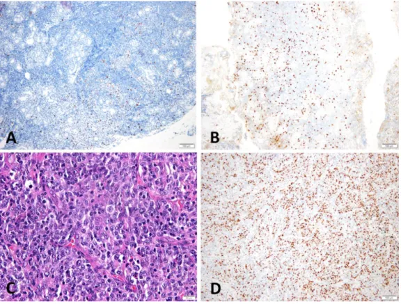
Figure 2. Images of Ki-67 index in case5 and diffuse large B cell lymphoma transformed case
(A, B) Immunohistochemistry stain for Ki-67 shows nuclear positivity in 5% of tumor cells initially, but nuclear positivity increases to 30% when there is recurrence in the eyelid. (Ki-67, A; x100, B; x100). (C, D) Transformed cells are scattered between small lymphoid cells and the Ki-67 stain shows increment (H&E, A; x400, Ki-67, B;
x100).
Although most cases of MALT lymphoma express IgM and develop from a T-helper type 1 (Th1) inflammatory environment, MALT lymphomas expressing other class-switched immunoglobulins, including IgG4 develop, from a T-helper type 2 (Th2) inflammatory environment [43]. Several studies have been performed on IgG4- expressing MALT lymphoma among cutaneous lymphomas because of the high proportion of positive cases. A Th2 immune response with an increased number of regulatory T cells predisposed this condition, although the ratio of CD4/CD8-positive T cells differed between studies [44,45]. Moreover, in a study by Brenner et al., IgG4
lymphoma slightly differed regarding the outcomes. In 5 (10%) of 50 patients positive for IgG4, Lee et al. found characteristic histological features such as extensive plasma cell infiltration and dense fibrosis. Most of these patients had lymphoma of the lacrimal gland (4 of 5 cases) and showed a lower response rate to initial treatment [46]. However, Sumii et al. showed that the standard treatment for marginal zone B-cell lymphoma is appropriate for IgG4 producing MALT lymphomas in a cases series of 7 patients, including 5 cases of ocular adnexal MALT lymphomas [47]. In our study, 4 cases (8.5%) were positive for IgG4, in which 3 cases (75%) had patchy patterned plasma cells infiltration, 2 cases (50%) had sclerosis as indicated earlier (1 mild, 1 moderate to severe), and 2 cases were in the lacrimal gland, 1 in the conjunctiva, and 1 was diffuse. All patients received RT and achieved complete remission. In a study by Kubota et al., 10 cases (9%) were positive for IgG4, 10 (100%) were associated with sclerosis, and 8 (80%) were associated with reactive follicles [48]. The relatively small percentage of sclerosis in our study might because of not only a small number of cases but also cases involving the conjunctiva.
Some researchers have suggested that MALT lymphoma can develop in the background of IgG4-related disease, and Ohno et al. suggested that IgG4-associated MALT lymphoma may have a unique pathogenesis [49-51].
Further studies are needed to elucidate the detailed pathogenesis and prognosis of this disease.
The positive result of the IgH gene rearrangement study was only found in 15 (42.9%) cases in the entire group and 6 (31.6%) and 9 (56.3%) cases in the conjunctiva and non-conjunctiva group, respectively. It was consistent with previous studies that the positive rate was present in 25%–70% of MALT lymphoma cases by polymerase chain reaction with a considerable false-negative rate [39]. The reasons for false-negative rate include limited sensitivity, related to normal polyclonal background, improper primer annealing and occurrence of somatic hypermutations in rearranged Ig genes, which are particularly found in germinal center and postgerminal center- derived B-cell malignancies [52]. Other suggested causes are clones expressing incomplete rearrangements, or exceedingly rarely, chromosomal translocations involving the Ig locus, or fixation or sampling error [53].
Underlying or adjacent inflammation can result in false negatives which are included in sampling errors. In our opinion, the negative result of the IgH gene rearrangement study should not preclude the diagnosis of MALT lymphoma, and it should be considered carefully along with histology and IHC studies. In addition, it should be
sites, in case 5 which the primary site was the right lacrimal gland and there was recurrence in the eyelid and lung, and another recurred case described above which recurred in right cheek and lung. There is a possibility that an association between the result of polyclonality and treatment response exists, which should be studied in the future.
The practical value of the IgH gene rearrangement study can be questioned since it is only positive in 42.9% of patients and is not related to response to treatment. But it is stated that the majority of lymphoid malignancy (>
98%) contains identically or clonally rearranged Ig, so despite the drawbacks, in about 5% to 10% of cases where distinguishing between reactive lymphoproliferation and malignancy is complicated, it can still be assumed to be beneficial [52].
Our study had several limitations. First, there was bias associated with the retrospective nature. Second, a small number of cases were analyzed because of the rare prevalence of ocular adnexa MALT lymphoma. Six cases among 47 cases and 3 cases each in the conjunctiva and non-conjunctiva subgroups experienced recurrence. Third, sampling error or reproducibility problems could be an issue. But careful examination was performed, few photographs were taken, and for the Ki-67 index, two pathologists separately counted the positive cells and reached an agreement. Care was taken to exclude reactive cells including the germinal center.
The major strength of our study is that it analyzes relatively complete data of pathological as well as clinical characteristics in ocular adnexal MALT lymphoma, which is a rare disease, at a single tertiary institution with a sufficient follow-up period.
Conclusion
In conclusion, active treatment is recommended even in the early stages of ocular adnexal MALT lymphoma, especially when the Ki-67 index and serum LDH levels are high. Additionally, for accurate diagnosis, the polyclonal result seen in the IgH gene rearrangement study should not be solely considered in order to exclude the diagnosis of MALT lymphoma; the result should be interpreted in combination with other findings. This will help accurately diagnose ocular adnexal MALT lymphoma and investigate related predictive and prognostic
References
1. Shields CL, Chien JL, Surakiatchanukul T, Sioufi K, Lally SE, Shields JA. Conjunctival Tumors: Review of Clinical Features, Risks, Biomarkers, and Outcomes--The 2017 J. Donald M. Gass Lecture. Asia Pac J Ophthalmol (Phila) 2017;6:109-20.
2. Verdijk RM. Lymphoproliferative Tumors of the Ocular Adnexa. Asia Pac J Ophthalmol (Phila) 2017;6:132-42.
3. Olsen TG, Holm F, Mikkelsen LH, Rasmussen PK, Coupland SE, Esmaeli B, et al. Orbital Lymphoma- An International Multicenter Retrospective Study. Am J Ophthalmol 2019;199:44-57.
4. Oh DE, Kim YD. Lymphoproliferative diseases of the ocular adnexa in Korea. Arch Ophthalmol 2007;125:1668-73.
5. Ferry JA, Fung CY, Zukerberg L, Lucarelli MJ, Hasserjian RP, Preffer FI, et al. Lymphoma of the ocular adnexa: A study of 353 cases. Am J Surg Pathol 2007;31:170-84.
6. Kirkegaard MM, Rasmussen PK, Coupland SE, Esmaeli B, Finger PT, Graue GF, et al. Conjunctival Lymphoma--An International Multicenter Retrospective Study. JAMA Ophthalmol 2016;134:406-14.
7. Graue GF, Finger PT, Maher E, Della Rocca D, Della Rocca R, Lelli GJ, Jr., et al. Ocular adnexal lymphoma staging and treatment: American Joint Committee on Cancer versus Ann Arbor. Eur J Ophthalmol 2013;23:344-55.
8. Jeon YW, Yang HJ, Choi BO, Jung SE, Park KS, O JH, et al. Comparison of Selection and Long-term Clinical Outcomes Between Chemotherapy and Radiotherapy as Primary Therapeutic Modality for Ocular Adnexal MALT Lymphoma. EClinicalMedicine 2018;4-5:32-42.
9. Cerhan JR, Habermann TM. Epidemiology of Marginal Zone Lymphoma. Ann Lymphoma 2021;5.
10. Stefanovic A, Lossos IS. Extranodal marginal zone lymphoma of the ocular adnexa. Blood 2009;114:501-10.
11. Parikh RR, Moskowitz BK, Maher E, Della Rocca D, Della Rocca R, Culliney B, et al. Long-term outcomes and patterns of failure in orbital lymphoma treated with primary radiotherapy. Leuk Lymphoma 2015;56:1266-70.
IE ocular adnexal mucosa-associated lymphoid tissue lymphomas: long-term results. Radiat Oncol 2020;15:25.
13. Cheson BD, Pfistner B, Juweid ME, Gascoyne RD, Specht L, Horning SJ, et al. Revised response criteria for malignant lymphoma. J Clin Oncol 2007;25:579-86.
14. Lagoo AS, Haggerty C, Kim Y, Hammons M, Neufeld K, Redher C, et al. Morphologic features of 115 lymphomas of the orbit and ocular adnexa categorized according to the World Health Organization classification: are marginal zone lymphomas in the orbit mucosa-associated lymphoid tissue-type lymphomas? Arch Pathol Lab Med 2008;132:1405-16.
15. Deshpande V, Zen Y, Chan JK, Yi EE, Sato Y, Yoshino T, et al. Consensus statement on the pathology of IgG4-related disease. Mod Pathol 2012;25:1181-92.
16. Kwon M, Lee JS, Lee C, Yoon DH, Sa HS. Prognostic factors for relapse and survival among patients with ocular adnexal lymphoma: validation of the eighth edition of the American Joint Committee on Cancer (AJCC) TNM classification. Br J Ophthalmol 2021;105:279-84.
17. Asadi-Amoli F, Nozarian Z, Bonaki HN, Mehrtash V, Entezari S. Clinicopathologic Assessment of Ocular Adnexal Lymphoproliferative Lesions at a Tertiary Eye Hospital in Iran. Asian Pac J Cancer Prev 2016;17:3727-31.
18. Forkasiewicz A, Dorociak M, Stach K, Szelachowski P, Tabola R, Augoff K. The usefulness of lactate dehydrogenase measurements in current oncological practice. Cell Mol Biol Lett 2020;25:35.
19. Bayraktar S, Bayraktar UD, Stefanovic A, Lossos IS. Primary ocular adnexal mucosa-associated lymphoid tissue lymphoma (MALT): single institution experience in a large cohort of patients. Br J Haematol 2011;152:72-80.
20. Thieblemont C, Cascione L, Conconi A, Kiesewetter B, Raderer M, Gaidano G, et al. A MALT lymphoma prognostic index. Blood 2017;130:1409-17.
21. Lu R, Jiang M, Chen Z, Xu X, Hu H, Zhao X, et al. Lactate dehydrogenase 5 expression in Non-Hodgkin lymphoma is associated with the induced hypoxia regulated protein and poor prognosis. PLoS One 2013;8:e74853.
22. Albano D, Bosio G, Camoni L, Farina M, Re A, Tucci A, et al. Prognostic role of baseline (18) F-FDG PET/CT parameters in MALT lymphoma. Hematol Oncol 2019;37:39-46.
23. Hoffmann M, Kletter K, Becherer A, Jager U, Chott A, Raderer M. 18F-fluorodeoxyglucose positron emission tomography (18F-FDG-PET) for staging and follow-up of marginal zone B-cell lymphoma.
Oncology 2003;64:336-40.
24. Albano D, Bosio G, Giubbini R, Bertagna F. 18F-FDG PET/CT and extragastric MALT lymphoma: role of Ki-67 score and plasmacytic differentiation. Leuk Lymphoma 2017;58:2328-34.
25. Masuda Y, Takeuchi K, Kodama T, Fujisaki T, Imaizumi Y, Otsuka E, et al. Treatment-associated outcomes of patients with primary ocular adnexal MALT lymphoma after accurate diagnosis. Int J Clin Oncol 2019;24:1620-8.
26. Hindso TG, Esmaeli B, Holm F, Mikkelsen LH, Rasmussen PK, Coupland SE, et al. International multicentre retrospective cohort study of ocular adnexal marginal zone B-cell lymphoma. Br J Ophthalmol 2020;104:357-62.
27. Lee JL, Kim MK, Lee KH, Hyun MS, Chung HS, Kim DS, et al. Extranodal marginal zone B-cell lymphomas of mucosa-associated lymphoid tissue-type of the orbit and ocular adnexa. Ann Hematol 2005;84:13-8.
28. Desai A, Joag MG, Lekakis L, Chapman JR, Vega F, Tibshirani R, et al. Long-term course of patients with primary ocular adnexal MALT lymphoma: a large single-institution cohort study. Blood 2017;129:324-32.
29. Harada K, Murakami N, Kitaguchi M, Sekii S, Takahashi K, Yoshio K, et al. Localized ocular adnexal mucosa-associated lymphoid tissue lymphoma treated with radiation therapy: a long-term outcome in 86 patients with 104 treated eyes. Int J Radiat Oncol Biol Phys 2014;88:650-4.
30. James R. Cook PGI, Andreas Chott, Shigeo Nakamura, Hans Konrad Müller-Hermelink, Nancy Lee Harris, Steven H. Swerdlow. Tumours of Haematopoietic and Lymphoid Tissues 2017 (Beta). 2019.
31. Coupland SE, Hellmich M, Auw-Haedrich C, Lee WR, Anagnostopoulos I, Stein H. Plasmacellular differentiation in extranodal marginal zone B cell lymphomas of the ocular adnexa: an analysis of the neoplastic plasma cell phenotype and its prognostic significance in 136 cases. Br J Ophthalmol 2005;89:352-9.
32. Legouffe E, Rodriguez C, Picot MC, Richard B, Klein B, Rossi JF, et al. C-reactive protein serum level is a valuable and simple prognostic marker in non Hodgkin's lymphoma. Leuk Lymphoma 1998;31:351-
7.
33. Hart PC, Rajab IM, Alebraheem M, Potempa LA. C-Reactive Protein and Cancer-Diagnostic and Therapeutic Insights. Front Immunol 2020;11:595835.
34. Lu J, Wu Y, Li B, Luo X, Zhang W, Zeng Y, et al. Predictive value of serological factors, maximal standardized uptake value and ratio of Ki67 in patients diagnosed with non-Hodgkin's lymphoma. Oncol Lett 2020;20:47.
35. Herishanu Y, Perry C, Braunstein R, Metser U, Goor O, Rogowski O, et al. Early-mid treatment C- reactive protein level is a prognostic factor in aggressive non-Hodgkin's lymphoma. Eur J Haematol 2007;79:150-4.
36. Jung SK, Paik JS, Park GS, Cho SG, Yang SW. Refractory follicular conjunctival lesions: overlook as just inflammation or not? Br J Ophthalmol 2019;103:1660-5.
37. Collina F, De Chiara A, De Renzo A, De Rosa G, Botti G, Franco R. Chlamydia psittaci in ocular adnexa MALT lymphoma: a possible role in lymphomagenesis and a different geographical distribution. Infect Agent Cancer 2012;7:8.
38. Ueda S, Usui Y, Nagai T, Diaz-Aguilar D, Nagao T, Goto H. Immunophenotypic profiles for distinguishing orbital mucosa-associated lymphoid tissue lymphoma from benign lymphoproliferative tumors. Jpn J Ophthalmol 2017;61:354-60.
39. Mannami T, Yoshino T, Oshima K, Takase S, Kondo E, Ohara N, et al. Clinical, histopathological, and immunogenetic analysis of ocular adnexal lymphoproliferative disorders: characterization of malt lymphoma and reactive lymphoid hyperplasia. Mod Pathol 2001;14:641-9.
40. Qu XL, Hei Y, Kang L, Yang XJ, Wang Y, Lu XZ, et al. Establishment of a combination scoring method for diagnosis of ocular adnexal lymphoproliferative disease. PLoS One 2017;12:e0160175.
41. Klavdianou O, Kondylis G, Georgopoulos V, Palioura S. Bilateral benign reactive lymphoid hyperplasia of the conjunctiva: a case treated with oral doxycycline and review of the literature. Eye Vis (Lond) 2019;6:26.
42. Petit B, Chaury MP, Le Clorennec C, Jaccard A, Gachard N, Moalic-Judge S, et al. Indolent lymphoplasmacytic and marginal zone B-cell lymphomas: absence of both IRF4 and Ki67 expression identifies a better prognosis subgroup. Haematologica 2005;90:200-6.
43. van Maldegem F, van Dijk R, Wormhoudt TA, Kluin PM, Willemze R, Cerroni L, et al. The majority of cutaneous marginal zone B-cell lymphomas expresses class-switched immunoglobulins and develops in a T-helper type 2 inflammatory environment. Blood 2008;112:3355-61.
44. Carlsen ED, Swerdlow SH, Cook JR, Gibson SE. Class-switched Primary Cutaneous Marginal Zone Lymphomas Are Frequently IgG4-positive and Have Features Distinct From IgM-positive Cases. Am J Surg Pathol 2019;43:1403-12.
45. Brenner I, Roth S, Puppe B, Wobser M, Rosenwald A, Geissinger E. Primary cutaneous marginal zone lymphomas with plasmacytic differentiation show frequent IgG4 expression. Mod Pathol 2013;26:1568- 76.
46. Lee MJ, Kim N, Choe JY, Khwarg SI, Jeon YK, C{Lee h, H. K., et al. Clinicopathological Analysis of Ocular Adnexal Extranodal Marginal Zone B-Cell Lymphoma with IgG4-Positive Cells. PLoS One 2015;10:e0131458.
47. Sumii Y, Asada N, Sato Y, Ohshima KI, Makita M, Yoshimoto Y, et al. Treatment outcomes of IgG4- producing marginal zone B-cell lymphoma: a retrospective case series. Int J Hematol 2020;112:780-6.
48. Kubota T, Moritani S, Yoshino T, Nagai H, Terasaki H. Ocular adnexal marginal zone B cell lymphoma infiltrated by IgG4-positive plasma cells. J Clin Pathol 2010;63:1059-65.
49. Nakayama R, Matsumoto Y, Horiike S, Kobayashi S, Nakao R, Nagoshi H, et al. Close pathogenetic relationship between ocular immunoglobulin G4-related disease (IgG4-RD) and ocular adnexal mucosa- associated lymphoid tissue (MALT) lymphoma. Leuk Lymphoma 2014;55:1198-202.
50. Ohno K, Sato Y, Ohshima K, Takata K, Miyata-Takata T, Takeuchi M, et al. A subset of ocular adnexal marginal zone lymphomas may arise in association with IgG4-related disease. Sci Rep 2015;5:13539.
51. Go H, Kim JE, Kim YA, Chung HK, Khwarg SI, Kim CW, et al. Ocular adnexal IgG4-related disease:
comparative analysis with mucosa-associated lymphoid tissue lymphoma and other chronic inflammatory conditions. Histopathology 2012;60:296-312.
52. van Dongen JJ, Langerak AW, Bruggemann M, Evans PA, Hummel M, Lavender FL, et al. Design and standardization of PCR primers and protocols for detection of clonal immunoglobulin and T-cell receptor gene recombinations in suspect lymphoproliferations: report of the BIOMED-2 Concerted Action BMH4-CT98-3936. Leukemia 2003;17:2257-317.
53. Bertoni F, Cotter FE, Zucca E. Molecular genetics of extranodal marginal zone (MALT-type) B-cell lymphoma. Leuk Lymphoma 1999;35:57-68.
Abstract
Background: Extranodal marginal zone lymphoma of the mucosa-associated lymphoid tissue(MALT) type is the most common subtype of ocular adnexal lymphoma. Despite its excellent prognosis, some have not shown complete remission(CR). We aimed to determine clinicopathologic differences in the treatment responder group but did not show complete remission.
Methods: This study retrospectively reviewed the data of 47 patients who were diagnosed with ocular adnexal MALT lymphoma at Ulsan University Hospital between March 2002 and August 2018. Patients who were followed up <6 months were excluded. Clinicopathologic features were analyzed. Among the 47 patients, 33 patients achieved CR, and 14 patients achieved non-CR during the mean follow-up period of 42.6 months(range 7–109 months). In the univariable analysis, serum lactate dehydrogenase(LDH) levels were found to be higher in the non-CR group. In the multivariable analysis, the application of treatment including radiotherapy(RT) or chemotherapy was associated with CR(odds ratio 6.347, 95% confidence interval 1.040–38.724, p=0.045). In subgroup analysis according to the site of involvement, none of the variables differed significantly between the two groups, the group including only conjunctiva, and the group including non-conjunctiva. Six(12.8%) cases had recurred in total and cases with a recurring site other than the primary site had higher Ki-67 labeling index.
Conclusions: Our study suggests several histologic features in ocular adnexal MALT lymphoma and differences according to the sites. Although belonging to early stages, the non-CR rate was high in patients with high serum LDH levels and treatment including radiotherapy or chemotherapy was essential. Thus, considering active treatment is recommended
Keywords: MALT lymphoma; Ocular adnexa; Conjunctiva; Prognostic factor