INTRODUCTION
Extranodal lymphoma of mucosa-associated lymphoid tis- sue (MALT) was first described by Isaacson and Wright in 1983 (1) and was recently reclassified as “extranodal marginal zone B-cell lymphoma (MZBL) of MALT-type”according to the REAL (2) and WHO classifications (3). Primary MZBLs of MALT-type have been described in a variety of sites, includ- ing gastrointestinal tract (1), thyroid gland (4) , lung (5), sali- vary gland (6), and ocular adnexa (7) associated with clinical setting of autoimmune diseases or chronic inflammation, usu- ally Sjogren’s syndrome, Hashimoto’s thyroiditis, and Heli- cobacter pylori infection. Thymic MZBL of MALT-type is an extremely rare disease with only 22 cases having been report- ed in the literature to date and the largest series has included 15 cases (8-19). In this paper, I report a case of low-grade MZBL of MALT-type in the thymus of a patient with Sjo- gren’s syndrome and rheumatoid arthritis.
CASE REPORT
A 43-yr-old Korean woman presented with a long history
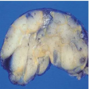
of Sjogren’s syndrome and rheumatoid arthritis. She had com- plained of dry eyes, dry mouth, and arthralgia for about 16 yr and also complained of both parotid swelling that had devel- oped 3 months before. The symmetrical swelling of the wrist, metacarpophalangeal, and proximal interphalangeal joints was accompanied by morning stiffness and pain. On admission, antinuclear antibody (ANA) revealed a speckled pattern and showed positive reaction for SS-A/Ro antibody and negative for the SS-B/La antibody. Rheumatoid factor was positive (147.3 IU/mL). A lobulated anterior mediastinal mass was detected by computerized tomography (Fig. 1) and the thy- mus was resected through median sternotomy. There were no symptoms of myasthenia gravis. The resected mass was con- fined within the thymus and did not show invasion to the surrounding structures. The specimen consisted of a solid, smooth, lobulated, encapsulated mass that measured 7 cm in maximum diameter and weighed 79 g. The cut surface was pale pink to tan, and showed homogenous appearance with some small cysts (Fig. 2). The tissue was fixed in 10% neutral buffered formalin and embedded in paraffin. Sections were stained with hematoxylin and eosin and periodic acid-Schiff.
Histologically, the normal architecture of the thymus was dif- fusely effaced by a dense infiltration of lymphoid cells. Reac-
Jin-Man Kim
Department of Pathology, Chungnam National University School of Medicine, Daejeon, Korea
Address for correspondence Jin-Man Kim, M.D.
Department of Pathology, Chungnam National University School of Medicine, 6 Munhwa-1-dong, Jung-gu, Daejeon 301-131, Korea
Tel : +82.42-580-8237, Fax : +82.42-581-5233 E-mail: [email protected]
897 J Korean Med Sci 2003; 18: 897-900
ISSN 1011-8934
Copyright � The Korean Academy of Medical Sciences
Primary Extranodal Marginal Zone B-cell Lymphoma of Mucosa-Associated Lymphoid Tissue-type in the Thymus of a Patient with Sjogren's Syndrome and Rheumatoid Arthritis
Primary thymic marginal zone B-cell lymphoma (MZBL) of mucosa-associated lymphoid tissue (MALT)-type is a very rare disease with distinct clinicopathologic features. I herein report a rare case of primary thymic MZBL of MALT-type arising in the thymus in a patient with Sjogren’s syndrome and rheumatoid arthritis. A mediastinal mass was detected by computerized tomography in a 43-yr-old Korean woman with a history of Sjogren’s syndrome and rheumatoid arthritis and the thy- mus was resected through median sternotomy. The solid and nodular tumor (7×6
×3 cm) was confined in the thymus. Histologically, the lymphoid infiltrate com- prised monotonous centrocyte-like cells with monocytoid cells, small lymphocytes, and plasma cells. Prominent lymphoepithelial lesions were formed by centrocyte-like cells infiltrating the Hassall’s corpuscles. Immunohistochemically, the tumor cells were positive for CD20, CD79a, and bcl-2 and negative for CD3, CD5, CD10, CD23, and bcl-6. IgA and kappa light chain restriction were also found in plasma cells in the tumor. Sjogren’s syndrome and rheumatoid arthritis are known to be associated with MALT lymphoma and were considered to play an important role in the devel- opment of malignant lymphoma in this patient.
Key Words : Lymphoma, B-cell; Lymphoma Mucosa-Associated Lymphoid Tissue; Sjogren’s Syn- drome; Arthritis; Rheumatoid; Thymus Neoplasms
Received : 12 November 2002 Accepted : 18 December 2002 . .
. .
. .
. .
. .
. .
. .
. .
898 J.-M. Kim
tive lymphoid follicles with germinal centers could be iden- tified within the tumor (Fig. 3A). The microscopic cysts filled with eosinophilic proteinaceous fluid were lined by attenuat- ed epithelium infiltrated by centrocyte-like (CCL) cells (Fig.
3B). The lymphoid infiltrate comprised monotonous CCL cells with monocytoid cells and plasma cells (Fig. 3C, D). There were considerable number of plasma cells and small lympho- cytes. A few large transformed cells were also recognized (Fig.
Fig. 1.CT scan shows a relatively well-demarcated lobulated solid mass in the anterior mediastinum.
Fig. 2.The cut surface of the thymic tumor shows pale pink to tan, solid, homogeneous and lobulated tissue with some small cysts.
Fig. 3.The low-power view shows totally effaced architecture with diffuse lymphoid infiltration and interspersed reactive lymphoid fol- licles (A, H&E, ×200). Focal variable-sized cysts containing eosi- nophilic fluid (B, H&E, ×100). Neoplastic cells are predominantly composed of centrocyte-like (CCL) cells, small lymphocytes, and plasma cells (C, H&E, ×400). Sheets of CCL cells with monocy- toid feature (D, H&E, ×400).
A B
C D
Fig. 4.Infiltration and expansion of Hassall’s corpuscles by neoplas- tic cells forming a lymphoepithelial lesion is noted (A, H&E, ×400) and highlightened by an immunostaining for cytokeratin (B,×400).
Thymic lymphoma showing positive reaction for CD20 (C, ×400).
Some plasma cells show cytoplasmic staining for kappa light chain (D,×400), whereas lambda light chain is negative (E,×400).
A B
C D E
3C). Prominent lymphoepithelial lesions (LEL), which were highlightened by cytokeratin staining, were formed by cen- trocyte-like cells infiltrating and expanding the Hassall’s cor- puscles (Fig. 4A, B). Immunohistochemical studies of paraffin sections were performed with the use of LSAB kit (DAKO, Carpinteria, CA, U.S.A.). The primary antibodies for the kappa light chain, lambda light chain, and CD3 were purchased from DiNona, Korea and primary antibodies for the CD20, CD79a, bcl-2, bcl-6, CD5, CD23, CD10, and cytokeratin were pur- chased from DAKO. Immunohistochemically, the CCL cells were positive for CD20, CD79a, and bcl-2 indicating B-cell phenotype (Fig. 4C) and negative for CD3, CD5, CD10, CD23, and bcl-6. Kappa light chain restriction was also found in pl- asma cells in the tumor confirming monoclonality of B-cell proliferation (Fig. 4D, E). The tumor cells showed IgA heavy- chain type. A variable number of CD3-positive T cells were admixed with CCL cells. Postoperative adjuvant chemother- apy or radiation therapy was not given. The patient has been followed up for 11 months without evidence of recurrent tumor.
DISCUSSION
I herein report a case of primary extranodal MZBL of MALT-type in the thymus associated with Sjogren’s syndrome and rheumatoid arthritis. MZBL of MALT-type is character- ized by an indolent clinical course and has been described in a variety of sites (1, 4-7) associated with clinical setting of au- toimmune diseases and chronic inflammation, usually Sjogren’s syndrome, Hashimoto’s thyroiditis, and Helicobacter pylori infec- tions which suggests proliferation of the lymphoma cells de- pend on the presence of antigen-driven T cells.
Thymic MZBL of MALT-type is an extremely rare disease with only 22 cases having been reported in the literature to date, including one Korean case (8-19). According to Inagaki et al. (19) who systematically investigated 15 cases of thymic MALT lymphoma, this tumor entity revealed prevalence in Asians, marked female predilection, strong association with autoimmune diseases, especially Sjogren’s syndrome, mostly IgA phenotype, and consistent lack of API2-MLT1 gene fu- sion, a recently reported MALT lymphoma-specific gene abnor- mality. They proposed that thymic MALT lymphoma is a dis- tinct subgroup of MALT lymphoma. The association with rheumatoid arthritis was reported in only one case (11). No cases associated with both Sjogren’s syndrome and rheuma- toid arthritis as in my case were reported.
B-cell lymphomas other than MALT lymphoma arise in the thymus, especially mediastinal large B-cell lymphoma, which has been included in the REAL (2) and WHO classifications (3). In contrast to MALT lymphoma which shows an indolent course, this tumor is a highly aggressive lymphoma with a poor prognosis. The mixture of epithelial elements and small lymphocytes could be confused with more common thymoma.
In type B thymomas, the lymphoid cells usually consist of
small immature lymphocytes of T-cell immunophenotype, whereas in the thymic MALT lymphoma there is an admix- ture of small lymphocytes, CCL cells, a few blastoid cells, and plasma cells showing mostly B-cell immunophenotype.
In summary, I described herein is a thymic low-grade ex- tranodal MZBL of MALT-type in a patient with both Sjogren’s syndrome and rheumatoid arthritis. It is possible that the ly- mphoma arose on the underlying basis of long standing auto- immune diseases. Pathologists should be aware that MZBL of MALT-type may occasionally involve thymus. In a small biopsy, the presence of mixture of small lymphoid cells and epithelial cells may closely mimic a thymoma. Morphologic features of CCL cells, plasmacytoid cells, lymphoepithelial lesions as well as immunophenotype or molecular studies are helpful for a definitive diagnosis.
REFERENCES
1. Isaacson P, Wright DH. Malignant lymphoma of mucosa-associated lymphoid tissue. A distinctive type of B-cell lymphoma. Cancer 1983;
52: 1410-6.
2. Harris NL, Jaffe ES, Stein H, Banks PM, Chan JK, Cleary ML, Del- sol G, De Wolf-Peeters C, Falini B, Gatter KC. A revised European- American classification of lymphoid neoplasms: a proposal from the International Lymphoma Study Group. Blood 1994; 84: 1361-92.
3. Isaacson PG, Berger F, Muller-Hermelink HK, Nathwani BN, Piris MA, Swerdlow SH, Harris NL. In: Extranodal marginal zone B-cell lymphoma of mucosa-associated lymphoid tissue (MALT lymphoma).
World Health Organization classification of tumours; Tumours of haematopoietic and lymphoid tissues, Jaffe ES, Harris NL, Stein H, Vardiman JW, editors, Lyon: IARC Press, 2001; 157-60.
4. Hyjek E, Isaacson PG. Primary B cell lymphoma of the thyroid and its relationship to Hashimoto’s thyroiditis. Hum Pathol 1988; 19: 1315-26.
5. Addis BJ, Hyjek E, Isaacson PG. Primary pulmonary lymphoma: a re-appraisal of its histogenesis and its relationship to pseudolym- phoma and lymphoid interstitial pneumonia. Histopathology 1988;
13: 1-17.
6. Hyjek E, Smith WJ, Isaacson PG. Primary B-cell lymphoma of sali- vary glands and its relationship to myoepithelial sialadenitis. Hum Pathol 1988; 19: 766-76.
7. Wotherspoon AC, Hardman-Lea S, Isaacson PG. Mucosa-associated lymphoid tissue (MALT) in the human conjunctiva. J Pathol 1994;
174: 33-7.
8. Isaacson PG, Chan JK, Tang C, Addis BJ. Low-grade B-cell lymphoma of mucosa-associated lymphoid tissue arising in the thymus. A thymic lymphoma mimicking myoepithelial sialadenitis. Am J Surg Pathol 1990; 14: 342-51.
9. Takagi N, Nakamura S, Yamamoto K, Kunishima K, Takagi I, Suya- ma M, Shinoda M, Sugiura T, Oyama A, Suzuki H. Malignant lym- phoma of mucosa-associated lymphoid tissue arising in the thymus of a patient with Sjogren’s syndrome. A morphologic, phenotypic, and genotypic study. Cancer 1992; 69: 1347-55.
10. Di Loreto C, Mariuzzi L, De Grassi A, Beltrami CA. B cell lymphoma
Extranodal Marginal Zone B-cell Lymphoma of MALT-type 899
. .
. .
. .
. .
. .
. .
of the thymus and salivary gland. J Clin Pathol 1996; 49: 595-7.
11. Yokose T, Kodama T, Matsuno Y, Shimosato Y, Nishimura M, Mukai K. Low-grade B cell lymphoma of mucosa-associated lymphoid tis- sue in the thymus of a patient with rheumatoid arthritis. Pathol Int 1998; 48: 74-81.
12. Yamasaki S, Matsushita H, Tanimura S, Nakatani T, Hara S, Endo Y, Hara M. B-cell lymphoma of mucosa-associated lymphoid tissue of the thymus: a report of two cases with a background of Sjogren’s syn- drome and monoclonal gammopathy. Hum Pathol 1998; 29: 1021-4.
13. Kim DH, Nam ES, Yi JG, Shin HS, Kim IS. Mucosa-associated lym- phoid tissue (MALT) lymphoma of the thymus. Case report and lit- erature review. Int J Surg Pathol 1998; 6: 229-34.
14. McCluggage WG, McManus K, Qureshi R, McAleer S, Wotherspoon AC. Low-grade B-cell lymphoma of mucosa-associated lymphoid tissue (MALT) of thymus. Hum Pathol 2000; 31: 255-9.
15. Lorsbach RB, Pinkus GS, Shahsafaei A, Dorfman DM. Primary ma- rginal zone lymphoma of the thymus. Am J Clin Pathol 2000; 113: 784- 91.
16. Nagasaka T, Lai R, Harada T, Chen YY, Chen WG, Arber DA, Weiss LM. Coexisting thymic and gastric lymphomas of mucosa-associated lymphoid tissues in a patient with Sjogren syndrome. Arch Pathol Lab Med 2000; 124: 770-3.
17. Moriyama E, Yokose T, Kodama T, Matsuno Y, Hojo F, Takahashi K, Nagai K, Nishiwaki Y, Ochiai A. Low-grade B-cell lymphoma of mucosa-associated lymphoid tissue in the thymus of a patient with pulmonary amyloid nodules. Jpn J Clin Oncol 2000; 30: 349-53.
18. Kamimura K, Nakamura N, Ishibashi T, Maruyama Y, Abe M. Somat- ic hypermutation of immunoglobulin heavy chain variable region genes in thymic marginal zone B-cell lymphoma of MALT type of a patient with Sjogren’s syndrome. Histopathology 2002; 40: 294-6.
19. Inagaki H, Chan JKC, Ng JWM, Okabe M, Yoshino T, Okamoto M, Ogawa H, Matsushita H, Yokose T, Matsuno Y, Nakamura N, Nagasa- ka T, Ueda R, Eimoto T, Nakamura S. Primary thymic extranodal marginal-zone B-cell lymphoma of mucosa-associated lymphoid tissue type exhibits distinctive clinicopathological and molecular features.
Am J Pathol 2002; 160: 1435-43.
900 J.-M. Kim
. .
. .
. .