74
C A S E
REPO RT
J Korean Thyroid Assoc Vol. 1, No. 1, May 2008논문접수일: 2008년 5월 3일 / 심사완료일: 2008년 5월 19일
교신저자: 김철호, 경기도 수원시 영통구 원천동 산 5, 442-721, 아주대학교 의과대학 이비인후과학교실
Tel: 031-219-5269, Fax: 031-219-5264, E-mail: [email protected]
미만성 대세포성 B세포 림프종과 MALT 림프종이 혼재된 갑상선 림프종 1예
아주대학교 의과대학 이비인후과학교실1, 병리학교실2
신향애1, 한재호2, 이종주1, 김철호1
A Case of Mixed Diffuse Large B-Cell Lymphoma and MALT Lymphoma of Thyroid Gland
Hyang Ae Shin, MD1, Jae Ho Han, MD, PhD2, Jong Joo Lee, MD1 and Chul-Ho Kim, MD, PhD1 Department of Otolaryngology1; Pathology2, Ajou University School of Medicine, Suwon, Korea
Primary thyroid lymphoma is a rare thyroid tumor, representing approximately 2∼3% of all non-Hodgkin's lymphoma (NHL) and 2∼8% of all thyroid malignancies. Thyroid lymphomas typically occur in middle- to older-aged women. Pathologically, most thyroid lymphomas are non-Hodgkin's lymphomas of B-cell origin whereas Hodgkin's and T-cell thyroid lymphomas occur rarely. Diffuse large B-cell lymphoma (DLBCL) is the most common histologic type followed by mucosa-associated lymphoid tissue (MALT) lymphoma. MALT lymphomas classically arise in a background of longstanding autoimmune thyroiditis. We report a case of mixed diffuse large B-cell lymphoma and MALT lymphoma of the thyroid gland accompanied by Hashimoto's thyroiditis with a review of the literature.
Key Words: Thyroid gland, Hashimoto's thyroiditis, Mucosa-associated lymphoid tissue lymphoma, Diffuse large B-cell lymphoma
서 론
원발성 갑상선 림프종은 모든 비호지킨성 림프종의 2∼3% 정도를 차지하고, 모든 갑상선 악성종양의 2∼
8%를 차지하는 비교적 드문 질환으로 알려져 있다.1,2) 전형적으로 중년 및 노년에서 호발하고, 남성보다 여 성에서 3∼4배 호발한다.1,3) 미만성 대세포성 B세포 림 프종(Diffuse large B-cell lymphoma)이 50%로 가장 흔 하고, mucosa-associated lymphoid tissue (MALT) 림프 종이 23%로 두 번째로 흔하다고 보고되었다.3) MALT 림프종은 B 세포 기원의 비호지킨성 림프종으로, 주로 위장관계에 흔히 발생하며 그 외 갑상선, 타액선, 안구 부속물, 피부, 간, 결막, 전립선에도 발생한다.3) 갑상선 에 발생한 MALT 림프종은 만성 염증이나 자가면역
질환에 의한 림프구 침윤과정에서 발생하는 것으로 생 각되고 있어 하시모토 갑상선염과의 연관성은 잘 알려 져 있다.4)
저자들은 특별히 갑상선과 연관된 질환의 기왕력은 없었으나 1개월 이내에 급속히 증대된 갑상선 종괴를 주소로 내원한 환자에서 조직검사 결과 갑상선에 발생 한 하시모토 갑상선염의 배경하에 미만성 대세포성 B 세포 림프종과 MALT 림프종이 혼재된 갑상선 림프종 으로 진단된 1예를 경험하였기에 문헌고찰과 함께 보 고하는 바이다.
증 례
62세 남자환자로 약 1개월 전에 우연히 좌측 경부에 종괴가 만져진 후 급속히 증대되어 내원하였다. 애성,
신향애 외
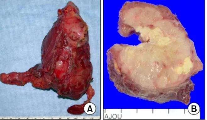
Vol. 1, No. 1, 2008 75 Fig. 3. (A) Gross photo, (B) Cut surface of the specimen shows pink brown lymph node-like solid tissue with vague nodularity and multifocal necrosis.
Fig. 2. FDG-PET/CT images demonstrate abnormal intense uptake in the left thyroid gland and nodal uptake.
Fig. 1. Preoperative axial neck CT with contrast shows 6×5 cm sized marked enlarged left thyroid gland deviating trachea and esophagus with multifocal hypoattenuated portion sug- gesting necrosis. Small enlarged lymph nodes along left internal jugular chain are noted.
통증, 연하곤란, 호흡곤란 등의 증상은 특별히 호소하 지 않았다. 환자는 가족력상 어머니가 고혈압인 것 외 에 특이사항은 없었다. 경부 진찰 소견상 좌측 전경부 에 딱딱하고 고정되어 만져지는 약 10×6 cm 크기의 무통성 거대종괴가 만져졌다. 후두내시경 검사상 성대 의 움직임은 정상소견이었다. 일반혈액검사, 뇨검사 및 심전도 검사에서 특이사항은 없었고, 갑상선 기능 검 사상 TSH, 유리T4, T3는 정상범위였고, thyroglobulin 463.7 ng/ml (정상범위 0∼40), antithyroglobulin antibody 328 U/ml (정상범위 0∼100)은 증가된 소견을 보였다.
단순흉부촬영에서 특이소견 보이지 않았고, 경부 전산 화 단층촬영에서 종괴는 좌측 악하선 하연에서 흉곽입 구까지 확장된 10×6×5 cm 크기로 갑상선 협부까지 침범하였고 경계는 비교적 명확하였고 주변조직에 침 범은 없었으나 기도와 식도 그리고 주변혈관들이 종괴 때문에 밀려서 관찰되었고 종괴의 내부에 일부 괴사
소견이 의심되었고, 경정맥을 따라 1 cm 크기의 림프 절이 관찰되었다(Fig. 1). FDG-PET/CT 스캔상 좌측 갑상선에서 pSUV 20 이상의 FDG 섭취증가소견과 좌 측 경부 림프절에서도 섭취증가소견을 보였다(Fig. 2).
갑상선 종괴의 세침 흡인 검사에서 중간 크기의 림프 구들로 구성되어 MALT 림프종이 의심되었다. 환자는 갑상선에 발생한 MALT 림프종 의심 하에 확진 및 급 속히 커진 종괴를 제거하기 위해 좌측 갑상선 절제술 을 시행하였다. 침범이 의심되는 주변의 림프절을 같 이 절제하였고, 좌측 반회후두신경은 확인하여 보존하 였다. 수술로 떼어낸 좌측 갑상선 종괴는 크기가 13×6.5×6 cm이고 무게는 210 gm이었고 절단면은 옅 은 핑크 갈색의 결절성 병변이 갑상선 전체에서 관찰 되었고 다수의 국소적 괴사소견도 관찰되었다(Fig. 3).
조직학적 검사상 종괴의 대부분은 핵소체가 뚜렷하고 수포성 핵을 가지는 큰 세포들이 미만성으로 침윤하고 있었고, 종괴의 변연부에는 정상 갑상선 실질에 림프 구들이 빽빽히 침윤하고, 배중심을 포함하는 림프소포 까지 형성하여 하시모토 갑상선염 소견을 보였고, 일 부에서는 중심구모양 세포가 갑상선 상피세포를 침윤 하여 만드는 림프상피병소가 관찰되어 하시모토 갑상 선염의 배경하에 미만성 대세포성 B세포 림프종과 MALT 림프종이 혼재된 갑상선 림프종으로 진단되었 다(Fig. 4). 면역조직화학검사(Immunohistochemistry) 상에서도 이에 합당한 소견을 보였다. 갑상선 주위림 프절에서 림프종 침습 소견을 보였고, 이하선 림프절 은 반응성 비후로 확인되었다. 환자는 Ann-Arbor stage IIE로 수술 후 혈액종양내과로 전과되어 CHOP (Cyto- xan, Doxorubicin, Vincristine, Predenisolone)+Rituxi- mab regimen을 이용한 화학요법을 6 cycle 시행하였고 완전 관해 후 현재 재발의 증거 없이 특별한 치료 없이
갑상선 림프종 1예
76 J Korean Thyroid Assoc
Fig. 4. Microscopic findings of the thyroid mass shows mixed diffuse large B-cell lymphoma and MALT lymphoma in a background of Hashimoto's thyroiditis. (A) Hashimoto's thyroiditis (H&E, ×40). Microscopic finding shows marked lymphocytic infiltration and a few reactive lymphoid follicles. (B) MALT lymphoma (H&E, ×400). The tumor cells are medium-sized lymphoid cells with irregular nuclei, inconspicuous nucleoli, resembling those of centrocytes. The atypical lymphoid cells infiltrates into follicular epithelium showing lymphoepithelial lesions. (C) Diffuse large B-cell lymphoma (H&E, ×400). The tumor cells are large with cleaved or round vesicular nuclei occasional prominent nucleoli and moderately abundant cytoplasm.
외래 추적 관찰 중이다.
고 찰
갑상선 림프종은 대부분 B세포 기원의 비호지킨성 림프종으로 호지킨성 림프종 및 T세포 기원의 림프종 은 드물다.5) 갑상선의 만성염증이나 자가 면역성 갑상 선염의 경우 일반인에 비해 40∼80배 높은 상대적 위 험도를 보이고, 평균적으로 20∼30년 후에 발생한다.5) 갑상선에는 원래 림프조직이 존재하지 않지만 하시모 토 갑상선염과 같은 자가면역질환에 의해 후천적으로 발생한 림프조직은 B세포 여포가 존재하고 갑상선 상 피에 B세포의 침윤이 있으며, 형질세포의 분화가 나타 나는 점에서 MALT와 유사하다.4) 갑상선 림프종과 하 시모토 갑상선염과의 연관성은 20∼100%까지 보고되 었다.6) MALT 림프종은 하시모토 갑상선염의 배경하 에서 흔히 생기고 서서히 진행한다. 미만성 대세포성 B세포 림프종은 조직학적으로 MALT 림프종과 미만 성 대세포성 B세포 림프종사이에 이행대(transitional area)가 존재하고 갑상선 종괴가 급속히 증대된다는 점 에서 MALT 림프종이 미만성 대세포성 B세포 림프종 으로 전환(transformation)하는 것으로 이해되고 있 다.1,6) 이 증례의 경우 특별히 갑상선과 연관된 질환의 기왕력은 없었으나 조직검사상 하시모토 갑상선염의 소견을 보이고 MALT 림프종에서 미만성 대세포성 B 세포 림프종로 전환되어 가는 전형적인 예라고 하겠다. 갑상선 림프종의 임상증상으로는 급속히 커지는 갑상 선 종대가 가장 흔하며 그 외에 갑상선 종대로 인한 주위조직의 압박증상으로 호흡곤란, 연하곤란, 애성, 통증 등이 환자의 30∼50%에서 있을 수 있으며,1) 발열,
야간 발한, 체중감소 등의 림프종의 전형적인 B 증상 은 드물다. 갑상선 종대는 비교적 단단하고 결절성으 로 촉지되며, 일반적으로 한쪽 엽을 침범하는 경우가 좀 더 많다. 이 증례는 갑상선 종대로 인한 주위조직의 압박증상은 특별히 호소하지 않았으나 전산화 단층 촬 영상 기도와 식도 그리고 주변혈관들이 종괴 때문에 밀려서 관찰될 정도의 거대 종괴였다. 갑상선 림프종 의 진단으로 세침 흡인 검사는 갑상선암의 진단에 매 우 효과적이고 민감한 것으로 알려져 있지만 MALT 림프종과 하시모토 갑상선염이 병발해 있는 경우 종양 성 병변과 반응성 병변이 공존해있어 이들의 감별진단 은 쉽지가 않다. 비교적 미만성 대세포성 B세포 림프 종과 같은 고도 림프종의 진단은 쉬우나 MALT 림프 종의 진단은 어려운 것으로 보고하고 있다.7) 추가검사 로 세침 흡인 검사와 더불어 flow cytometry를 함께 실 시했을 때 진단율을 높일 수 있다.8) Flow cytometry상 미만성 대세포성 B세포 림프종은 CD19, CD20, CD45 에 양성소견을 보이는 반면 MALT 림프종은 표면 면 역글로불린을 발현하고 CD5, CD10, CD23에 음성소견 을 보인다.9) 세침 흡인 검사는 갑상선 림프종의 선별검 사로써 유용하게 시행될 수 있으나 확진 및 아형을 알 기 위해서는 개방성 조직 생검을 통한 병리조직 검사 와 면역조직화학 검사가 추천된다.9) 방사선학적 검사 로는 초음파 검사, 갑상선 스캔, 전산화 단층 촬영, 자 기공명촬영, FDG PET/CT 스캔 등이 이용되고 있으 며, 갑상선 초음파 검사상에는 남아있는 갑상선 조직 에 비해 확연하게 저에코 영상으로 보이며, 전산화 단 층 촬영상에서 주변 구조물들을 침범하지는 않으면서 주변 근육과 비슷한 조영증강을 보이는 균일한 밀도의 갑상선 종괴로 보인다.2) 이 증례에서는 종괴 내부에 괴
신향애 외
Vol. 1, No. 1, 2008 77 사로 의심되는 소견이 보여 비균일한 밀도의 갑상선
종괴였다. FDG PET/CT 스캔상 FDG 섭취가 크게 증 가된다. FDG PET/CT 스캔은 갑상선 림프종의 병기 결정, 치료에 대한 반응 및 추적관찰을 하면서 재발여 부를 보는데 유용하다.10)
갑상선 림프종의 치료는 종양의 조직학적 소견, 종 양의 크기, 병기, 다른 질환의 합병여부에 따라서 결정 한다. 항암화학요법, 방사선치료, 수술적 치료 및 이들 의 병합요법이 있다. State IE (갑상선 피막내에 국한된 경우)의 MALT 림프종은 수술이나 방사선 치료의 단 독요법이 추천되고 있으나 아직 논란의 여지는 있다.11) 미만성 대세포성 B세포 림프종이나 미만성 대세포성 B세포 림프종과 혼재된 MALT 림프종의 경우에는 방 사선 치료와 항암치료의 병합요법이 추천된다.9) 수술 적 치료의 역할은 시간이 지남에 따라 변하고 있으나 개방성 조직 생검을 통한 확진을 위해서, 서서히 진행 하는 종양의 국소관리를 위해서, 또한 갑상선 종괴의 크기가 빠르게 증가하여 압박증상을 유발할 경우 이를 해소하는데 중요한 역할을 하고 있다.12) 그러나 수술로 기도압박증상을 해소할 수 있지만, 항암방사선 병합요 법으로도 충분히 해결할 수 있다고 하여,13) 아직 논란 의 여지가 있다. 이 증례의 경우는 조직병리학적 확진 및 급속히 커진 종괴 소견으로 인해 좌측 갑상선 절제 술로 치료하였으며, 항암요법을 추가로 시행하였다. 갑 상선 림프종은 적절한 치료로 좋은 결과를 가지나 임 상적 병기와 조직학적 소견에 따라 예후는 달라진다.
갑상선의 MALT 림프종은 좋은 예후를 보이나 미만성 대세포성 B세포 림프종이나 미만성 대세포성 B세포 림프종과 혼재된 림프종은 비교적 불량한 예후를 보인 다.1) Stage IE와 림프절 침범이 동반된 경우(Stage IIE) 에는 비교적 좋은 예후를 보이나, 갑상선 피막 침범이 있으면 III, IV 병기의 림프절 림프종(nodal lymphoma) 과 비슷한 불량한 예후를 보인다.14)
중심 단어: 갑상선, 하시모토 갑상선염, MALT 림프 종, 미만성 대세포성 B세포 림프종.
References
1) Derringer GA, Thompson LD, Frommelt RA, Bijwaard KE, Heffess CS, Abbondanzo SL. Malignant lymphoma of thyroid gland: A clinicopathologic study of 108 cases. Am J Surg Pathol 2000;24:623-39.
2) Kim HC, Han MH, Kim KH, Jae HJ, Lee SH, Kim SS, et al. Primary thyroid lymphoma: CT findings. Eur J Radiol 2003;46:233-9.
3) Thieblemont C, Mayer A, Dumontet C, Barbier Y, Callet- Bauchu E, Felman P, et al. Primary thyroid lymphoma is a heterogeneous disease. J Clin Endocrinol Metab 2002;87:105-11.
4) Hyjek E, Isaacson PG. Primary B cell lymphoma of the thyroid and its relationship to Hashimoto's thyroiditis. Hum Pathol 1988;19:1315-26.
5) Pedersen RK, Pedersen NT. Primary non-Hodgkin's lympho- ma of the thyroid gland: a population based study. Histo- pathology 1996;28:25-32.
6) Ansocombe AM, Wright DH. Primary malignant lymphoma of the thyroid-a tumor of mucosa associated lymphoid tissue:
review of seventy six cases. Histopathology 1985;9:81-97.
7) Sangalli G, Serio G, Zampatti C, Lomuscio G, Colombo L.
Fine needle aspiration cytology of primary lymphoma of the thyroid: a report of 17 cases. Cytopathology 2001;12:257-63.
8) Bangerter M, Brudler O, Heinrich B, Griesshamnuer M. Fine needle aspiration cytology and flow cytometry in the diagnosis and subclassification of non-Hodgkin's lymphoma based on the World Health Organization classification. Acta Cytol 2007;51:
390-8.
9) Mack LA, Pasieka JL. An evidence-based approach to the treatment of thyroid lymphoma. World J Surg 2007;31:978-86.
10) Zijlstra JM, Lindauer-van der Werf G, Hoekstra OS, Hooft L, Riphagen II, Huijgens PC. 18F-fluorodeoxyglucose positron emission tomography for post-treatment evaluation of malignant lymphoma: a systematic review. Haematologica 2006;91:522-9.
11) Zelenetz AD, Advani RH, Buadi F, Cabanillas F, Caligiuri MA, Czuczman MS, et al. Non-Hodgkin's lymphoma. Clinical practice guidelines in oncology. J Natl Compr Canc Netw 2006;4:258-310.
12) Widder S, Pasieka JL. Primary thyroid lymphomas. Curr Treat Options Oncol 2004;5:307-13.
13) Matsuzuka F, Miyauchi A, Katayama S, Narabayashi I, Ikeda H, Kuma K, et al. Clinical aspects of primary thyroid lymphoma: diagnosis and treatment based on our experience of 119 cases. Thyroid 1993;3:93-9.
14) Burke JS, Butler JJ, Fuller LM. Malignant lymphomas of the thyroid: a clinical pathologic study of 35 patients including ultrastructual observations. Cancer 1977;39:1587-602.