© 2011 The Korean Academy of Medical Sciences.
This is an Open Access article distributed under the terms of the Creative Commons Attribution Non-Commercial License (http://creativecommons.org/licenses/by-nc/3.0) which permits unrestricted non-commercial use, distribution, and reproduction in any medium, provided the original work is properly cited.
pISSN 1011-8934 eISSN 1598-6357
Marginal Zone B-cell Lymphoma of MALT in Small Intestine Associated with Amyloidosis: A Rare Association
A 62-yr-old man presented with a 5-yr history of intermittent abdominal distention and pain. These symptoms persisted for several months and subsided without treatment. A diagnosis of suspected small bowel lymphoma was made based on plain radiograph and computerized tomogram findings, and he was referred to our institution for further evaluation. Segmental resection of the small intestine was performed and the diagnosis of marginal zone B-cell lymphoma associated with amyloidosis was made. This is the first case of marginal zone B-cell lymphoma of mucosa-associated lymphoid tissue (MALT) in the small intestine associated with amyloidosis in Korea.
Key Words: Gastrointestinal Tract; Lymphoma; Lymphoma, B-Cell, Marginal Zone;
Amyloidosis Sanghui Park, Hyun Yee Cho,
Seung Yeon Ha, Dong Hae Chung, Na Rae Kim, and Jung Suk An Department of Pathology, Gil Medical Center, Gachon University, School of Medicine and Science, Incheon, Korea
Received: 12 November 2010 Accepted: 28 February 2011 Address for Correspondence:
Hyun Yee Cho, MD
Department of Pathology, Gil Medical Center, Gachon University, School of Medicine and Science, 774 Namdong-daero, Namdong-gu, Incheon 405-760, Korea
Tel: +82.32-460-3850, Fax: +82.32-460-3073 E-mail: [email protected]
DOI: 10.3346/jkms.2011.26.5.686 • J Korean Med Sci 2011; 26: 686-689
CASE REPORT
Oncology & Hematology
INTRODUCTION
The extent of amyloid deposition is greatest in the small intes- tine (1, 2). Autopsy demonstrated that 31% of patients with sys- temic amyloidosis experienced amyloid deposition in the small intestine (1, 2), although localized amyloidosis of the small in- testine remains very rare. Only four cases on localized small bowel amyloidosis have been reported (3-6). Amyloid deposits can mimic a tumor, and have occasionally been associated with gastrointestinal lymphoma (7-11), but localized amyloidosis associated with small intestine lymphoma is extremely rare. Only three cases reports have been published on small bowel local- ization of amyloidosis associated with lymphoma (7, 8, 10). Clin- copathologic features of previously reported cases were summa- rized in Table 1.
We present an unusual case of intestinal marginal zone B-cell lymphoma of mucosa-associated lymphoid tissue (MALT) with concurrent localized amyloidosis. To the best of our knowledge, this is the first case report on localized intestinal amyloidosis associated with intestinal marginal zone B-cell lymphoma of MALT in Korea.
CASE DESCRIPTION
A 62 yr-old man presented with a 5-yr history of intermittent abdominal pain associated with abdominal distention which waxed and waned without treatment on June 4, 2010. Ten days
before presenting to our institution, he was urgently admitted to a local hospital with abdominal pain. Computerized tomogra- phy (CT) scan taken there revealed multi-focal enhancing small bowel wall thickening associated with skipped lesion sparing of the terminal ileum and the ileocecal valve. Inflammatory bowel disease was suggested as a preliminary diagnosis. He was sub- sequently referred to our hospital for further evaluation. On ex- amination, he appeared ill but remained apyrexial. Abdominal examination elicited hypogastric guarding, pelvic tenderness and some rebound tenderness. Initial laboratory studies revealed severe thrombocytopenia of 4 × 10³/mm2 without leukocytosis.
Diagnostic and therapeutic segmental resection of the small in- testine was subsequently performed on 14th June, 2010. Intra- operatively, the jejunum exhibited 30 cm segmental wall thick- ening, and a localized mass-like lesion was found in the ileum which caused luminal obstruction and multifocal bowel wall segmental thickening. Moreover, one enlarged mesenteric lymph node was identified and subsequently sent to our department for frozen diagnosis. Segmental resection of jejunum (about 40 cm in length) and ileum (about 1 m in length) was performed.
The surgical specimen was fixed in 10% buffered formalin. Tis- sue samples were taken, processed for routine histology and embedded in paraffin. Five micrometer-thick sections were cut and stained with hematoxylin and eosin, Congo Red with and without KMnO4 (12). Immunohistochemistry was conducted with the avidin-biotin-peroxidase complex methods using anti- body CD3 (Dako, Glostrup, Denmark, prediluted), CD20 (Dako,
Park S, et al. • Marginal Zone B-cell Lymphoma in Small Intestine Associated with Amyloidosis
http://jkms.org 687
DOI: 10.3346/jkms.2011.26.5.686
prediluted), CD23 (Dako, prediluted), CD5 (Dako, prediluted), cyclin D1 (Dako, prediluted), kappa (Dako, prediluted), lambda (Dako, prediluted), Ki-67 (Dako, prediluted). Unstimulated iso- lated bone marrow cells were cultured for 24 hr and G-banded according to standard procedures. Metaphases were analyzed and karyotyped according to the nomenclature system proposed by the International System for Human Cytogenetic Nomencla- ture, 1995.
The gross specimen showed two segments of small intestine, measuring 1 m and 40 cm in length, respectively. The diameter and thickness of the intestinal segments varied greatly in size.
The appearance of the overlying mucosa revealed a short linear and transverse ulceration in an irregular and thickened wall.
Sections of the involved intestinal segment showed two differ- ent processes: first, we observed a homogeneous acidophilic substance (Fig. 1A, B). A Congo Red stain demonstrated doubly refractive property with polarized light (Fig. 1B, inset). Second, a diffuse proliferative lymphoid infiltrate spread through the in- testinal wall up to the serosa (Fig. 1C). The lymphoid compo- nent was comprised mainly of small lymphocytes with round nuclei, often with a small central nucleoli, clumped chromatin with scanty basophilic cytoplasm admixed with plasma cells (Fig. 1D). Smaller lymphocytes without discernable cytoplasm and some large cells with oval and vesicular nuclei with promi- nent nucleoli. In addition, a monocytoid component was also found. No obvious lymphoepithelial lesion was identified and Table 1. Clinicopathologic features of previously reported small bowel amyloidosis associated with lymphoma
Cases Year Age/Sex Site Type of lymphoma
Arista Nasr J et al. (7) 1993 80/M Entire gastrointestinal tract Unknown
Caulet S et al. (8) 1995 47/F Small intestine Marginal zone B-cell lymphoma of MALT
Goteri G et al. (10) 1998 45/M Small intestine Immunoproliferative small intestinal disease with
high-grade transformation
Present case 2010 62/M Small intestine Marginal zone B-cell lymphoma of MALT
MALT, mucosa-associated lymphoid tissue.
A B
C D
Fig. 1. Histologic features of a resection specimen of small intestine. (A) The intestinal wall was thickened by a double process. Here, amorphous acidophilic material in the submucosa and intestinal wall were mixed with lymphoid infiltrate (H&E, scanning view). (B) High power view (H&E, × 40) and apple-green bi-refringence under polarized light is seen Congo-red staining of amyloid (inset). (C) Diffuse proliferative lymphoid infiltrate through the intestinal wall up to the serosa (H&E, scanning view). (D) The lymphoid infiltrate was composed mainly of small lymphoid cells (H&E, × 400).
Park S, et al. • Marginal Zone B-cell Lymphoma in Small Intestine Associated with Amyloidosis
688 http://jkms.org DOI: 10.3346/jkms.2011.26.5.686
plasma cells with Dutcher bodies were occasionally seen. The excised mesenteric lymph node showed extensive amyloid de- posits intermixed by small aggregates of lymphoid cells (Fig. 2).
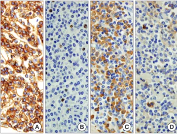
Most lymphocytes were stained positively with CD20 (Fig. 3A) but negative for CD3, CD5, CD23, and cyclin D1. The Ki-67 label- ing index was low (less than 5%) (Fig. 3B). Kappa chain restric- tion was found in the plasma cells (Fig. 3C, D). There was no clin- ical evidence of systemic amyloidosis and renal function remain- ed normal. Bone marrow biopsy showed no evidence of lym- phoma or amyloid deposition whereas echocardiogram, rectal biopsy and abdominal fat aspirates were not performed. Cyto- genetic study performed on bone marrow cells showed chromo- somal abnormality. The karyotype was 46XY, inv(2)(p25q13)[14]/
46,XY[16]. The serum total protein level was within the normal limits, and serum electrophoresis showed an IgM kappa mono- clonal gammopathy. The patient received four cycles of chemo- therapy (R-CVP) over 4 months and he is currently in remission.
DISCUSSION
The association of amyloidosis and gastrointestinal lymphoma is very rare and to our knowledge it has only been described in 8 reports (7, 8, 10, 11). Moreover, lymphadenopathy secondary to amyloid deposition in non-Hodgkin’s lymphoma is also rare.
On reviewing the Korean literature, we could not identify a re- port on such an association in intestinal lymphoma. Only one Korean case reported amyloidosis in the bone marrow associ- ated with lymphoma but the histologic type could not be deter- mined, because monotonous B-cells were only present in the bone marrow and only positive for CD20 (13). Our case is very unusual because localized amyloidosis was associated with low grade B-cell intestinal lymphoma.
This case represented a diagnostic challenge, because the am- Fig. 2. Histologic features of mesenteric lymph node biopsy. Amyloid deposits are dense admixed with some aggregates of lymphoid cells. Islands of lymphoid infiltration are composed of small lymphoid cells, some of which show plasmacytic differentiation (H&E, scanning view and × 100-inset).
Fig. 3. The immunohistochemical findings. The lymphoid infiltrate was positive for CD20, (A) Ki-67 labeling index was low (less than 5%), (B) kappa light chain-producing plasma cells were detected, (C) with the exclusion of lambda light chain-producing plasma cells, (D) (immunoperoxidase, × 400).
A B C D
yloid could have displaced lymphomatous proliferation and produced a tumoral mass. Some area showed definite lympho- tous infiltration, but some area showed extensive amyloid depo- sition with scarce lymphocytic infiltration. Moreover, one en- larged mesenteric lymph node was sent for frozen diagnosis. At that time, the diagnosis of lymphoma was not made, as the lymph node showed extensive amyloid deposits with interspersed ag- gregates of lymphoid cells. The detection of amyloid deposits in a lymph node biopsy should therefore raise the possibility of concurrent lymphoma. There are several case reports on local- ized amyloidosis in the gastrointestinal tract. Usually, these cases are diagnosed by endoscopic biopsy. As our case demonstrated, histological findings varied with the site of intestinal segments.
Therefore, multiple endoscopic biopsies are recommended to exclude the possibility of hidden intestinal lymphoma associat- ed with amyloidosis.
The amyloid had the typical microscopic appearance of ex- tracellular sheets or large masses of eosinophilic, amorphous, hyaline substance. This substance was Congo red-positive. How- ever, we did not perform anti-amyloid A or P component anti- body for the AA or AL subtype.
On the basis of the above clinicopathologic findings, we are unable to explain the presence of amyloid in this case. It has been suggested that intense, prolonged antigenic stimulation can lead to marked plasma cell differentiation with abnormal immuno- globulin production and amyloid deposition. However, this hy- pothesis does not fully explain our findings, because plasmacyt- ic differentiation was not prominent in this case.
The diagnosis of marginal zone B-cell lymphoma may be ex- cluded, because marginal zone B-cell lymphoma is rare in the small intestine. In addition, amyloidosis and monoclonal gam- mopathy is very rare in this subtype. Other low grade B-cell lym- phoma should be included in the differential diagnoses. The di-
Park S, et al. • Marginal Zone B-cell Lymphoma in Small Intestine Associated with Amyloidosis
http://jkms.org 689
DOI: 10.3346/jkms.2011.26.5.686
agnosis of marginal zone B-cell lymphoma is based on both mor- phological and immunophenotypic findings.
In conclusion, we report the first case of marginal zone B-cell lymphoma of MALT in the small intestine associated with amy- loidosis in Korea. This unusual association should be borne in mind when diagnosing gastrointestinal tract amyloidosis, espe- cially with endoscopic biopsy.
REFERENCES
1. Briggs GW. Amyloidosis. Ann Intern Med 1961; 55: 943-57.
2. Legge DA, Carlson HC, Wollaeger EE. Roentgenologic appearance of systemic amyloidosis involving gastrointestinal tract. Am J Roentgenol Radium Ther Nucl Med 1970; 110: 406-12.
3. Baldewijns M, Ectors N, Verbeeck G, Janssens J, De Schepper J, Ponette E, Geboes K, Desmet V. Intermittent subobstruction and cholestasis as complications of duodenal amyloid tumours. Gastroenterol Clin Biol 1995; 19: 218-21.
4. Hamaya K, Kitamura M, Doi K. Primary amyloid tumors of the jejunum producing intestinal obstruction. Acta Pathol Jpn 1989; 39: 207-11.
5. Hauben E, Fierens H, Heylen H, Van Marck E. Localized amyloid tumour of the duodenum: a case report. Acta Gastroenterol Belg 1997; 60: 304-5.
6. Peny MO, Debongnie JC, Haot J, Van Gossum A. Localized amyloid tu- mor in small bowel. Dig Dis Sci 2000; 45: 1850-3.
7. Arista Nasr J, Lome-Maldonado C. Diffuse small lymphoplasmacytic lymphoma of the GI tract associated with massive intestinal amyloido- sis. Rev Invest Clin 1993; 45: 71-5.
8. Caulet S, Robert I, Bardaxoglou E, Noret P, Tas P, Le Prise Y, Launois B, Ramee MP. Malignant lymphoma of mucosa associated lymphoid tis- sue: a new etiology of amyloidosis. Pathol Res Pract 1995; 191: 1203-7.
9. Das K, Ghoshal UC, Jain M, Rastogi A, Tiwari S, Pandey R. Primary gas- tric lymphoma and Helicobacter pylori infection with gastric amyloido- sis. Indian J Gastroenterol 2005; 24: 220-2.
10. Goteri G, Ranaldi R, Pileri SA, Bearzi I. Localized amyloidosis and gas- trointestinal lymphoma: a rare association. Histopathology 1998; 32:
348-55.
11. Ranaldi R, Goteri G, Santinelli A, Rezai B, Pileri S, Poggi S, Bearzi I. Cen- trocytic-like lymphoma associated with localized amyloidosis of the large intestine. Virchows Arch 1994; 425: 327-30.
12. Wright JR, Calkins E, Humphrey RL. Potassium permanganate reaction in amyloidosis. A histologic method to assist in differentiating forms of this disease. Lab Invest 1977; 36: 274-81.
13. Kim SY, Bang BK, Park CW, Kim KW, Yun SR, Han CM, Park YH, Ahn SJ, Park SY, Kim HJ, Suh KS, Park KK. An unusual case of AA type amy- loidosis in lymphoma. Korean J Nephrol 1999; 18: 808-14.