- 63 - 대한두경부종양학회지, 제33권 제2호, 2017. pp.63-66
Korean Journal of Head & Neck Oncology, Vol.33, No.2
http://doi.org/10.21593/kjhno/2017.33.2.63 ISSN 1229-5183(Print) / ISSN 2586-2553(Online)
부이하선에 발생한 점막관련 림프조직 림프종 1예
홍석정1⋅이미지2⋅김승우1+
중앙보훈병원 이비인후과1, 병리과2
A Case of Mucosa Associated Lymphoid Tissue Lymphoma in Accessory Parotid Gland
Seok Jung Hong, MD1, Mi Ji Lee, MD2, Seung Woo Kim, MD1+
Department of Otolaryngology-Head and Neck Surgery1 and Pathology,2 Veterans Health Service Medical Center, Seoul, Korea
= Abstract =
The tumor in accessory parotid gland (APG) is rarely occurred and its incidence is about 7.7% of all parotid gland neoplasms, but has a higher frequency of malignancy than major salivary glands. The mucoepidermoid carci- noma is the most common malignancy in APG, while B-cell lymphoma is less than 2%. It is often appeared as superficial mass in mid-cheek area. This lesion requires differential diagnosis with epidermoid cyst, lipoma, neurogenic tumors, Stensen’s duct stone, lymphadenopathy and hemangioma etc. The mucosa associated lymphoid tissue (MALT) lymphoma, which is also termed extra-nodal marginal zone B-cell lymphoma tends to be localized disease for long time and has a relatively indolent course. We recently encountered a 69-year-old man with super- ficial solitary mass on the right cheek area that finally diagnosed as MALT lymphoma in APG. We report the rare and unique case with brief literature review.
Key W ords:Accessory parotid gland⋅Lymphoma⋅MALT.
R eceived R e v i s e d A ccepted
: September 21, 2017 : October 12, 2017 : October 23, 2017
+Corresponding author: Seung Woo Kim, MD
Department of Otolaryngology-Head and Neck Surgery, Veterans Health Service Medical Center, 53, Jinhwangdo-ro 61-gil, Gangdong-gu, Seoul, Korea. 05368
Tel: +82-2-2225-1384 Fax: +82-2-2225-1385 E-mail: [email protected]
서 론
부이하선은 이하선의 전방으로 해부학적으로 분리되 어 있는 타액선 조직으로 교근의 근막 위에 이하선관을 따라 존재하고, 이하선과 조직학적으로 유사하다.1)부이 하선 종양은 타액선 종물 가운데 가장 드물며, 다른 타액 선보다 악성의 빈도가 높다고 알려져 있다.1,2) 이 곳에서 발생하는 악성 종양으로는 점액표피 암종이 가장 흔하 고, 그 외에도 선낭 암종, 편평세포 암종 등의 보고가
있다.3,4) B세포 림프종은 부이하선에 발생하는 종양의 2% 미만으로 매우 드물다.2)저자들은 우측 협부의 표재 성 종물을 주소로 내원한 69세 남자 환자에서 점막관련 림프조직 림프종으로 최종 진단된 매우 드문 증례를 치 험하여 문헌고찰과 함께 보고한다.
증 례
69세 남자 환자가 내원 4개월 전부터 시작된 우측 협부 의 무통성 및 표재성 종물을 주소로 내원하였다. 과거력 상 고혈압 및 협심증 등이 있었고, 음주력은 주 1회 소주 1병 정도였고, 흡연력은 없었다. 신체검사에서 우측 협부 에 약 1.5 × 1.5 cm크기의 원형의 비교적 딱딱하고, 주변 과의 경계는 비교적 명확하지만, 아래의 구조물에 고정 되어 있는 종물이 촉지되었다(Fig. 1). 다른 경부 종물은 관찰되지 않았다. 전신상태를 평가하는 ECOG (Eastern Cooperative Oncology Group) 활동도는 0점이었고, 발열,
- 64 -
Fig. 1. External photograph shows a 1.5x1.5 cm sized mass on right cheek area (black arrowheads).
Fig. 3. Intraoperative finding. The main mass was almost com- pletely extracted.
A B
Fig. 2. Radiologic findings. Transverse scan of neck ultrasonography show 1.3x1.8x0.7 cm sized hypoechoic ovoid mass with well-defined margin in subcutaneous layer (arrow)(A). Enhanced axial neck CT scan show homogenous enhanced mass on right cheek area (arrow)(B).
A B
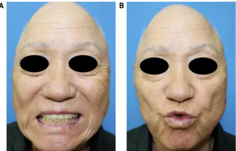
Fig. 4. Postoperative facial photographs. They show normal facial expression including lip movements (A and B).
발한 및 체중 감소 등의 증상은 없었다. 젖산탈수소효소 를 포함한 모든 혈액학적 검사에서 정상 범위였다.
영상학적 평가를 위하여 시행한 초음파 검사에서 교근 의 천층에 위치하는 1.3 × 1.8 × 0.7 cm크기의 경계가 뚜 렷한 저에코 음영을 보이는 난원형의 종물이 관찰되었다
(Fig. 2A). 경부 전산화 단층 촬영에서는 우측 협부에 주 변과 경계가 명확하면서 균질하고 미세하게 조영 증강되 는 종물 소견이었다(Fig. 2B). 초음파 유도 하 세침흡인 검사에서는 소량의 림프구만 관찰되었다. 이상의 소견 을 종합하여, 표피 낭종, 신경기원 종양, 결절성 근막염,
- 65 -
A B C
D E
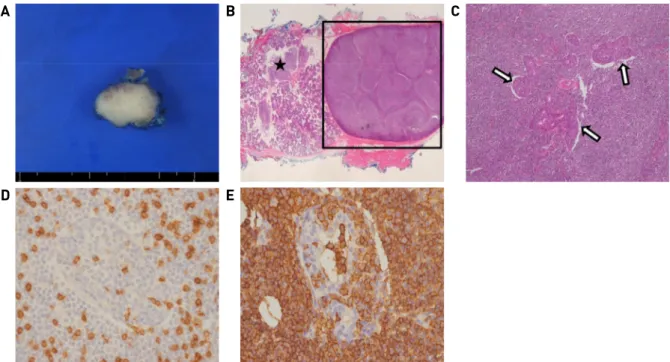
Fig. 5. Histopathological findings. The surgical specimen shows that 2.2x1.5 cm sized mass with grayish white firm cut surface (A). Nodular infiltration of lymphoid cells (square) with periductal lymphoid infiltration (black star) are observed in low-power view(H&E, X10)(B). Lymphoepithelial lesion shows lymphocytic invasion into ductal epithelium (arrows)(H&E, X100)(C). Reactive CD3-positive T-cells are scattered(X400)(D). Neoplastic CD20-positive B-cells are heavily infiltrated. Their invasion into ductal epi- thelial cells is highlighted by immunohistochemistry (X400)(E).
섬유종 및 염증성 가성종양 등의 가능성을 염두에 두고, 확진을 위하여 절제 생검을 시행하였다. 환자의 나이 등 을 고려하여 국소마취 하에 종물의 직상방 피부의 주름 선에 절개를 가하고, 피하층에 위치하는 종물을 절제하 였다. 종물의 심부 박리 시 교근과 유착이 심하고, 심한 출혈이 있었다(fig. 3). 수술 후 3일째 안면신경 마비를 포함한 특별한 합병증 없이 퇴원하였다(fig. 4). 최종 조 직 검사상 종물의 절단면은 균일한 양상의 회백색을 띤 결절성 병변이었고(Fig. 5A), 이하선관 주변에 림프구 침 윤과 이를 둘러싸는 타액선 조직들이 관찰되었고, 림프 상피성 병변이 형성되었다(Fig. 5B and C). 또한, 면역 조직화학 염색에서는 CD20과 Bcl2에 양성, cyclin D1에 는 음성을 보였다(Fig. 5D and E). 이상의 소견들을 종합 하여 부이하선에 발생한 림프절 외 변연부 B세포 림프종 으로 최종 진단하였다. 혈액종양내과로 전과되어 병기 평가를 위하여 양전자 방출 단층촬영, 흉부 및 복부 전산 화 단층촬영 등을 시행하였으나 다른 부위로의 전이는 없었으며, 골수 생검에서도 암세포는 관찰되지 않아 최 종 병기는 부이하선에 국한된 Ann Arbor IE로 평가되었다.
이후 CVP (Cytoxan 1395mg, Vincristine 2mg, Prednisolone 50mg) 요법으로 3회의 항암치료와 총 24Gy의 방사선 치 료를 시행 받았으며, 8개월이 지난 현재 재발 소견 없이 추적 관찰 중이다.
고 찰
부이하선은 21~56%의 빈도로 존재하는 비교적 흔한 해 부학적 변이이지만, 종양의 발생은 전체 이하선 종양의 7.7% 미만으로 극히 드물다.1,2)가장 흔하게 발생하는 양 성 종양은 다형선종이고 악성 종양은 점액표피 암종으로, 이는 이하선과 조직학적으로 비슷하기 때문이다.2) 점막 관련 림프조직 (mucosa associated lymphoid tissue, MALT) 림프종은 두경부 영역에서는 타액선에서 주로 발생하지 만 전체 타액선 종양 중 3.1% 미만으로 매우 드물고,5)이 하선 또는 설하선 등에서 보고된 예는 있으나,5,6)부이하 선에 원발성으로 발생한 경우는 국내에서는 보고된 바가 없다. PubMed, Embase®및 Cochrane Library 등에서 검색 어 “accessory parotid gland and lymphoma”를 통한 영문 논문 검색에서는 4례의 부이하선 B세포 림프종에 대한 보고가 있었다.2)
MALT 림프종 혹은 림프절 외 변연부 B세포 림프종은 비호지킨 림프종의 아형으로 가장 호발하는 부위는 위장 관이고, 그 외에 타액선, 갑상선, 간, 결막, 안구, 폐, 신장, 전립선 등에서 발생할 수 있다.5) 타액선은 위장관과는 달리 정상적인 점막관련 림프조직이 존재하지는 않지만, 소량의 림프조직을 함유하고 있기 때문에, C형 간염, 헬 리코박터 감염, 쇼그렌 증후군 및 류마티스 관절염 등의 자가 면역 질환이 림프구 증식 및 악성변화를 일으켜
- 66 - MALT 림프종이 생길 수 있다고 알려져 있다.5,6)그러나 본 증례와 같이 감염 요인이나 자가 면역 질환이 없이 발생할 수도 있다.
부이하선 종양의 주 임상 양상은 이하선관의 주행과 일치하는 중협부 (mid-cheek area) 의 무통성 종물로 나타 나고, 표피 낭종, 지방종, 혈관종, 타석, 림프절 병증 등과 의 감별 진단을 고려해볼 수 있다.1,3) MALT 림프종은 비교적 병변이 국한되고 진행이 느린 무통성의 점막 하 부종으로 나타나고 B증상이 동반되기도 한다.6)표피 낭 종은 외상에 의해 발생할 수 있고, 지방종은 표면이 부드 럽고 긴 시간 동안 크기가 변하지 않는 종물일 때 의심해 볼 수 있다.3)또한, 혈관종은 경계가 불분명한 부드러운 무통성 종물이 크기가 변할 수 있고, 타석은 이하선관의 폐색 증상이 나타날 수 있고, 영상 검사로 확인할 수 있 다.7)림프절 병증은 특징적으로 수 일에서 수 주의 짧은 기간 동안 통증이 있고 크기가 변하는 종물이다.7)
부이하선 종물에 대한 영상 검사는 주변 구조물과의 관계 및 병변의 범위를 평가할 수 있고, 수술 전 세침흡인 세포 검사는 진단에 도움을 준다.8)하지만, 외과적 절제 의 필요 여부를 결정할 수 없고 타액선에 발생한 MALT 림프종의 경우에 진단적 가치는 적다.5) 본 증례의 경우 영상 검사에서는 비특이적 소견이었고, 세침흡인 검사 에서도 소량의 림프구만 확인되어 진단을 내릴 수 없었 다. 본 증례처럼 대부분 술 후 병리 결과로 진단된다.
MALT 림프종의 조직학적 소견은 중심세포양 세포, 단 핵구 모양 B세포, 형질 세포와 작은 림프구의 미만성 침 윤이 특징이며, 변연대 세포들이 림프절 외 상피 또는 관 조직을 침범하여 림프 상피성 병변을 형성한다.9)또 한, 면역조직화학 염색에서 CD20, 21, Bcl2 및 IgM 등에 대해서 양성 소견을 보이며, CD5, 10 및 Cyclin D1 등에 서 음성 소견을 나타낸다.9)
부이하선 종물의 치료는 수술적 치료가 원칙이며, 일 반적으로 부이하선은 안면신경의 관골협부분지를 따라 존재하기 때문에 안면신경 마비 및 이하선관 누공과 같 은 합병증을 줄이기 위해 직접 절개에 의한 종물의 제거 보다는 이하선 천엽절제술을 추천한다.10)본 증례에서는 종물의 크기가 비교적 작고, 이하선과 비교적 떨어져 있 었기 때문에, 조직학적 확진을 위해서 절제 생검을 시행 하였다. MALT 림프종의 치료는 Ann Arbor 분류에 의한 병기에 따라 달라지는데 병기 I, II는 방사선 치료를 주로 시행하고, 병기 III, IV는 항암치료를 시행한다.11) 하지만, 두경부 MALT 림프종은 국소 치료만 하는 경우에는 재 발과 다발성 파종 가능성이 높기 때문에, 낮은 병기에서 도 항암화학요법과 같은 전신 치료를 시행하기도 한다.12)
본 증례의 경우도 다학제 진료를 통하여 항암방사선 동 시요법을 시행하였다.
저자들은 본 증례를 통해 주변의 염증 소견 없이 중협 부의 표재성 종물이 존재하는 경우에 반드시 부이하선에 서 발생한 종양을 감별진단에 포함해야 한다는 교훈을 얻었다.
중심 단어:부이하선⋅점막관련 림프조직 림프종⋅타액선.
References
1) Luksic I, Suton P, Rogic M, Dokuzovic S. Accessory parotid gland tumours: 24 years of clinical experience. Int J Oral Maxillofac Surg. 2012;41:1453-1457.
2) Newberry TR, Kaufmann CR, Miller FR. Review of accessory parotid gland tumors: pathologic incidence and surgical management. Am J Otolaryngol. 2014;35:48-52.
3) Das S, Nayak UK, Buggavetti R, Sekhar S. Adenoid cystic carci- noma of accessory parotid gland: A case report. J Oral Maxillofac Surg. 2016;74:1097.e1091-1095.
4) Sakurai K, Urade M, Kishimoto H, Takahashi Y, Hozumi S, Yanagisawa T. Primary squamous cell carcinoma of accessory parotid gland duct epithelium: Report of a case. Oral Surg Oral Med Oral Pathol Oral Radiol Endod. 1998;85:447-451.
5) Kim SY, Nam WJ, Kim TH, Lee SH. A case of extranodal mar- ginal zone B-cell lymphoma in both parotid glands. Korean J Head Neck Oncol. 2017;33:65-71.
6) Cho KJ, Kim JP, Woo SH, Park JJ. A case of mucosa-associated lymphoid tissue lymphoma in sublingual glands without auto- immune disease. Korean J Otorhinolaryngol-Head Neck Surg.
2016;59:458-461.
7) Dell’ Aversana Orabona G, Abbate V, Piombino P, Iaconetta G, Califano L. Midcheek mass: 10 year of clinical experience. J Craniomaxillofac Surg. 2014;42:e353-358.
8) Yang X, Ji T, Wang LZ, Yang WJ, Hu YJ, Zhong LP, et al.
Clinical management of masses arising from the accessory paro- tid gland. Oral Surg Oral Med Oral Pathol Oral Radiol Endod.
2011;112:290-297.
9) Kwak SG, Baek HH, Kim YJ, Kim SW. A case of MALT lympho- ma of buccal area. Korean J Otorhinolaryngol-Head Neck Surg.
2015;58:287-289.
10) Sun G, Hu Q, Tang E, Yang X, Huang X. Diagnosis and treat- ment of accessory parotid-gland tumors. J Oral Maxillofac Surg.
2009;67:1520-1523.
11) Shin HA, Kahng H, Hwang E, Kim CH. A case of multifocal MALT lymphoma in salivary glands. Korean J Otorhinolaryngol- Head Neck Surg. 2008;51:1166-1169.
12) Wenzel C, Fiebiger W, Dieckmann K, Formanek M, Chott A, Raderer M. Extranodal marginal zone B-cell lymphoma of muco- sa-associated lymphoid tissue of the head and neck area: High rate of disease recurrence following local therapy. Cancer.
2003;97:2236-2241.