• 검색 결과가 없습니다.

A Case of Synchronous Lung Adenocarcinoma and Extranodal Marginal Zone B-Cell Lymphoma of Mucosa-Associated Lymphoid Tissue (MALT) Type

N/A
N/A
Protected

Academic year: 2021

Share "A Case of Synchronous Lung Adenocarcinoma and Extranodal Marginal Zone B-Cell Lymphoma of Mucosa-Associated Lymphoid Tissue (MALT) Type"

Copied!
6
0
0

로드 중.... (전체 텍스트 보기)

전체 글

(1)

(MALT) Type

Chi Young Jung, M.D. 1 , Kun Young Kwon, M.D. 2

Departments of 1 Internal Medicine and 2 Pathology, Keimyung University School of Medicine, Daegu, Korea

Extranodal marginal zone B-cell lymphoma of mucosa-associated lymphoid tissue type (extranodal MZL) is a distinct subgroup of non-Hodgkin's lymphoma. Pulmonary extranodal MZL is a rare entity and accounts for less than 0.5%

of primary pulmonary malignancies. Only a few cases of simultaneous occurrence of lung cancer and pulmonary extranodal MZL have been reported. A 60-year-old woman was referred to our hospital with a pulmonary nodule.

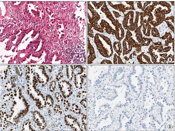
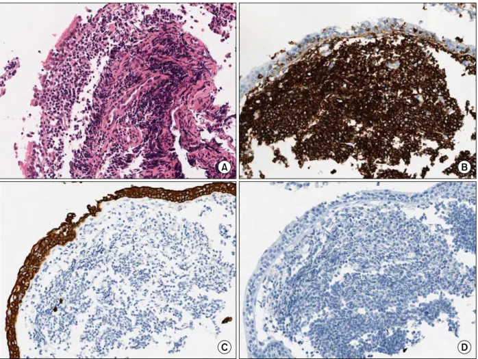
She was diagnosed with lung adenocarcinoma by percutaneous needle biopsy. The protrusions into the left main bronchus were found by accident while performing bronchoscopy during lung cancer evaluation. The bronchial lesions were diagnosed as extranodal MZL. Although the patient underwent surgical resection for the lung adenocarcinoma, the pulmonary extranodal MZL was left untreated; it was monitored during follow-up visits. To our knowledge, this is the first report of synchronous lung adenocarcinoma and primary extranodal MZL of the main bronchus.

Key Words: Lymphoma, B-Cell, Marginal Zone; Adenocarcinoma of Lung; Lung Neoplasms

Address for correspondence: Chi Young Jung, M.D.

Department of Internal Medicine, Keimyung University School of Medicine, 56, Dalseong-ro, Jung-gu, Daegu 700-712, Korea

Phone: 82-53-250-8095, Fax: 82-53-250-7434 E-mail: jcy2475@dsmc.or.kr

Received: Apr. 10, 2012 Revised: Apr. 24, 2012 Accepted: May 18, 2012

CC

It is identical to the Creative Commons Attribution Non-Commercial License (http://creativecommons.org/licenses/by-nc/3.0/).

Introduction

Extranodal marginal zone B-cell lymphoma of muco- sa-associated lymphoid tissue (MALT) type (extranodal MZL), also called MALT lymphoma, is a distinct sub- group of non-Hodgkin's lymphoma (NHL) that con- stitutes about 5% of all NHLs 1,2 . Although the stomach is involved most frequently, the lung is one of the most common sites among non-gastrointestinal sites in extra- nodal MZL 3,4 . More than two thirds of primary pulmo- nary NHLs are extranodal MZLs but they account for less than 0.5% of all primary lung neoplasms 5-7 .

The simultaneous occurrence of gastric adenocarci- noma and gastric extranodal MZL has often been docu- mented 8 . However, only a few cases of simultaneous lung cancer and pulmonary extranodal MZL have been reported 9-11 . Also, it is extremely rare that primary extra- nodal MZL occurs in the large airways 12 . To our knowl- edge, only one case of lung cancer coexisting with ex- tranodal MZL of the large airway has been reported 9 . We report a case of synchronous lung adenocarcinoma and primary extranodal MZL of the left main bronchus.

Case Report

A 60-year-old woman was referred to our hospital with a pulmonary nodule in the right upper lung seen on chest radiograph during a routine health checkup.

She had been treated curatively with surgical resection

for papillary thyroid carcinoma 3 years ago. She had

never smoked. There was no history of respiratory ill-

ness such as tuberculosis. Her vital signs were normal,

and physical examination revealed no abnormalities.

(2)

Figure 1. (A) Chest radio- graph shows a nodule in the right upper lung. (B) Chest computed tomo- graphic scan shows a 2.4×1.8 cm sized nodule in the anterior segment of the right upper lobe.

Figure 2. Fluorine 18-labeled fluorodeoxyglucose-positron emission tomography-computed tomographic scan shows a hypermetabolic nodule in the right upper lobe (SUVmax, 11.2).

Figure 3. Bronchoscopic findings show a few small varia- ble-sized protrusions in the proximal left main bronchus.

Laboratory data were within the normal range except a tumor marker, carcinoembryonic antigen, which had a level of 6.07 ng/mL. Pulmonary function testing re- vealed no ventilatory defect.

A chest radiograph showed a nodule in the right up- per lung (Figure 1A). A chest computed tomographic (CT) scan showed a peripherally located 2.4×1.8 cm sized solid nodule in the anterior segment of the right upper lobe (Figure 1B). Fluorine 18-labeled fluorodeox- yglucose (FDG)-positron emission tomography-CT scan revealed abnormal FDG uptake in the right upper lobe lung nodule and in the right hilar lymph node (Figure 2). She underwent CT image-guided percutaneous core needle biopsy of the nodule in the right upper lobe.

Additionally, a flexible bronchoscopic examination was

performed to identify the endobronchial lesion. At bron- choscopy, a few small variable-sized protrusions were observed in the proximal left main bronchus and a bi- opsy was performed (Figure 3).

The needle biopsy specimens of the right lung nod-

ule showed non-mucinous adenocarcinoma with a lepi-

dic and acinar growth pattern (Figure 4A). Immunohis-

tochemical stains were positive for cytokeratin (CK) 7

and thyroid transcription factor-1, and negative for CK20

(Figure 4B∼D). Histopathology of the bronchoscopic

biopsy specimens of the left main bronchus lesions

showed diffuse proliferation of atypical lymphoid cells

beneath the mucosal epithelium (Figure 5A). Immuno-

(3)

Figure 4. The computed tomography-guided percutaneous needle biopsy specimens of the right lung nodule. (A) Lung tumor tissue shows non-mucinous adenocarcinoma with a lepdic and acinar growth pattern and stromal infiltration.

Immunohistochemical stains were positive for (B) cytokeratin (CK) 7 and (C) thyroid transcription factor-1, and negative for (D) CK20 (A∼D, ×200).

histochemical stains showed diffuse strong positive for CD20, and were negative for pan cytokeratin and CD10 (Figure 5B∼D). The proliferation fraction (Ki67) was very low (<5%). The bronchial lesion was diagnosed as extranodal MZL. A bone scan and a brain magnetic resonance imaging study found no other abnormal lesion. A gastroduodenoscopy revealed no mucosal abnormality.

She underwent right upper lobectomy and lymph node dissection for the lung adenocarcinoma. However, she did not receive specialized therapy for extranodal MZL of the left main bronchus. Since then she has been observed regularly.

Discussion

In 1983, Isaacson and Wright 13 first described two cases of MALT lymphoma arising in the gastrointestinal tract. Recently, it has been reclassified as extranodal marginal zone B-cell lymphoma of MALT type in the revised European-American classification of lymphoid neoplasms and the World Health Organization classi- fication 2,4,7 .

Extranodal MZL is an extranodal lymphoma with 'low

grade' features that infiltrates the marginal zone of re-

active B-cell follicles. Extranodal MZL arises from the

MALT of the stomach most frequently and also from

lung, salivary gland, and thyroid gland 2,4,7 .

(4)

Figure 5. The bronchoscopic biopsy specimens of the left main bronchial lesions. (A) The bronchial mucosa shows diffuse proliferation of atypical lymphoid cells beneath the mucosal epithelium. The neoplastic lymphoid cells show diffuse strong positive for (B) CD20, but negative for (C) pan cytokeratin and (D) CD10 (A∼D, ×200).

Pulmonary extranodal MZL is a rare entity and ac- counts for less than 0.5% of primary pulmonary malig- nancies, and less than 1% of all lymphomas 5-7 . Lung ad- enocarcinoma usually arises from peripheral small bron- chi or alveolar epithelial cells and is increasing in in- cidence globally.

Synchronous primary malignancy and extranodal MZL in the same anatomical sites, such as stomach and lung is very unusual 8-11 . To our knowledge, there is only one other report showing the coexistence of lung cancer and pulmonary extranodal MZL of the large airway 9 . How- ever, in that case, the histology of lung cancer was squ- amous cell carcinoma 9 . The present case is the first re- port of the simultaneous occurrence of lung adeno- carcinoma and pulmonary extranodal MZL of the large

airway.

The pathogenesis of extranodal MZL may be asso- ciated with chronic immune stimulation driven by in- fection or autoimmune disorder. The strong association between Helicobacter pylori and gastric extranodal MZL is known 2,4 . However, the etiology of pulmonary extra- nodal MZL is unclear 7 .

The simultaneous occurrence of adenocarcinoma and extranodal MZL in the stomach is well established. H.

pylori seems to be one of the common causes of both diseases 8 .

The cause of the synchronous lesions in our case is

uncertain. We postulate that a pulmonary extranodal

MZL may coincide with a lung adenocarcinoma by

accident. Little is known about the mechanism of the

(5)

netic or epigenetic alterations participating in the devel- opment of both the lung adenocarcinoma and extra- nodal MZL. One case report speculated that smoking could lead to both lung cancer and extranodal MZL 9 . Another report suggested that the API2-MALT1 fusion gene or factors associated with the gene may play a role in the development of both diseases 11 . In the current case, the patient had never smoked, and no examina- tion for genetic change was performed. Further studies will be required to clarify the relationship between lung adenocarcinoma and pulmonary extranodal MZL. It is necessary to accumulate more cases with both malig- nancies.

Approximately half of the patients are asymptomatic at presentation, with abnormal radiologic findings being discovered on chest radiograph. Symptomatic patients present with nonspecific pulmonary symptoms, includ- ing cough, dyspnea, chest pain and hemoptysis. B symptoms are uncommon and observed in only a few patients 4,7,14,15 . In this current case, the patient with lung cancer was asymptomatic and the protrusions into the left main bronchus were found by accident while per- forming bronchoscopy during lung cancer evaluation.

The major radiographic patterns of pulmonary extra- nodal MZL have been reported as nodules, consol- idation, ground-glass opacity, and centrilobular nodules with linear branching opacities (tree-in-bud sign) 4,7,15,16 . In the one published report on CT findings, single or multiple nodules or areas of consolidation were the main patterns, while none exhibited involvement of the main bronchus 16 . In the most recent Korean study, of the 61 enrolled patients, only two had masses in the main bronchus 14 . Therefore, our case is unusual be- cause the patient presented with endobronchial pro- trusions without lung parenchymal lesions on bron- choscopy.

Although diagnosis can be achieved by broncho- scopic or transbronchial biopsy or by percutaneous nee-

clei and abundant cytoplasm. Reactive follicles are usu- ally observed. Lymphoepithelial lesions, in which tumor cells infiltrate the bronchial, bronchiolar, and alveolar epithelium, are characteristic but not pathognomonic 2,4,7 . The tumor cells are positive for B cell markers (CD19, CD20, CD22, CD79a) and complement receptors (CD21 and CD35), and are usually negative for CD5, CD10, and CD23. The proliferation fraction (Ki67) is usually very low (<10%). Immunophenotyping studies are use- ful in confirming diagnosis, particularly when done on small biopsy specimens, and in differentiating this ma- lignancy from diffuse large B cell lymphoma, small lym- phocytic lymphoma, mantle cell lymphoma and fol- licular lymphoma 2,4,7,13 .

There is controversy about the optimal treatment of extranodal MZL; treatment ranges from observation only to surgery, radiotherapy or chemotherapy alone or in combination. Surgical resection and radiotherapy are preferred for localized cases and chemotherapy may be considered for bulky or disseminated cases. However, treatment protocols have not yet been established. If the patient is asymptomatic, the treatment may be a watch- and-wait policy instead of attempting immediate cura- tive treatment 4,7,14,15 .

Pulmonary extranodal MZL is an indolent disease and has a favorable prognosis. The five-year survival rate is about 90% 4,7,14 . Extrapulmonary lesions and lymph node involvement are poor prognostic factors 14 .

In the present case, although the patient underwent surgical resection for the lung adenocarcinoma, the pul- monary extranodal MZL was left untreated, and moni- tored during follow-up visits.

Here, we report a unique case of pulmonary extra-

nodal MZL of main bronchus coexisting with lung

adenocarcinoma. The patient was asymptomatic and

was diagnosed with pulmonary extranodal MZL by

chance during investigation of an endobronchial lesion.

(6)

References

1. Armitage JO, Weisenburger DD. New approach to clas- sifying non-Hodgkin's lymphomas: clinical features of the major histologic subtypes. Non-Hodgkin's Lympho- ma Classification Project. J Clin Oncol 1998;16:2780-95.

2. Harris NL, Jaffe ES, Stein H, Banks PM, Chan JK, Cleary ML, et al. A revised European-American classification of lymphoid neoplasms: a proposal from the Interna- tional Lymphoma Study Group. Blood 1994;84:1361- 92.

3. Zucca E, Conconi A, Pedrinis E, Cortelazzo S, Motta T, Gospodarowicz MK, et al. Nongastric marginal zone B-cell lymphoma of mucosa-associated lymphoid tissue. Blood 2003;101:2489-95.

4. Jaffe ES, Harris NL, Stein H, Vardiman JW. World Health Organization classification of tumours: pathol- ogy and genetics of tumours of haematopoietic and lymphoid tissues. Lyon: IARC Press; 2001.

5. Ahmed S, Siddiqui AK, Rai KR. Low-grade B-cell bron- chial associated lymphoid tissue (BALT) lymphoma.

Cancer Invest 2002;20:1059-68.

6. Koss MN. Malignant and benign lymphoid lesions of the lung. Ann Diagn Pathol 2004;8:167-87.

7. Travis WD, Brambilla E, Müller-Hermelink HK, Harris CC. World Health Organization classification of tu- mours: pathology and genetics of tumours of the lung, pleura, thymus and heart. Lyon: IARC Press; 2004.

8. Wotherspoon AC, Isaacson PG. Synchronous adeno- carcinoma and low grade B-cell lymphoma of mucosa associated lymphoid tissue (MALT) of the stomach.

Histopathology 1995;27:325-31.

9. Suzuki T, Akizawa T, Suzuki H, Kitazume K, Omine M, Mitsuya T. Primary tracheal mucosa-associated lym-

phoid tissue lymphoma accompanying lung cancer.

Common tumorigenesis or coincidental coexistence?

Jpn J Thorac Cardiovasc Surg 2000;48:817-9.

10. Chanel S, Burke L, Fiche M, Molina T, Lerochais JP, Icard P, et al. Synchronous pulmonary adenocarci- noma and extranodal marginal zone/low-grade B-cell lymphoma of MALT type. Hum Pathol 2001;32:129-32.

11. Ichihara E, Tabata M, Takigawa N, Sato Y, Kondo E, Aoe M, et al. Synchronous pulmonary MALT lympho- ma and pulmonary adenocarcinoma after metachro- nous gastric MALT lymphoma and gastric adenocar- cinoma. J Thorac Oncol 2008;3:1362-3.

12. Solomonov A, Zuckerman T, Goralnik L, Ben-Arieh Y, Rowe JM, Yigla M. Non-Hodgkin's lymphoma present- ing as an endobronchial tumor: report of eight cases and literature review. Am J Hematol 2008;83:416-9.

13. Isaacson P, Wright DH. Malignant lymphoma of muco- sa-associated lymphoid tissue. A distinctive type of B-cell lymphoma. Cancer 1983;52:1410-6.

14. Oh SY, Kim WS, Kim JS, Kim SJ, Kwon HC, Lee DH, et al. Pulmonary marginal zone B-cell lymphoma of MALT type--what is a prognostic factor and which is the optimal treatment, operation, or chemotherapy?:

Consortium for Improving Survival of Lymphoma (CISL) study. Ann Hematol 2010;89:563-8.

15. Ahmed S, Kussick SJ, Siddiqui AK, Bhuiya TA, Khan A, Sarewitz S, et al. Bronchial-associated lymphoid tis- sue lymphoma: a clinical study of a rare disease. Eur J Cancer 2004;40:1320-6.

16. Bae YA, Lee KS, Han J, Ko YH, Kim BT, Chung MJ,

et al. Marginal zone B-cell lymphoma of bronchus-as-

sociated lymphoid tissue: imaging findings in 21 pa-

tients. Chest 2008;133:433-40.

수치

Figure  1.  (A)  Chest  radio- radio-graph  shows  a  nodule  in  the  right  upper  lung
Figure 4. The computed tomography-guided percutaneous needle biopsy specimens of the right lung nodule
Figure  5.  The  bronchoscopic  biopsy  specimens  of  the  left  main  bronchial  lesions

참조

관련 문서

a prospective analysis of 50 cases. Preoperative staging of non-small-cell lung cancer with positron-emission tomography. Staging of non- small-cell lung cancer

After first field tests, we expect electric passenger drones or eVTOL aircraft (short for electric vertical take-off and landing) to start providing commercial mobility

1 John Owen, Justification by Faith Alone, in The Works of John Owen, ed. John Bolt, trans. Scott Clark, "Do This and Live: Christ's Active Obedience as the

Development of Design Information Template for Nuclear Power Plants for Electromagnetic Pulse ( EMP ) Effect Analysis. Minyi Kim, Hosun Ryu, Songhae Ye, and

12 As such, using finite element modeling, the effect of stress distribution around the internal non-submerged type implants on marginal bone resorption

connection type abutment had larger marginal gap than the external-hex connection type abutment, it is more difficult to make customized abutment of the internal-hex

(Saturated zone is the zone in which the voids in the soil and rocks are filled with water at a pressure greater than atmospheric pressure).. (Unsaturated zone is the zone

These results of the histologic distribution of lamina propria, adipose tissue, and glandular tissue of the hard palatal mucosa and the topography of the greater palatine