저작자표시-비영리-변경금지 2.0 대한민국 이용자는 아래의 조건을 따르는 경우에 한하여 자유롭게
l 이 저작물을 복제, 배포, 전송, 전시, 공연 및 방송할 수 있습니다. 다음과 같은 조건을 따라야 합니다:
l 귀하는, 이 저작물의 재이용이나 배포의 경우, 이 저작물에 적용된 이용허락조건 을 명확하게 나타내어야 합니다.
l 저작권자로부터 별도의 허가를 받으면 이러한 조건들은 적용되지 않습니다.
저작권법에 따른 이용자의 권리는 위의 내용에 의하여 영향을 받지 않습니다. 이것은 이용허락규약(Legal Code)을 이해하기 쉽게 요약한 것입니다.
Disclaimer
저작자표시. 귀하는 원저작자를 표시하여야 합니다.
비영리. 귀하는 이 저작물을 영리 목적으로 이용할 수 없습니다.
변경금지. 귀하는 이 저작물을 개작, 변형 또는 가공할 수 없습니다.
Tachycardia Burden in Stroke Unit
is Associated with Functional
Outcome after Ischemic Stroke
i Abstract
Tachycardia Burden in Stroke Unit is Associated with Functional Outcome after Ischemic Stroke
Han-Gil Jeong Graduate Program of Translational Medicine Department of Medicine The Graduate School Seoul National University
Background: Stroke unit care is associated with decrease in mortality and improvement in neurological outcome in patients with acute stroke. Heart rate is a commonly monitored variable in the stroke unit. However, little is known about tachycardia burden in the stroke unit and its association with outcome. In this study, we investigate the effects of tachycardia burden in the stroke unit on functional outcome in patients with acute ischemic stroke.
Methods We collected data from 246 patients with acute ischemic stroke admitted to our stroke unit between July 2013 and June 2014. Tachycardia burden was defined as duration of heart rate over 95 per minute divided by the total monitoring time, using the heart rate data sampled every 1 minute. We divided the study population into quartiles of tachycardia burden and analyzed their association with poor 3-month functional outcome (modified Rankin Scale
ii score of ≥3).
Results: Among included patients (age, 67.4± 12.8; male, 53.7%), tachycardia burden was 0.7% (median, interquartile range [0.1%-5.7%]). The patients with higher tachycardia burdens were older, more likely to have higher stroke severity, cardioembolic etiology, atrial fibrillation, fever, pneumonia, higher initial glucose level and higher white blood cell count. As compared with the lowest quartile (<0.1%), the highest quartile of tachycardia burden (≥6.0%) was significantly associated with poor outcome (adjusted odds ratio, 5.10; 95%
confidence interval, 1.38-18.90; P=0.01) after adjustment for covariates.
Conclusions: Patients with increased tachycardia burden during stroke unit stay have poor functional outcome. Countermeasures against worsening factors might be utilized for patients with increased tachycardia burden.
Key words: Tachycardia, Heart rate, Stroke units, Acute, Ischemic stroke, Outcome
Student Number: 2014-21160
iii
Contents
Abstract in English---i
Contents---iii
Introduction---1
Methods---2
Results---4
Discussion---6
References---10
Tables and Figures---14
Abstract in Korean---21
1
Introduction
Stroke unit (SU) care has been shown to render significant benefit to patient with acute stroke by decreasing mortality and morbidity. (1-5) Early detection and timely management of neurological and medical complications may account for beneficial effects of SU care. (6-8) In addition to blood pressure,
electrocardiogram (ECG), respiration or oxygen saturation, and heart rate are commonly monitored physiologic variables in the SU. (9-11) However, clinical utility of continuous heart rate monitoring still remains to be elucidated in the setting of acute ischemic stroke. Tachycardia can be triggered by various clinical scenarios including enhanced catecholamine release, fever, volume depletion, sepsis, anemia, hypoxia or anxiety. (12) Increased tachycardia burden is
associated with major cardiac event in critically ill, cardiac high risk patients. (13) Moreover, prolonged elevated heart rate is significantly associated with poor functional outcome and major cardiopulmonary events in patients with subarachnoid hemorrhage (SAH). (14) However, it is not clear whether
tachycardia is associated with functional outcome in patients with acute ischemic stroke. In this study, we sought to investigate the association of tachycardia burden during stroke unit stay and 3-month functional outcome after ischemic stroke.
2
Methods
Collection of patients’ data
A total of 356 consecutive patients with acute ischemic stroke and transient ischemic attack (TIA) (≤7 days after onset) were admitted to stroke unit between July 2013 and June 2014. Patients with enough data for analysis (SU monitoring time > 12 hours) were included. Therefore, a third of patients (n=105) were excluded. We also excluded 5 patients who had no information of modified Rankin Scale (mRS) score at 3 months after stroke due to loss of follow-up.
Finally, a total of 246 patients were included for analysis. The analysis was performed retrospectively using a prospectively collected stroke registry
database. This study was approved by the Institutional Review Board at the Seoul National University Hospital [IRB approval No. H-1212-087-450]
Definition of clinical information
We collected baseline demographic, clinical and laboratory information for all study participants, including age, sex, initial systolic and diastolic blood pressure, history of previous stroke, and cardiovascular risk factors. (15-17) Stroke
characteristics included National Institutes of Health Stroke Scale (NIHSS) score at admission, thrombolytic treatment, pneumonia in the first two weeks of admission, and mRS score at 3 months after stroke. TIA was defined as stroke
3
symptoms lasting less than<24 hours. Stroke subtype was categorized using Trial of ORG 10172 in Acute Stroke Treatment classification. (18)
Monitoring Data Acquisition
A high resolution data acquisition system (BedmasterEX, Excel Medical
Electronics) was used to acquire digital data including blood pressure, heart rate and body temperature in the stroke unit via GE Dash 4000 monitor. Physiologic variables were sampled every 1 minute. Heart rate of > 95 beats per minute was defined as an occurrence of tachycardia based on previous study. (13, 14)
Tachycardia burden was defined as duration of heart rate over 95/min divided by the total monitoring time. Fever was defined as body temperature elevation over 37.5 °C. (19, 20)
Statistical analyses
Poor functional outcome was defined as 3-month mRS score of 3 to 6.
Differences between continuous variables were analyzed using the t test and differences between categorical variables were analyzed using the χ2 test or Fisher exact test, as appropriate. Logistic regression analysis was used to
evaluate the association between tachycardia burden and functional outcome at 3 months. Significance levels were set at a P value of <0.05. Statistical analyses were performed using R statistical software (R, version 3.1.1, R Project).
4
Results
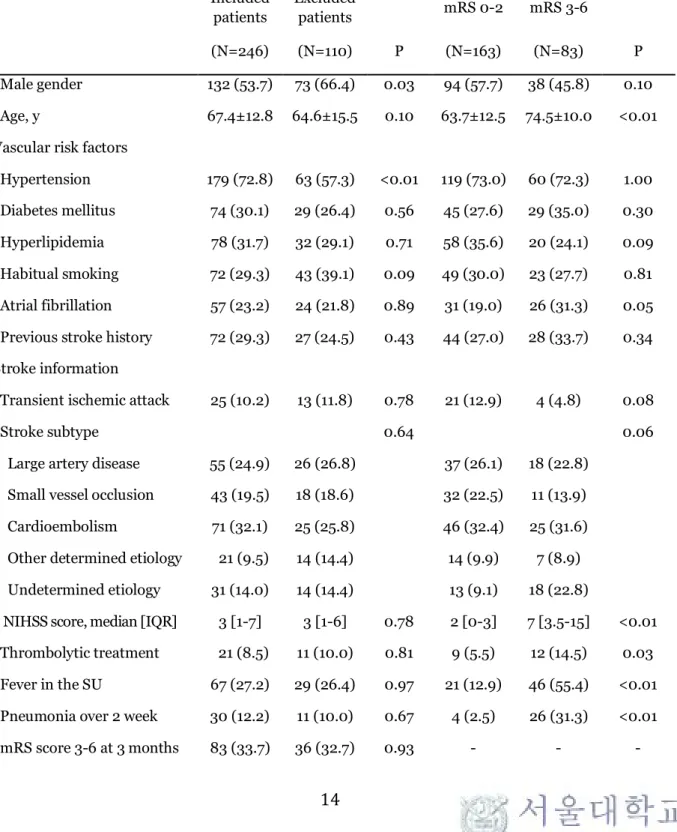
A total of 246 consecutively admitted patients were included. Excluded patients (n=110) were more likely to be male (66.4% vs. 53.7%, P=0.03) with less
hypertension (57.3% vs. 72.8%, P<0.01) compared to the study subjects. All other baseline characteristics were not different between the two groups (Table 1).
Mean age of study patients was 67.4 (standard deviation; 12.8) with higher proportion of male (53.7%). The risk factors included hypertension in 179 (72.8%) patients, diabetes mellitus in 74 (30.1%) patients, hyperlipidemia in 78 (31.7%) patients, habitual smoking in 72 (29.3%) patients, and atrial fibrillation (AF) in 57 (23.2%) of patients. Twenty-five patients (10.2%) had transient ischemia attack. The median NIHSS score at admission was 3 (interquartile range, 1-7) and 8.5% (n=21) of patients received thrombolytic treatment. Sixty- seven (27.2%) patients had fever in the stroke unit and 30 (12.2%) patients had pneumonia in the first two weeks of admission.
Association between tachycardia and functional outcome
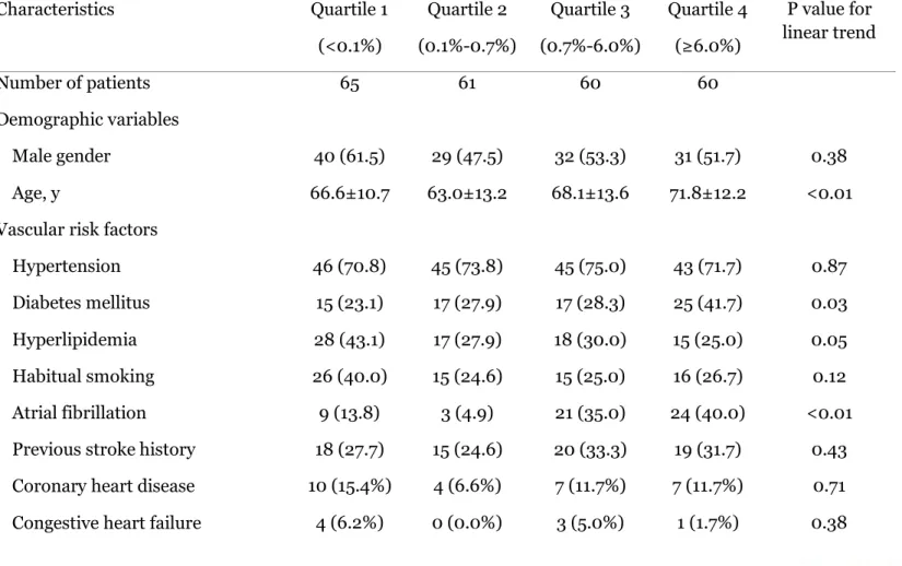
Tachycardia burden, the duration of heart rate over 95/min divided by the total monitoring time, was classified into quartiles (median, 0.7%; interquartile range, 0.1%-5.7%) (Table 2). The patients with the highest tachycardia burden (≥6.0%) were older, more likely to have diabetes, AF, cardioembolic etiology with higher
5
stroke severity at admission, stayed longer in the SU. In addition, they were more likely to have fever in the SU, pneumonia in the first two weeks of admission, and poor functional outcome (mRS 3-6). They were more likely to have higher white blood cell count, initial fasting glucose, HbA1c, but lower hematocrit.
As expected, the patients with the poor functional outcome were older (74.5 years vs. 63.7 years, P<0.01), had higher initial stroke severity (median NIHSS 7 vs. 2, P <0.01), higher initial fasting glucose (112 mg/dL vs. 101 mg/dL, P=0.02), higher proportion of fever (55.4% vs. 12.9%, P<0.01) and pneumonia (31.3% vs. 2.5%, P<0.01), lower hematocrit (36.9% vs. 39.8%, P<0.01), and longer total monitoring time (70.5± 43.4 hours vs. 42.7± 24.5 hours, P<0.01).
(Table 1)
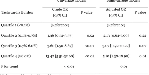
Increased tachycardia burden was associated with an increased odd for poor outcome (mRS 3-6) (odds ratio 1.36, 3.60, and 13.42 in 2nd, 3rd, and 4th quartiles; P for trend, <0.01). As compared with the lowest quartile (<0.1 %), the fourth quartile of tachycardia burden (≥6.0%) was significantly associated with the poor outcome (adjusted odds ratio, 5.10; 95% confidence interval, 1.38-18.90;
P=0.01) after adjusting for confounding variables with bivariate P-values <0.05 (age, transient ischemia attack, stroke subtype, initial stroke severity, AF,
thrombolytic treatment, fever, pneumonia, initial fasting glucose, hematocrit and total monitoring time). (Table 3; Figure)
6
Discussion
In this study, we found that increased tachycardia burden during stroke unit stay was associated with overall mortality and disability in patients with acute
ischemic stroke. About 68% of the patients in the highest quartile of tachycardia burden (≥6.0%) had poor functional outcome.
The association between tachycardia and functional outcome in patients with acute stroke is controversial. One study showed elevated heart rate in subacute phase after stroke is a risk indicator for mortality and poor functional outcome. (21) However, it was unknown whether tachycardia is important in stroke patients with acute phase when heart related change is more important in prognosis. (22, 23) A recent study on acute stroke patients showed tachycardia is frequent phenomenon, while its association to poor functional outcome is not significant. (23) However, the study evaluated merely the occurrence of
tachycardia within 24 hours as ‘yes or no’, thus could not assess overall burden of tachycardia during stroke unit stay. However, our study analyzed heart rate data with 1-minute resolution. Therefore, we could evaluate tachycardia burden as the percentage of duration of tachycardia per total monitoring time from continuous ECG monitoring using high resolution data acquisition system. With these strength, we think that our study provides more accurate information on the relationship between tachycardia and clinical outcome.
The factors associated with poor outcome in patients with stroke were old age, initial stroke severity, stroke progression, recurrence, and medical
7
complications such as pneumonia. (7, 24-26) Likewise, the patients with higher tachycardia burden also were more likely to be old, had higher initial NIHSS score and higher incidence of fever and pneumonia. However, tachycardia burden still showed significant association with poor functional outcome after adjusting for covariates described above.
Well-known causes of tachycardia such as sympathetic hyperactivity, pain, agitation, volume depletion, anemia, heart failure and chronic pulmonary disease are frequent in the acute stroke population. (12) Unfortunately,
identifying the main cause of tachycardia which drives the association with poor outcomes is not possible in our study. In addition, it is not clear whether
tachycardia itself has a direct negative effect on functional outcome or
tachycardia just reflects poor clinical conditions. It is likely that multiple factors may be contributing to this association of tachycardia and poor outcomes. Our finding that tachycardia was strongly associated with poor functional outcome after controlling for potential factors associated poor outcomes suggests that tachycardia is at least a surrogate marker for multiple neurologic and/or medical worsening. One possibility is that pathologic sympathetic activation during the acute phase of stroke is may be a predictor of future cardiovascular and
cerebrovascular events. (27) Several studies have shown that elevated heart rate is associated with plaque rupture (28) and recurrent myocardial infarction with coronary artery disease. (29)
There are several limitations in our study. First, our study is a
retrospective study which excluded a significant numbers of patients due to lack
8
of data for analysis, therefore, there is a chance for selection bias. However, excluded patients were not different except for sex and hypertension compared to study population, suggesting that the chances of selection bias are not high.
Second, the association of tachycardia and early neurologic deterioration could not be assessed in our study. The lack of information on the specific time point of neurologic worsening and tachycardia occurrence limits the interpretation and further studies are needed to investigate timing of tachycardia and neurologic symptoms. Third, we tried to adjust pneumonia and fever as a possible confounder of fever in analyzing data, however, many unmeasured possible causes of tachycardia may still exist. Volume depletion may be one of the
triggering factors of tachycardia in unstable patients. However, our patients were treated aggressively with hydration with normal saline. Therefore, the chances of hypovolemia in our patients are not high. Fourth, there is still a chance that more severe cardioembolic stroke patient had higher tachycardia burden due to
frequent AF rapid ventricular response (RVR). In order to solve this issue, we adjusted AF in analyzing the association between tachycardia and functional outcome, and the correlation was still valid even after adjusting covariates.
In conclusion, an increased tachycardia burden during stroke unit stay is associated with poor 3-month functional outcome, which underscores
importance of continuous heart rate monitoring in the SU. Moreover, it could be inferred from our results that countermeasures against worsening factors might be utilized for patients with increased tachycardia burden. Future clinical studies
9
are needed analyzing occurrence of early neurologic deterioration and its association with increased tachycardia burden.
10
References
1. Indredavik B, Bakke F, Solberg R, Rokseth R, Haaheim LL, Holme I.
Benefit of a stroke unit: a randomized controlled trial. Stroke. 1991;22(8):1026- 31.
2. Langhorne P, Williams B, Gilchrist W, Howie K. Do stroke units save lives? The Lancet. 1993;342(8868):395-8.
3. Jørgensen HS, Nakayama H, Raaschou HO, Larsen K, Hübbe P, Olsen TS. The Effect of a Stroke Unit: Reductions in Mortality, Discharge Rate to Nursing Home, Length of Hospital Stay, and Cost A Community-Based Study.
Stroke. 1995;26(7):1178-82.
4. Indredavik B, Bakke F, Slørdahl S, Rokseth R, Håheim L. Stroke unit treatment 10-year follow-up. Stroke. 1999;30(8):1524-7.
5. Candelise L, Gattinoni M, Bersano A, Micieli G, Sterzi R, Morabito A.
Stroke-unit care for acute stroke patients: an observational follow-up study. The Lancet. 2007;369(9558):299-305.
6. Davalos A, Toni D, Iweins F, Lesaffre E, Bastianello S, Castillo J.
Neurological deterioration in acute ischemic stroke Potential predictors and associated factors in the European Cooperative Acute Stroke Study (ECASS) I.
Stroke. 1999;30(12):2631-6.
7. Rohweder G, Ellekjaer H, Salvesen O, Naalsund E, Indredavik B.
Functional outcome after common poststroke complications occurring in the first 90 days. Stroke. 2015;46(1):65-70.
8. Stroke Unit Trialists’ Collaboration. Organised inpatient (stroke unit) care for stroke. Cochrane Database Syst Rev. 2013;9:CD000197.
9. Cavallini A, Micieli G, Marcheselli S, Quaglini S. Role of monitoring in management of acute ischemic stroke patients. Stroke. 2003;34(11):2599-603.
11
10. Ciccone A, Celani MG, Chiaramonte R, Rossi C, Righetti E. Continuous versus intermittent physiological monitoring for acute stroke. Cochrane Database Syst Rev. 2013:CD008444.
11. Silva Y, Puigdemont M, Castellanos M, Serena J, n i, Suñer RM, et al.
Semi-intensive monitoring in acute stroke and long-term outcome.
Cerebrovascular diseases. 2004;19(1):23-30.
12. Michaud GF, Stevenson WG. Supraventricular Tachyarrhythmias; in Kasper D, Fauci A, Hauser S, Longo D, Jameson J, Loscalzo J (eds): Harrison's principles of internal medicine, 19th edn. New York, McGraw-Hill Professional, 2015.
13. Sander O, Welters ID, Foëx P, Sear JW. Impact of prolonged elevated heart rate on incidence of major cardiac events in critically ill patients with a high risk of cardiac complications. Critical Care Medicine. 2005;33(1):81-8.
14. Schmidt JM, Crimmins M, Lantigua H, Fernandez A, Zammit C, Falo C, et al. Prolonged elevated heart rate is a risk factor for adverse cardiac events and poor outcome after subarachnoid hemorrhage. Neurocritical care.
2014;20(3):390-8.
15. American Diabetes Association. Diagnosis and classification of diabetes mellitus. Diabetes care. 2010;33(Supplement 1):S62-S9.
16. Expert Panel on Detection, Evaluation, and Treatment of High Blood Cholesterol in Adults. Executive summary of the third report of the National Cholesterol Education Program (NCEP) expert panel on Detection, Evaluation, and Treatment of high blood cholesterol in adults (Adult Treatment Panel III).
JAMA. 2001;285(19):2486
17. Whitworth JA. 2003 World Health Organization (WHO)/International Society of Hypertension (ISH) statement on management of hypertension. J Hypertens. 2003;21(11):1983-92.
12
18. Adams HP, Bendixen BH, Kappelle LJ, Biller J, Love BB, Gordon DL, et al. Classification of subtype of acute ischemic stroke. Definitions for use in a multicenter clinical trial. TOAST. Trial of Org 10172 in Acute Stroke Treatment.
Stroke. 1993;24(1):35-41.
19. Reith J, Jorgensen H, Pedersen P, Nakamaya H, Jeppesen L, Olsen T, et al. Body temperature in acute stroke: relation to stroke severity, infarct size, mortality, and outcome. The Lancet. 1996;347(8999):422-5.
20. Wang Y, Lim LL-Y, Levi C, Heller RF, Fisher J. Influence of admission body temperature on stroke mortality. Stroke. 2000;31(2):404-9.
21. Bohm M, Cotton D, Foster L, Custodis F, Laufs U, Sacco R, et al. Impact of resting heart rate on mortality, disability and cognitive decline in patients after ischaemic stroke. European heart journal. 2012;33(22):2804-12.
22. Kallmunzer B, Bobinger T, Kopp M, Kurka N, Arnold M, Hilz MJ, et al.
Impact of heart rate dynamics on mortality in the early phase after ischemic stroke: a prospective observational trial. Journal of stroke and cerebrovascular diseases. 2015;24(5):946-51.
23. Ritter MA, Rohde A, Heuschmann PU, Dziewas R, Stypmann J, Nabavi DG, et al. Heart rate monitoring on the stroke unit. What does heart beat tell about prognosis? An observational study. BMC neurology. 2011;11:47.
24. Adams H, Davis P, Leira E, Chang K-C, Bendixen B, Clarke W, et al.
Baseline NIH Stroke Scale score strongly predicts outcome after stroke A report of the Trial of Org 10172 in Acute Stroke Treatment (TOAST). Neurology.
1999;53(1):126-.
25. Andersen KK, Andersen ZJ, Olsen TS. Predictors of early and late case- fatality in a nationwide danish study of 26 818 patients with first-ever ischemic stroke. Stroke. 2011;42(10):2806-12.
13
26. Koennecke H-C, Belz W, Berfelde D, Endres M, Fitzek S, Hamilton F, et al. Factors influencing in-hospital mortality and morbidity in patients treated on a stroke unit. Neurology. 2011;77(10):965-72.
27. Sander D, Winbeck K, Klingelhöfer J, Etgen T, Conrad B. Prognostic relevance of pathological sympathetic activation after acute thromboembolic stroke. Neurology. 2001;57(5):833-8.
28. Heidland UE, Strauer BE. Left ventricular muscle mass and elevated heart rate are associated with coronary plaque disruption. Circulation.
2001;104(13):1477-82.
29. Fox K, Ford I, Steg PG, Tendera M, Robertson M, Ferrari R. Heart rate as a prognostic risk factor in patients with coronary artery disease and left- ventricular systolic dysfunction (BEAUTIFUL): a subgroup analysis of a randomised controlled trial. The Lancet. 2008;372(9641):817-21.
14
Tables
Table 1. Demographics of study patients
Included patients
Excluded
patients mRS 0-2 mRS 3-6
(N=246) (N=110) P (N=163) (N=83) P
Male gender 132 (53.7) 73 (66.4) 0.03 94 (57.7) 38 (45.8) 0.10 Age, y 67.4± 12.8 64.6± 15.5 0.10 63.7± 12.5 74.5± 10.0 <0.01 Vascular risk factors
Hypertension 179 (72.8) 63 (57.3) <0.01 119 (73.0) 60 (72.3) 1.00 Diabetes mellitus 74 (30.1) 29 (26.4) 0.56 45 (27.6) 29 (35.0) 0.30 Hyperlipidemia 78 (31.7) 32 (29.1) 0.71 58 (35.6) 20 (24.1) 0.09 Habitual smoking 72 (29.3) 43 (39.1) 0.09 49 (30.0) 23 (27.7) 0.81 Atrial fibrillation 57 (23.2) 24 (21.8) 0.89 31 (19.0) 26 (31.3) 0.05 Previous stroke history 72 (29.3) 27 (24.5) 0.43 44 (27.0) 28 (33.7) 0.34 Stroke information
Transient ischemic attack 25 (10.2) 13 (11.8) 0.78 21 (12.9) 4 (4.8) 0.08
Stroke subtype 0.64 0.06
Large artery disease 55 (24.9) 26 (26.8) 37 (26.1) 18 (22.8) Small vessel occlusion 43 (19.5) 18 (18.6) 32 (22.5) 11 (13.9) Cardioembolism 71 (32.1) 25 (25.8) 46 (32.4) 25 (31.6) Other determined etiology 21 (9.5) 14 (14.4) 14 (9.9) 7 (8.9) Undetermined etiology 31 (14.0) 14 (14.4) 13 (9.1) 18 (22.8)
NIHSS score, median [IQR] 3 [1-7] 3 [1-6] 0.78 2 [0-3] 7 [3.5-15] <0.01 Thrombolytic treatment 21 (8.5) 11 (10.0) 0.81 9 (5.5) 12 (14.5) 0.03 Fever in the SU 67 (27.2) 29 (26.4) 0.97 21 (12.9) 46 (55.4) <0.01 Pneumonia over 2 week 30 (12.2) 11 (10.0) 0.67 4 (2.5) 26 (31.3) <0.01
mRS score 3-6 at 3 months 83 (33.7) 36 (32.7) 0.93 - - -
15
Tachycardia burden <0.01
Quartile 1 (<0.1%) 56 (34.4) 9 (10.8)
Quartile 2 (0.1%-0.7%) 50 (30.7) 11 (13.3)
Quartile 3 (0.7%-6.0%) 38 (23.3) 22 (26.5)
Quartile 4 (≥6.0%) 19 (11.7) 41 (49.4)
Total monitoring time, h 52.1± 34.6 - - 42.7± 24.5 70.5± 43.4 <0.01 mRS denotes modified Rankin Scale; NIHSS, National Institutes of Health Stroke Scale;
IQR, interquartile range. mRS scores were missed 7 cases in the excluded patients due to loss of follow up.
16
Table 2. Characteristics of acute ischemic stroke patients in our stroke unit, according to quartiles of tachycardia burden.
Characteristics Quartile 1
(<0.1%)
Quartile 2 (0.1%-0.7%)
Quartile 3 (0.7%-6.0%)
Quartile 4 (≥6.0%)
P value for linear trend
Number of patients 65 61 60 60
Demographic variables
Male gender 40 (61.5) 29 (47.5) 32 (53.3) 31 (51.7) 0.38
Age, y 66.6± 10.7 63.0± 13.2 68.1± 13.6 71.8± 12.2 <0.01
Vascular risk factors
Hypertension 46 (70.8) 45 (73.8) 45 (75.0) 43 (71.7) 0.87
Diabetes mellitus 15 (23.1) 17 (27.9) 17 (28.3) 25 (41.7) 0.03
Hyperlipidemia 28 (43.1) 17 (27.9) 18 (30.0) 15 (25.0) 0.05
Habitual smoking 26 (40.0) 15 (24.6) 15 (25.0) 16 (26.7) 0.12
Atrial fibrillation 9 (13.8) 3 (4.9) 21 (35.0) 24 (40.0) <0.01 Previous stroke history 18 (27.7) 15 (24.6) 20 (33.3) 19 (31.7) 0.43 Coronary heart disease 10 (15.4%) 4 (6.6%) 7 (11.7%) 7 (11.7%) 0.71 Congestive heart failure 4 (6.2%) 0 (0.0%) 3 (5.0%) 1 (1.7%) 0.38
17 Stroke information
Transient ischemic attack 8 (12.3) 10 (16.4) 3 (5.0) 4 (6.7) 0.10
Stroke subtype 0.05
Large artery disease 18 (31.6) 14 (27.5) 16 (28.1) 7 (12.5) Small vessel occlusion 11 (19.3) 15 (29.4) 9 (15.8) 8 (14.3)
Cardioembolism 15 (26.3) 10 (19.6) 23 (40.4) 23 (41.1)
Other determined etiology 5 (8.8) 6 (11.8) 4 (7.0) 6 (10.7) Undetermined etiology 8 (14.0) 6 (11.8) 5 (8.8) 12 (21.4)
NIHSS score, median [IQR] 2 [0-4] 2 [0-4] 3 [1-8] 7 [2-14] <0.01
Thrombolytic treatment 5 (7.7) 3 (4.9) 7 (11.7) 6 (10.0) 0.40
Fever 6 (9.2) 4 (6.6) 22 (36.7) 35 (58.3) <0.01
Pneumonia 1 (1.5) 1 (1.6) 8 (13.3) 20 (33.3) <0.01
mRS score 3-6 at 3 months 9 (13.8) 11 (18.0) 22 (36.7) 41 (68.3) <0.01 Laboratory information
Systolic blood pressure, mmHg 153± 29 157± 31 160.6± 30.2 152.6± 31.1 0.82 Diastolic blood pressure, mmHg 80± 14 87± 16 87.7± 15.1 85.2± 15.5 0.05 Initial fasting glucose, mg/dL 99± 32 102± 27 105± 32 114± 40 0.01
18
HbA1c, % 6.0± 0.7 6.1± 0.8 6.2± 1.1 6.4± 0.9 0.01
Total cholesterol, mg/dL 167± 40 172± 43 172± 45 164± 39 0.66
White blood cell count, /μL 7926± 3331 7871± 3085 7725± 2786 9177± 3237 0.05
Hematocrit, % 39.4± 4.9 41.0± 4.0 37.6± 4.9 37.1± 5.7 <0.01
Ejection fraction, %† 61.2± 7.0 61.9± 6.2 59.9± 5.7 59.9± 7.2 0.13 Medications to control heart rate
Diltiazem 0 (0.0%) 0 (0.0%) 0 (0.0%) 11 (18.3%) <0.01
Digoxin 1 (1.5%) 0 (0.0%) 2 (3.3%) 3 (5.0%) 0.12
Beta blockers 8 (12.3%) 0 (0.0%) 9 (15.0%) 4 (6.7%) 0.87
Tachycardia burden, % 0.0± 0.0 0.3± 0.2 2.3± 1.4 34.2± 26.8 <0.01 Total monitoring time, h 42.1± 24.8 47.3± 27.7 49.1± 28.5 70.8± 47.1 <0.01 mRS denotes modified Rankin Scale; NIHSS, National Institutes of Health Stroke Scale; IQR, interquartile range.
†Transthoracic echocardiography was performed in 93% of the patients (n=228 of 246) during admission period.
19
Table 3. Associations between tachycardia burden and poor 3-month functional outcome
Univariate models Multivariable models* Tachycardia Burden Crude OR
[95% CI] P value Adjusted OR
[95% CI] P value
Quartile 1 (<0.1%) (Reference) (Reference)
Quartile 2 (0.1%-0.7%) 1.36 [0.52-3.57] 0.52 2.13 [0.64-7.09] 0.22 Quartile 3 (0.7%-6.0%) 3.60 [1.50-8.67] <0.01 3.07 [0.92-10.22] 0.07 Quartile 4 (≥6.0%) 13.42 [5.51-32.68] <0.01 5.10 [1.38-18.90] 0.01
P for trend < 0.01 0.01
OR denotes odds ratio; CI, confidence interval
* Multivariable models were adjusted for age, transient ischemic attack, stroke subtype, atrial fibrillation, initial stroke severity, thrombolytic treatment, fever, pneumonia, initial fasting glucose, hematocrit and total monitoring time.
20
Figures and Figure legends
Figure. Distribution of modified Rankin Scale score according to the quartile of tachycardia burden
21
22