저작자표시-비영리-변경금지 2.0 대한민국 이용자는 아래의 조건을 따르는 경우에 한하여 자유롭게
l 이 저작물을 복제, 배포, 전송, 전시, 공연 및 방송할 수 있습니다. 다음과 같은 조건을 따라야 합니다:
l 귀하는, 이 저작물의 재이용이나 배포의 경우, 이 저작물에 적용된 이용허락조건 을 명확하게 나타내어야 합니다.
l 저작권자로부터 별도의 허가를 받으면 이러한 조건들은 적용되지 않습니다.
저작권법에 따른 이용자의 권리는 위의 내용에 의하여 영향을 받지 않습니다. 이것은 이용허락규약(Legal Code)을 이해하기 쉽게 요약한 것입니다.
Disclaimer
저작자표시. 귀하는 원저작자를 표시하여야 합니다.
비영리. 귀하는 이 저작물을 영리 목적으로 이용할 수 없습니다.
변경금지. 귀하는 이 저작물을 개작, 변형 또는 가공할 수 없습니다.
A DISSERTATION FOR THE DEGREE OF MASTER
Modified Intraglandular Sialography by US-guided Contrast Injection in Dogs
개에서 초음파 유도하에 턱샘에
Iopromide 를 주입한 변형 침샘조영술
February, 2016
Major in Veterinary Clinical Sciences (Diagnostic Imaging)
Department of Veterinary Medicine The Graduate School
Seoul National University
Suyeon Kim
Modified Intraglandular Sialography by US-guided Contrast Injection in Dogs
개에서 초음파 유도하에 턱샘에
Iopromide 를 주입한 변형 침샘조영술
지도교수 최 민 철
이 논문을 수의학석사학위논문으로 제출함 2015 년 10 월
서울대학교 대학원
수의학과 임상수의학(수의영상의학)전공
김 수 연
김수연의 석사학위논문을 인준함 2015 년 12 월
위 원 장 윤 정 희 (인)
부위원장 최 민 철 (인)
위 원 서 강 문 (인)
1
ABSTRACT
Modified Intraglandular Sialography by US-guided Contrast Injection in Dogs
Supervised by
Professor Mincheol Choi
Suyeon Kim
Veterinary Clinical Sciences (Diagnostic Imaging) Department of Veterinary Medicine
The Graduate School
Seoul National University
2
Sialography is a useful diagnostic method in the investigation of disease involving the salivary glands and ducts.
However, the disadvantages of conventional method include the need for general anesthesia and the difficulty associated with locating the duct openings. The purpose of this study was to develop a simple and reliable sialography protocol in dogs and to determine the optimal dose of contrast medium into the mandibular gland under fluoroscopy.
Eight healthy dogs without salivary gland abnormalities were used. Ultrasound-guided percutaneous injection of iopromide (Ultravist 370®; SCHERING Co., Seoul, South Korea) into the left mandibular gland of all dogs was performed to determine the optimal dose and fluoroscopic imaging time. Fluoroscopic evaluation was performed from immediately before injection of iopromide until each dose was fully injected.
On the fluoroscopic images, mandibular gland and entire mandibular duct were identified in five dogs (62.5%). Mandibular gland and partial duct were identified in two dogs (25%), and only mandibular gland was visualized in a dog (12.5%). Of five dogs, entire duct was identified after 7.0 ml iopromide injection in two dogs (25%), and 9.0 ml injection in three dogs (37.5%), respectively.
The time from iopromide injection to the identification of mandibular duct ranged variously, from 6 to 27 seconds.
3
Based on the results of the present study, it was recommended that the fluoroscopy of mandibular glands and ducts should be performed immediately after (at least within 6 seconds) the contrast injection of 7.0 ml or more volume of iopromide into the mandibular gland under ultrasound guidance.
This simple and reliable method may be helpful to increase the detection sensitivity for abnormalities of salivary glands and guiding surgical approaches in dogs. Therefore, this technique may be an alternative technique to conventional sialography in dogs.
Key words: sialography, fluoroscopy, mandibular gland, dog, ultrasound guidance
Student number: 2014-21945
4
CONTENTS
INTRODUCTION ∙∙∙∙∙∙∙∙∙∙∙∙∙∙∙∙∙∙∙∙∙∙∙∙∙∙∙∙∙∙∙∙∙∙∙∙∙∙∙∙∙∙∙∙∙∙∙∙∙∙∙∙∙∙∙∙∙∙∙∙∙∙∙∙∙∙∙∙∙∙ 5
MATERIALS AND METHODS ∙∙∙∙∙∙∙∙∙∙∙∙∙∙∙∙∙∙∙∙∙∙∙∙∙∙∙∙∙∙∙∙∙∙∙∙∙∙∙∙∙∙∙∙∙∙∙∙∙∙∙∙∙ 8 1. Animals ∙∙∙∙∙∙∙∙∙∙∙∙∙∙∙∙∙∙∙∙∙∙∙∙∙∙∙∙∙∙∙∙∙∙∙∙∙∙∙∙∙∙∙∙∙∙∙∙∙∙∙∙∙∙∙∙∙∙∙∙∙∙∙∙∙∙∙∙ 8 2. Contrast media ∙∙∙∙∙∙∙∙∙∙∙∙∙∙∙∙∙∙∙∙∙∙∙∙∙∙∙∙∙∙∙∙∙∙∙∙∙∙∙∙∙∙∙∙∙∙∙∙∙∙∙∙∙∙∙ 10 3. Modified sialography using US-guided contrast injection into mandibular gland∙∙∙∙∙∙∙∙∙∙∙∙∙∙∙∙∙∙∙∙∙∙∙∙∙∙∙∙∙∙∙∙∙∙∙∙∙∙∙∙∙∙∙∙∙∙ 10 4. Fluoroscopy and image evaluation ∙∙∙∙∙∙∙∙∙∙∙∙∙∙∙∙∙∙∙∙∙∙∙∙∙∙∙∙ 14
RESULTS ∙∙∙∙∙∙∙∙∙∙∙∙∙∙∙∙∙∙∙∙∙∙∙∙∙∙∙∙∙∙∙∙∙∙∙∙∙∙∙∙∙∙∙∙∙∙∙∙∙∙∙∙∙∙∙∙∙∙∙∙∙∙∙∙∙∙∙∙∙∙∙∙∙∙∙∙∙∙∙ 15 1. Modified sialography by US-guided contrast injection into mandibular gland ∙∙∙∙∙∙∙∙∙∙∙∙∙∙∙∙∙∙∙∙∙∙∙∙∙∙∙∙∙∙∙∙∙∙∙∙∙∙∙∙∙∙∙∙∙ 15
2. Identification of the mandibular gland and duct under Fluoroscopy ∙∙∙∙∙∙∙∙∙∙∙∙∙∙∙∙∙∙∙∙∙∙∙∙∙∙∙∙∙∙∙∙∙∙∙∙∙∙∙∙∙∙∙∙∙∙∙∙∙∙∙∙∙∙∙∙∙∙∙∙ 18
3. Normal appearance of the mandibular gland and duct on the fluoroscopic images ∙∙∙∙∙∙∙∙∙∙∙∙∙∙∙∙∙∙∙∙∙∙∙∙∙∙∙∙∙∙∙∙∙∙∙∙∙∙ 20 DISCUSSION ∙∙∙∙∙∙∙∙∙∙∙∙∙∙∙∙∙∙∙∙∙∙∙∙∙∙∙∙∙∙∙∙∙∙∙∙∙∙∙∙∙∙∙∙∙∙∙∙∙∙∙∙∙∙∙∙∙∙∙∙∙∙∙∙∙∙∙∙∙∙∙∙∙ 23 CONCLUSION ∙∙∙∙∙∙∙∙∙∙∙∙∙∙∙∙∙∙∙∙∙∙∙∙∙∙∙∙∙∙∙∙∙∙∙∙∙∙∙∙∙∙∙∙∙∙∙∙∙∙∙∙∙∙∙∙∙∙∙∙∙∙∙∙∙∙∙∙∙∙∙ 29 REFERENCES ∙∙∙∙∙∙∙∙∙∙∙∙∙∙∙∙∙∙∙∙∙∙∙∙∙∙∙∙∙∙∙∙∙∙∙∙∙∙∙∙∙∙∙∙∙∙∙∙∙∙∙∙∙∙∙∙∙∙∙∙∙∙∙∙∙∙∙∙∙∙∙∙∙ 30 ABSTRACT IN KOREAN ∙∙∙∙∙∙∙∙∙∙∙∙∙∙∙∙∙∙∙∙∙∙∙∙∙∙∙∙∙∙∙∙∙∙∙∙∙∙∙∙∙∙∙∙∙∙∙∙∙∙∙∙∙∙∙∙∙∙ 35
5
INTRODUCTION
Disease involving the salivary glands and ducts in dogs and cats was reported overall incidence of 0.17% (Hammer et al., 2001).
Differential diagnoses of salivary glands and ducts pathological conditions include neoplasia, abscess, sialolithiasis, sialoangiectasis, sialandenitis, and salivary mucocele (Harvey et al., 1969). The clinical signs associated with salivary glands abnormalities include retching, gulping, nausea, vomiting or regurgitation, and painful hard enlarged salivary glands (Kelly et al., 1979; Mawby et al., 1991;
Chapman et al., 1992; Cooke et al., 1992; Brooks et al., 1995).
In one study, a histopathological diagnosis of neoplasia was made in 30% of salivary gland biopsies, indicating that tumors are a significant cause of disease in this tissue (Spangler et al., 1991). The mandibular and parotid salivary glands are most often affected and accounted for 75% to 80% of all salivary gland neoplasia (Carberry et al., 1988; Koestner et al., 1965). The tumor type most often reported is simple adenocarcinoma; other tumor types include squamous cell carcinoma, mucoepidermoid carcinoma, anaplastic carcinoma, and complex carcinoma (Carberry et al., 1988; Spangler et al., 1991; Koestner et al., 1965; Brunnert et
6
al., 1990; Carpenter et al., 1991; Wells et al., 1975). Also, sialocele or salivary mucocele, subcutaneous or submucosal collections of saliva (Glen et al., 1972), occur frequently in dogs. Potential for the development of sialocoeles in the dog include trauma to salivary ducts or glands, foreign bodies, sialoliths, and neoplasia.
However, in the majority of cases the actual causes are still unknown (Mulkey et al., 1971; Glen et al., 1972; Durtnell et al., 1977; Bellenger et al., 1992). On the other hand, sialolithiasis is a rare condition in dogs and has only been reported to affect the parotid salivary duct (Mulkey et al., 1971; Jeffreys et al., 1996;
Hunt et al., 1997; Dunning et al., 2003; Trumpatori et al., 2007).
There are several diagnostic techniques for examining the salivary glands and ducts, such as ultrasonography, contrast study, computed tomography (CT), and magnetic resonance imaging (MRI). Fluoroscopy in relation to the sialographic injection was suggested in the human fields, although the salivary glands and ducts were not visible in the survey radiographs (Waite et al., 1969; Schmitt et al., 1976). Especially, retrograde sialography is performed by injection of radiopaque dye onto a salivary duct through a cannula inserted in the duct orifice, is a useful diagnostic aid in the investigation of disease involving the salivary glands and ducts in both human and dogs. However, the disadvantages of this traditional method include the need for
7
general anesthesia and the difficulty associated with locating the duct openings (Harvey et al., 1969).
The purpose of this study was to develop a simple and reliable sialography protocol in dogs and to determine the optimal dose of contrast medium into the mandibular gland and fluoroscopic imaging time.
8
MATERIALS AND METHODS
1. Animals
Eight healthy beagles, unknown ages, with weights ranging from 8 to 13 kg were used. There were two neutered males, two spayed female and four males. They were housed in indoor cages and fed a balanced diet (Nutrena Active®, Agribrand Purina Co., Seoul, South Korea) with water ad libitum.
Dogs were fasted for at least 12 hours and the following protocol was performed. Just before the experiments, light sedation was induced by intravenous administration of acepromazine (0.1 mg/kg, Sedaject®, Samu Median Co., Seoul, South Korea) and the modified sialography was performed. Following the sialography, all dogs was administered cefaxin per oral (30 mg/kg, Cefaxin®, Koruspharm Co., Jecheon, South Korea) and physical examination was performed. The experimental protocol was approved by the Institute of laboratory Animal Resources Seoul National University (SNU-150903-5).
9
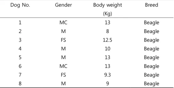
Table 1. Individual data of eight dogs used in this study
MC, castrated male; FS, spayed female; M, male; F, female
Dog No. Gender Body weight
(Kg)
Breed
1 MC 13 Beagle
2 M 8 Beagle
3 FS 12.5 Beagle
4 M 10 Beagle
5 M 13 Beagle
6 MC 13 Beagle
7 FS 9.3 Beagle
8 M 9 Beagle
10
2. Contrast media
Four different doses (3.0 ml, 5.0 ml, 7.0 ml, and 9.0 ml) of Iopromide (Ultravist 370®; SCHERING Co., Seoul, South Korea) were injected into the left mandibular gland of all dogs for determination of optimal dose and fluoroscopic imaging time.
Four doses were used at intervals of a week, therefore total experimental period for a dog were four weeks.
3. Modified sialography using US-guided contrast injection into the left mandibular gland
The beagle dogs were positioned in dorsal recumbency, the hair around the left mandibular salivary gland was clipped and skin was aseptically prepared. Survey lateral and dorsoventral radiographs were taken to confirm no remarkable findings around the left mandibular gland and to determine the optimal kVp and mAs (Figure 1). After identification of the left mandibular gland with ultrasound guidance using a 6-12MHz linear array ultrasound transducer (SONOVET 2000®, Medison Co., Seoul, South Korea), the four different doses of iopromide were injected into the left mandibular gland using a needle stylet of 26-gauge connected to
11
a 10 ml syringe. A neck region of each dog was pulled toward the head of the dog for extension of soft tissue around the mandibular gland. Identification of the left mandibular gland and insertion of a needle were performed under ultrasound guidance, and iopromide was injected into the left mandibular gland (Figure 2).
12
Figure 1. Survey radiographs of the skull. There were no remarkable findings around the left mandibular gland on the lateral (A) and dorsoventral (B) views.
13
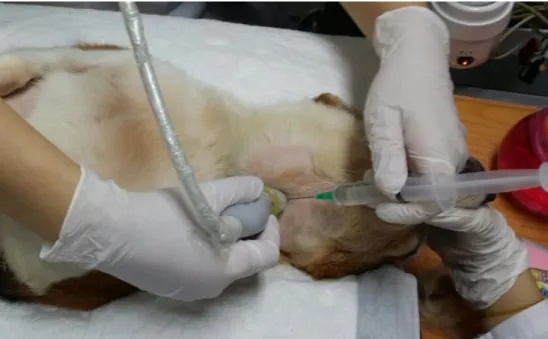
Figure 2. Injection of Iopromide into the left mandibular gland. A neck region of each dog was pulled toward the head of the dog for extension of soft tissue around the mandibular gland.
Identification of the left mandibular gland and insertion of a needle were performed under ultrasound guidance, and iopromide was injected into the left mandibular gland.
14
4. Fluoroscopy and imaging evaluation
Fluoroscopy (DC-525R/F®, Dong-A X-ray Co., Chungnam, South Korea) was performed at 60-90 kVp, 1 mA from immediately before injection of iopromide until each dose of iopromide was fully injected. The center of collimation was the left mandibular gland, and the ROI included whole mandible region to evaluate the salivary ducts.
Fluoroscopic images for each dog were evaluated independently after completion of the study. Evaluation of the images was performed for the presence of contrast medium in the left mandibular gland and duct, and for characterization of the mandibular duct course and branching.
15
RESULTS
1. Modified sialography by US-guided contrast injection into the left mandibular gland.
The mandibular glands were recognized as an oval or round-triangular shaped and showed homogeneously unilobulated appearance. The long diameter of mandibular gland ranged from 2.62 to 3.20 cm and the short diameter ranged from 1.40 to 2.70 cm (Table 2). When iopromide was injected into the left mandibular gland by ultrasound guidance, each injection was continued for 14~32 seconds, an injection of iopromide was dispersed around the needle tip and this area became hyperechoic.
The mandibular glands appeared bigger and showed increased echogenicity compared to the appearance before iopromide injection. If the contrast media was leaked around the mandibular gland, anechoic fluid was identified around it. Extranodal leakage did not occur in any dogs at iopromide volume of 3.0 ml, leakage of a small volume was apparent in four dogs using 5.0 ml and a moderate volume of iopromide leaked in all dogs using 7.0 ml and 9.0 ml (Figure 3).
16
Table 2. Long and short diameter of mandibular gland of each dogs on the ultrasonographic images
Dog No. Long diameter (cm) Short diameter (cm)
1 2.77 1.77
2 2.62 2.25
3 3.05 2.70
4 2.93 1.81
5 2.65 2.55
6 3.11 1.40
7 3.20 1.95
8 2.81 2.04
17
Figure 3. Ultrasonographic images of the left mandibular gland in a dog. The mandibular gland after injection of iopromide volume of 3.0 ml (B), 5.0 ml (C), and 7.0 ml (D) were increased in size and echogenicity compared to that before iopromide injection (A). The hyperechoic line (short white arrow) is the needle tip. Extranodal leakage of iopromide is seen as anechoic fluid around the mandibular gland (long arrows).
18
2. Identification of the mandibular gland and duct under fluoroscopy.
On the fluoroscopic images of total eight dogs, mandibular gland and entire mandibular duct were identified in five dogs (62.5%). Mandibular gland and partial duct were identified in two dogs (25%), and only mandibular gland was visualized in the other dog (12.5%). Of five dogs that entire mandibular duct was visualized, entire mandibular duct was identified after 7.0 ml iopromide injection in two dogs (25%), and after 9.0 ml injection in three dogs (37.5%). Also, of two dogs that partial mandibular duct was visualized, mandibular partial duct was identified after 3.0 ml iopromide injection in one dog, and after 5.0 ml injection in the other dog (Table 3).
The time from iopromide injection to the identification of mandibular duct ranged variously, from 6 to 27 seconds (6, 9, 17, 22 and 27 seconds, respectively).
19
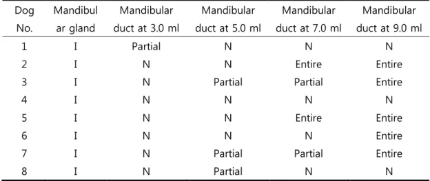
Table 3. Identification of the left mandibular gland and duct according to iopromide injection
Dog No.
Mandibul ar gland
Mandibular duct at 3.0 ml
Mandibular duct at 5.0 ml
Mandibular duct at 7.0 ml
Mandibular duct at 9.0 ml
1 I Partial N N N
2 I N N Entire Entire
3 I N Partial Partial Entire
4 I N N N N
5 I N N Entire Entire
6 I N N N Entire
7 I N Partial Partial Entire
8 I N Partial N N
I, identification; N, non-identified
20
3. Normal appearance of the mandibular gland and delineation of the mandibular duct on the fluoroscopic images
On the contrast fluoroscopic studies, the mandibular gland could be clearly visualized at the mandibular angle region in all eight dogs, which is invisible on the survey radiographs. The mandibular glands were an oval or a round-triangular shaped, the appearance of the mandibular gland on the contrast fluoroscopic images was similar to that on the ultrasound images. The mandibular duct demonstrated some variations in the course and diameter, and several branches emerge from the main duct. The mandibular duct leaves the gland, runs dorsally and curved cranially, and then lay in the middle aspect of the mandible until the opening, finally, opens into the mouth on the sublingual caruncle (Figure 4-5).
21
Figure 4. Fluoroscopic images before (A) and after injection of 5.0 ml (B), and 9.0 ml (C and D) into the left mandibular gland in a dog. After 5.0 ml injection of iopromide, the mandibular gland and partial duct (short white arrow) was visualized. And the entire duct (long white arrow) was visualized after 9.0 ml injection of iopromide.
22
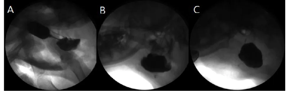
FIgure 5. Fluoroscopic images. In case of the first dog (A), mandibular gland and entire duct (white arrow) were visualized although the gland was not fully filled with contrast medium. In the second (B) and third (C) dogs, on the other hand, the entire ducts were not identified even though the mandibular gland was almost full of contrast medium.
23
DISCUSSION
Sialography has been used as a standard method since 1925 when Barsony introduced the technique of injecting radiopaque material into the duct system of the salivary glands (Akin et al., 1991).
Although few changes have occurred since that time, in 1904, Charpy first isolated glands, and in 1913 Arcelin attempted to use bismuth as a contrast medium. Payne was the first to suggest fluoroscopy in relation to the sialographic injection (Waite et al., 1969). Also, in 1969, contrast radiography has been used in the diagnosis of salivary gland disease in canines by Harvey (Tadjalli et al., 2004). All of the human and animal studies referred to above related to retrograde sialography, where the dye is injected through a cannula inserted into the duct opening.
Physiological (secretory) sialography has also been reported in the human fields, although not in the veterinary literature (Harvey et al., 1969).
However, there are some disadvantages of imaging using a conventional sialography such as difficulty in finding duct openings or taking a risk of anesthesia (Harvey et al., 1969). For overcoming these problems, modified sialography using ultrasound-guided percutaneous injection of contrast medium enables the evaluation of the salivary glands and ducts under fluoroscopy for contrast study.
In comparison to the conventional sialography, one of the
24
advantages of the modified sialography was free from anesthetic procedure, because a mild sedation and holding a dog by hands were enough to perform the sialography in the present study. Another advantage of the modified sialography was no need to make effort to find duct openings. Instead of complicated procedure, identification of salivary gland and injection of contrast medium were performed under ultrasound guidance. Thus, it is a non-invasive and simple technique to evaluate the salivary glands and ducts. To the author’s knowledge, this technique has not been reported in both human and veterinary studies.
The most important factor for successful sialography of dogs was the successful injection of iopromide into the mandibular gland.
Because of many structures around the mandibular gland, such as several lymph nodes, other salivary glands and muscles, injection of iopromide under palpation was very difficult. Also, the needle could penetrate the mandibular gland since it is not the deep-structure.
Therefore, neck of each dog should be pulled fully toward the head of dog for extension of soft tissue around the mandibular gland. And appropriate pressure should be applied to the probe to allow mandibular gland to be well identified under ultrasound.
When the injection of contrast medium, there was leakage of iopromide around the mandibular gland as relatively a large amount of iopromide should be injected to the mandibular gland. One possible reason for this may be the high pressure applied to the syringe due to high viscosity of iopromide. Also, this high pressure made it difficult to maintain the position of a needle within the mandibular gland, so the
25
needle could be moved away from the mandibular gland during injection. Although slow injection of contrast medium is advised to prevent this leakage, too slow rate of injection could lead to fewer successful evaluations on the sialography.
In the present study, iopromide was injected into the mandibular gland under ultrasound guidance not only for instead of invasive procedure, but also for determination of optimal dose of contrast medium for fluoroscopic imaging time. Since there is no studies using this modified method in the both human and veterinary fields so far, it was difficult to determine the optimal volume of contrast medium that could visualize the mandibular gland and duct. Based on the previous study about the mean mass of the mandibular gland (Tadjalli et al., 2004), modified sialography of dogs were performed by injection of iopromide with four different doses of 3.0 ml, 5.0 ml, 7.0 ml and 9.0 ml volume to each dogs in the present study.
Assessment of the enhancement effect by iopromide of four different doses may be sufficient for identification of mandibular gland and duct under fluoroscopy. The injected volume of iopromide that could visualize the mandibular duct was not that same in each dogs. In the present study, mandibular gland and entire mandibular duct were identified in five dogs (62.5%). Of five dogs, entire duct was identified after 7.0 ml iopromide injection in two dogs (25%) and after 9.0 ml injection in three dogs (37.5%). Therefore, 7.0 ml or more volume injection should be indicated for diagnostic imaging of mandibular gland and duct on the fluoroscopic images.
26
Because the time for clear delineation of mandibular duct in each 5 dogs was various from 6 seconds to 27 seconds in the present study (6, 9, 17, 22 and 27 seconds, respectively), it is recommended that the fluoroscopy of mandibular glands should be performed at the same time or immediately after the percutaneous injection of contrast medium into the mandibular gland under ultrasound guidance. One possible reason for this is the relationship between the histologic structure of the salivary glands and the position of injected needle (Ogata T., 1955).
Based on the anatomic and histologic structure of the salivary system, the secretory unit (salivary unit) consists of the acinus, myoepithelial cells, the intercalated duct, the striated duct, and the main excretory duct. The basic secretory units of salivary glands are clusters of cells called an acini. These cells secrete fluid that contains water, electrolytes, mucus and enzymes, all of which flow out of the acinus into collecting ducts. Small collecting ducts within salivary glands lead into larger ducts, eventually forming a single large duct that empties into the oral cavity (Bowen et al., 2002; Ogata et al., 1955).
In the present study, the position of the needle, in other words, how closer the needle from the main excretory duct might play a prominent role in visualizing mandibular ducts. Therefore, it could be assumed that the closer the needle injected adjacent to the large excretory duct, the faster and more clear the mandibular duct could be identified on the fluoroscopic images.
On the fluoroscopic images, mandibular gland and entire duct
27
were visualized in case of some dogs, although the gland was not fully filled with contrast medium. On the other hand, the entire ducts were not identified in case of others although the mandibular gland was almost full of contrast medium. Based on the anatomic and histologic structure of the salivary system mentioned above, it might also provide a basis that there is close relationship between the location of injected needle and the possibility of successful sialographic imaging. That is, the needle injected more adjacent the main excretory duct could assist the mandibular duct to be identified, although the gland was not fully filled with contrast medium.
Furthermore, according to the results of the present study, it is expectable that there would not be much difference in both fluoroscopic sialography protocol with ultrasound guidance and imaging evaluation of the other salivary glands and ductal system such as parotid glands or sublingual glands. Thus, this modified sialography could be applied to the salivary glands generally, as well as mandibular gland and duct.
The prevalence of adverse reactions to the contrast medium would appear to be very low (Cockrell et al., 1993). Nonionic and water-soluble iodinated contrast media is safe when small volume of contrast media leaked as they exert only minimal osmotic potential with low osmolality (Parry, 2009). Although contrast media could induce various symptoms that include urticaria, bronchospasm, angioedema and vasomotor collapse (Cockrell et al., 1993), no adverse effects were observed in this study based on the clinical signs and
28
physical examinations in all dogs.
This study has several limitations. Though the conventional sialographic technique is not so complicated, the application of modified sialographic technique requires a technical expertise for clear identification of the mandibular gland and duct, so that contrast media could be injected accurately into the mandibular gland. In addition, a nervous or uncooperative patient might make it extremely difficult to perform the modified sialographic procedure. Since just light sedation was induced in the present study, one of all eight dogs was excluded from imaging evaluation due to pretty excited condition. Also, because the histopathologic examination was not performed, possibility of the histologic damage or infection was not completely excluded in this study. Therefore, it is recommended that serial recheck after experiments should be performed in the further studies.
From the results of the present study, it was found that a simple and reliable fluoroscopic sialography protocol is effective in dogs. It was recommended that the fluoroscopy of mandibular glands should be performed immediately after (at least within 6 seconds) the percutaneous injection of 7.0 ml or more volume of iopromide into the mandibular gland under ultrasound guidance. This simple and reliable method may be helpful to increase the detection sensitivity for abnormalities of salivary glands and ducts and guiding surgical approaches in dogs. Therefore, this technique may be an alternative technique to conventional sialography in dogs.
29
CONCLUSION
The purpose of the present study was to develop a simple and reliable fluoroscopic sialography protocol in dogs and to determine the optimal dose of contrast medium into the mandibular gland and fluoroscopic imaging time. It was recommended that the fluoroscopy of mandibular glands should be performed immediately after (at least within 6 seconds) the percutaneous injection of 7.0 ml or more volume of iopromide into the mandibular gland under ultrasound guidance.
This non-invasive, simple and reliable method may be helpful to increase the detection sensitivity for abnormalities of salivary glands and ducts and guiding surgical approaches in dogs. Therefore, this technique may be an alternative technique to conventional sialography in dogs.
30
REFERENCES
Adam EJ, Willson SA, Corcoran MO, Hobsley M. The value of parotid sialography. British journal of surgery 70(2): 108-110, 1983.
Akin I, Esmer N, Gerceker M, Aytac S, Erden I, Akan H. Sialographic and ultrasonographic analyses of major salivary glands. Acta otolaryngol 111(3): 600-606, 1991.
Blair GS. Hydrostatic sialography: An analysis of a technique. Oral surgery, oral medicine, oral pathology 36(1): 116-130, 1973.
Brown JE. Interventional sialography and minimally invasive techniques in benign salivary gland obstruction. Seminars in ultrasound, CT and MRI 27(6): 465-475, 2006.
Cockrell DJ, Rout PGJ. An adverse reaction following sialography.
Dentomaxillofacial radiology 22(1): 41-42, 1993.
Dehghani SN, Tadjalli M, Seifali A. Sialography in horse: technique and normal appearance. Veterinarski arhiv 75(6): 531-540, 2005.
Eisenbud L, Cranin N. The role of sialography in the diagnosis and
31
therapy of chronic obstructive sialadentitis. Oral surgery, oral medicine, oral pathology 16(10): 1181-1199, 1963.
Ericson S. The importance of sialography for the determination of the parotid flow: The normal variation in salivary output in relation to the size of the gland at stimulation with citric acid. Acta oto-laryngologica 72(1): 437-444, 1971.
Hammer A, Getzy D, Ogilvie G, Upton M, Klausner J, Kisseberth WC.
Salivary gland neoplasia in the dog and cat: Survival times and prognostic factors. J Am Anim Hosp Assoc 37(5): 478-482, 2001.
Hasson O. Modern sialography for screening of salivary gland obstruction. Journal of oral and maxillofacial surgery 68(2): 276-280, 2010.
Harvey CE. Sialography in the dog. Veterinary radiology and ultrasound 10(1): 18-27, 1969.
Kalinowski M, Heverhagen JT, Rehberg E, Klose KJ, Wagner HJ.
Comparative study of MR sialography and digital subtraction sialography for benign salivary gland disorders. American journal of neuroradiology 23(9): 1485-1492, 2002.
Ogata T. The internal secretion of salivary gland. Endocrinologia
32
japonica 2(4): 247-261, 1955.
Payne RT. Sialography: its technique and applications. The british journal of surgery 19(73): 142-148, 1931.
Ritter MJ, Pfeil DJF, Stanley BJ, Hauptman JG, Walshaw R. Mandibular and sublingual sialocoeles in the dog: A retrospective evaluation of 41 cases, using the ventral approach for treatment. New Zealand veterinary journal 54(6): 333-337, 2006.
Rubaltelli L, Sponga T, Candiani F, Pittarello F, Andretta M. Infantile recurrent sialectatic parotitis: the role of sonography and sialography in diagnosis and follow-up. The british journal of radiology 60(720): 1211- 1214, 1987.
Ryan T, Welsh E, Mcgorum I, Yool D. Sublingual salivary gland sialolithiasis in a dog. Journal of small animal practice 49(5): 254-256, 2008.
Schmitt G, Lehmann G, Strotges MW, Wehmer W, Reinecke V, Teske HJ, Rottinger EM. The diagnostic value of sialography and scintigraphy in salivary gland diseases. The british journal of radiology 49: 326-329, 1976.
Schortinghuis J, Pijpe J, Spijkervet FKL, Vissink A. Retention of lipiodol
33
after parotid gland sialography. International journal of oral and maxillofacial surgery 38(4): 346-349, 2009.
Schroeder H, Berry WL. Salivary gland necrosis in dogs: a retrospective study of 19 cases. Journal of small animal practice 39(3): 121-125, 1998.
Siddiqui SJ. Sialolithiasis: an unusually large submandibular salivary stone. British dental journal 193(2): 89-91, 2002.
Tadjalli M, Dehghani SN, Basiri M. Sialography in dog: Normal appearance. Veterinarski Arhiv 74(3): 225-233, 2004.
Tadjalli M, Dehghani SN, Ghadiri M. Sialography of the goat parotid, mandibular and sublingual salivary glands. Small ruminant research 44(3): 179-185, 2002.
Tsioli V, Papazoglou LG, Basdani E, Kosmas P, Brellou G, Poutahidis T, Bagias S. Surgical management of recurrent cervical sialoceles in four dogs. Journal of small animal practice 54(6): 331-333, 2013.
Tsujii H. Quantitative dose-response analysis of salivary function following radiotherapy using sequential ri-sialography. International journal of radiation oncology biology physics 11(9): 1603-1612, 1985.
Varghese JC, Thornton F, Lucey BC, Walsh M, Farrell MA, Lee MJ. A
34
prospective comparative study of MR sialography and conventional sialography of salivary duct disease. American journal of roentgenology 173(6): 1497-1503, 1999.
Waite DE. Secretory sialography of the salivary glands. Oral surgery, oral medicine, oral pathology 27(5): 635-641, 1969.
Yousem DM, Kraut MA, Chalian AA. Major salivary gland imaging.
Radiology 216(1): 19-29, 2000.
Yoshimura Y, Inoue Y, Odagawa T. Sonographic examination of sialolithiasis. Journal of oral and maxillofacial surgery 47(9): 907-912, 1989.
Zbaren P, Ducommun JC. Diagnosis of salivary gland disease using ultrasound and sialography: a comparison. Clinical otolaryngology and Allied sciences 14(3): 189-197, 1989.
35
국 문 초 록
개에서 초음파 유도하에 턱샘에 Iopromide 를 주입한 변형
침샘조영술
지도교수 최 민 철
김 수 연
서울대학교 대학원
수의학과 임상수의학 (수의영상의학) 전공
36
침샘 조영술은 침샘과 침샘관을 포함하는 질병의 평가에 있어 유용한 진단법이다. 그러나 기존의 침샘 조영술은 전신마취를 필요로 하고 침샘관 개구부의 위치를 유지하는데 어려움이 있다. 본 연구는 개에서 적정 조영제의 용량을 턱샘에 주입하고 투시 촬영을 실시하는 간단하고 신뢰성 있는 침샘 조영술을 확립하고자 실시되었다.
턱샘에 이상이 없는 건강한 8 마리 개의 좌측 턱샘에 초음파 유도하에 네 가지 서로 다른 용량 (3.0 ml, 5.0 ml, 7.0 ml, 9.0 ml)의 iopromide 를 주입하였다. 적정 조영제의 용량과 투시 영상화 시간을 결정하기 위해서 Iopromide 의 주입 직전부터 각 용량의 조영제가 완전히 주입될 때까지 투시 촬영 및 영상 평가를 실시하였다.
투시 영상에서, 5 마리(62.5%) 에서는 턱샘과 턱샘관 전체가, 2 마리(25%) 에서는 턱샘과 턱샘관 일부가, 나머지 한 마리(12.5%) 에서는 턱샘만 영상화 되었다. 5 마리 중 2 마리(25%)는 iopromide 7.0 ml 주입 후에, 3 마리(37.5%)는 9.0 ml 주입 후에 턱샘관 전체가 영상화 되었다. 조영제 주입으로부터 턱샘관의 조영이 되기까지의 시간은 6 초부터 27 초까지 다양하게 나타났다.
그 결과 침샘관의 조영 효과면에서 선명한 침샘 조영술을 실시 할 수 있는 7.0 ml 또는 그 이상을 적정 조영제의 용량으로 결정하였으며, 투시 영상은 조영제를 주입 직후부터 촬영하는 것이 추천된다. 투시 영상에서, 조영 증강된 침샘 및 침샘관의 구조와 그 가지들을 명확히 확인할 수 있었다.
37
본 연구의 결과를 바탕으로, 이 방법은 침샘 및 침샘관을 묘사하기 위한 비침습적이며 간단하고 신뢰성 있으며 앞으로 개에서 기존의 침샘 조영술을 대체할 수 있을 것으로 생각된다.
주요어: 침샘 조영술, 투시, 턱밑 샘, 개, 초음파 유도 학번: 2014-21945