저작자표시-비영리-변경금지 2.0 대한민국 이용자는 아래의 조건을 따르는 경우에 한하여 자유롭게
l 이 저작물을 복제, 배포, 전송, 전시, 공연 및 방송할 수 있습니다. 다음과 같은 조건을 따라야 합니다:
l 귀하는, 이 저작물의 재이용이나 배포의 경우, 이 저작물에 적용된 이용허락조건 을 명확하게 나타내어야 합니다.
l 저작권자로부터 별도의 허가를 받으면 이러한 조건들은 적용되지 않습니다.
저작권법에 따른 이용자의 권리는 위의 내용에 의하여 영향을 받지 않습니다. 이것은 이용허락규약(Legal Code)을 이해하기 쉽게 요약한 것입니다.
Disclaimer
저작자표시. 귀하는 원저작자를 표시하여야 합니다.
비영리. 귀하는 이 저작물을 영리 목적으로 이용할 수 없습니다.
변경금지. 귀하는 이 저작물을 개작, 변형 또는 가공할 수 없습니다.
의학석사 학위논문
요역학적 평가에 근거한 난치성 야뇨증에 대한 내시경적 치료
Endoscopic treatment for refractory enuresis based on urodynamic assessment
2020년 7 월
서울대학교 대학원 의학과 비뇨의학 전공
정 규 환
A Thesis of the Master’s Degree
Endoscopic treatment for refractory enuresis based on urodynamic assessment
요역학적 평가에 근거한 난치성 야뇨증에 대한 내시경적 치료
July 2020
Gyoohwan Jung, MD Urology, College of Medicine
Graduate School
Seoul National University
요역학적 평가에 근거한 난치성 야뇨증에 대한 내시경적 치료
지도교수 박 관 진
이 논문을 의학석사 학위논문으로 제출함
2020 년 7 월 서울대학교 대학원 의학과 비뇨의학전공
정 규 환
정규환의 의학석사 학위논문을 인준함 2020 년 7 월
위 원 장 (인)
부위원장 (인)
위 원 (인)
학위논문 원문제공 서비스에 대한 동의서
본인의 학위논문에 대하여 서울대학교가 아래와 같이 학위논문 제공하는 것에 동의합니다.
1. 동의사항
① 본인의 논문을 보존이나 인터넷 등을 통한 온라인 서비스 목적으로 복제할 경우 저작물의 내용을 변경하지 않는 범위 내에서의 복제를 허용합니다.
② 본인의 논문을 디지털화하여 인터넷 등 정보통신망을 통한 논문의 일부 또는 전부의 복제․배포 및 전송 시 무료로 제공하는 것에 동의합니다.
2. 개인(저작자)의 의무
본 논문의 저작권을 타인에게 양도하거나 또는 출판을 허락하는 등 동의 내용을 변경하고자 할 때는 소속대학(원)에 공개의 유보 또는 해지를 즉시 통보하겠습니다.
3. 서울대학교의 의무
① 서울대학교는 본 논문을 외부에 제공할 경우 저작권 보호장치(DRM)를 사용하여야 합니다.
② 서울대학교는 본 논문에 대한 공개의 유보나 해지 신청 시 즉시 처리해야 합니다
논문 제목: 요역학적 평가에 근거한 난치성 야뇨증에 대한 내시경 치료
학위구분: 석사 ■ · 박사 □ 학 과: 의학과
학 번: 2015-23221 연 락 처: 02-2072-2428 저 작 자: 정 규 환 (인) 제 출 일: 2020년 7월 30일
서울대학교총장 귀하
Abstract
Gyoohwan Jung Department of Urology The Graduate School Seoul National University
Purpose:This study aimed to determine the urodynamic characteristics of refractory enuresis and explore whether they can be managed through differential endoscopic injection with botulinum toxin (BTX).
Subjects and Methods: A total of 27 patients with nonmonosymptomatic enuresis (NME) who showed no response after conservative treatment for more than 12 months were included herein. Patients then underwent videourodynamic study (VUDS) and received a differential endoscopic injection of BTX within the same day. Reduced capacity (RC), detrusor overactivity (DO), and bladder neck widening (BNW) were the three major abnormal findings assessed during the filling phase, while sphincter hyperactivity (SH) was the only abnormality assessed during the emptying phase. Intravesical or intrasphincteric injection of BTX was attempted according to VUDS findings. Follow-up was conducted 1, 3, 6, and 12 months after treatment.
Results: The median age was 10 (7–31) years. Although 19 and 8 patients had an overactive bladder or dysfunctional voiding prior to the procedure, respectively, more than half had a different diagnosis following VUDS. Those showing DO benefited from intravesical BTX injection, whereas those with only SH benefited from both intravesical and intrasphincteric injections.
Treatment resistance to BTX seemed to have been attributed to BNW. Time had no apparent effect on efficacy, which remained 6 months after the injection. More than 80% of the patients retained the benefits of injection after 1 year.
Conclusion: VUDS was useful in characterizing lower urinary tract dysfunction and determining appropriate treatment among patient with NME.
Sphincter dysfunction plays a major role in refractoriness to conventional treatment.
---
Keywords: enuresis, refractory, nonmonosymptomatic, urodynamics, botulinum toxin
Student number: 2015-23221
Contents
Abstract ...1
Contents...3
Legends of Tables and Figures ...4
Introduction ...5
Subjects and Methods ...9
Results...14
Discussion...18
Conclusion...22
References ...23
Abstract in Korean...32
Legends of Tables and Figures
Table 1. Clinical features of the 27 patients who underwent endoscopic injection following videourodynamic study ...27 Fig 1. Brief schematic diagram of this study...28 Fig 2. VUDS results for the included patients ...29 Fig 3. Treatments provided to patients and enuresis outcomes at the 1-year follow-up...30 Fig 4. Time courses for responses can differ according to the type of urethral movement during voiding ...31
Introduction
Enuresis can be a frequent and bothersome problem. Although most cases resolve spontaneously with age, some experience a protracted course and even persist into adulthood.1Cases that persist into adulthood may cause significant distress and potential social withdrawal among affected individuals.2 Given the significant individual variation in the natural resolution of such a problem, it is inappropriate to withhold treatment on the premise that children would eventually experience spontaneous resolution, especially in severe cases wherein spontaneous resolution is unlikely.1, 3
Once enuresis treatment is decided, the first step involves determining whether patients experience daytime symptoms caused by lower urinary tract dysfunction (LUTD).3, 4 The nature of enuresis can be categorized into monosymptomatic enuresis (ME) or nonmonosymptomatic (NME) based on the presence of LUTD. Treatment for NME is complicated given the presence of multiple relevant etiologies that need to be addressed individually in accordance with the pathophysiology. Hence, no specific guideline exists for the diverse features of NME. After successfully converting NME into ME through appropriate treatment, the latter could then be managed using the
established treatment. One study on patients with nonneurogenic voiding dysfunction revealed that most NME cases can be classified into either an overactive bladder (OAB) or dysfunctional voiding (DV).5
OAB is urinary urgency, usually accompanied by frequency and nocturia, with or without urinary incontinence, in the absence of urinary tractinfection (UTI) or other obvious pathology. Children with OAB usually have detrusor overactivity, but this label can only be applied with cystometric evaluation.
Urgency incontinence is the complaint of involuntary loss of urine associated with urgency and is thus applicable to many children with OAB.13
The child with dysfunctional voiding habitually contracts the urethral sphincter or pelvic floor during voiding and demonstrates a staccato pattern with or without an interrupted flow on repeat uroflow when EMG activity is concomitantly recorded. This is a term associated with a neurologically intact patient.13
Treatment has generally been successful even when based on data from noninvasive diagnostic measures. Occasionally, however, cases refractory to treatment emerge and often need invasive diagnostics to determine new and relevant pathologic findings that could guide treatment. Given the 60%–80%
efficacy of drugs and biofeedback for the management of OAB and DV,10 approximately 20%–40% of cases do not respond to treatment. Such odds in
controlling enuresis still remains unacceptable and constitutes another reason why managing NME is difficult.
Excluding constipation and psychologic problems that could influence the course of enuresis, the remaining possible causes of enuresis likely reside within the bladder. A urodynamic study in patients with enuresis may reveal a novel pathophysiology which previously undetectable with noninvasive diagnostics.6 The addition of fluoroscopy [videourodynamics (VUDS)] or needle electromyography may facilitate the correct identification of sphincter movement.7 Although VUDS can reveal the potential contribution of the etiologies, the odds of identifying relevant findings had been reported to be low.9A potential list of urodynamic pathologies include detrusor overactivity (DO), sphincter hyperactivity (SH) during voiding, bladder outlet obstruction (or bladder neck dysfunction), and intrinsic deficiency of sphincter function as previously reported.5, 6, 8Detrusor overactivity is the occurrence of involuntary detrusor contractions during filling cystometry. They may be spontaneous or provoked and produce a waveform of variable duration and amplitude.
Contractions may be phasic or terminal. Symptoms of urgency and/or urgency incontinence may or may not occur. Similar to the latest IUGA/ICS terminology, if a relevant neurological cause is present, then neurogenic
detrusor overactivity is noted, otherwise idiopathic detrusor overactivity is the preferred term.13
Botulinum toxin (BTX) has been applied to treat refractory OAB and DV in both children and adults. Given that most cases of refractory enuresis were found have either OAB or DV, BTX injection may be effective in treating enuresis and could facilitate the control of LUTD.11, 12Thus, we hypothesized that VUDS would reveal the underlying LUTD in patients with refractory enuresis, which would be effectively addressed by selective BTX injection.
This study aimed to determine the underlying urodynamic problems among patients with refractory enuresis and investigate the treatment course following endoscopic BTX injection. To correctly determine the site of injection, VUDS was performed beforehand.
Subjects and Methods
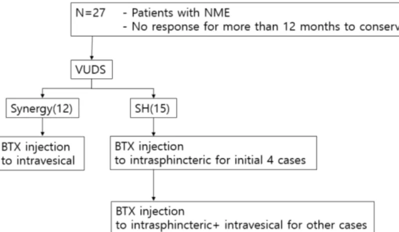
Figure 1 presents brief schematic diagram of this study.
Preoperative management
Since December 2016, patients in whom conventional treatment was ineffective were managed as previously described. Upon consultation, patients with enuresis were classified into ME or NME based on data obtained from a detailed history. Selective physical examination, including back examination and digital rectal examination, was conducted to rule out neurogenic problems.
To obtain objective findings for the confirmation of NME, determine the potential presence of lower urinary tract dysfunction (LUTD), and appropriately classify patients, 2-day frequency–volume charting, uroflowmetry with simultaneous electromyography, and postvoid residual urine measurement were conducted. Those who were diagnosed with NME were further classified into the OAB and DV group based on clinical features and noninvasive assessment. Those with OAB featured frequency and urgency in their history and small voided volume in their voiding diary. This study was approved by institutional review board.
All patients were initially provided standard urotherapy, including
timed voiding, and active bowel control regimen, for at least 1 month.
Thereafter, refractory patients received treatment according to their presumed diagnosis. Patients with ME were initially provided either an enuretic alarm or desmopressin together with standard urotherapy. Those with NME who had OAB received a combination of anticholinergics (solifenacin 5 mg/day) and desmopressin (0.2 mg tablet or 120–240 µg in Melt form) in conjunction with standard urotherapy. Those with NME who had DV received a 6-week course of pelvic floor muscle relaxation exercise using biofeedback focusing on bowel control together with ongoing standard urotherapy. Treatment efficacy, history, and voiding diary were assessed every 3 months. The persistence of LUTD and abnormal urine volume in the voiding diary led us to conclude that daytime problems had not been sufficiently addressed. Consequently, patients received more vigorous treatment for constipation. Updosing of anticholinergics was attempted among those with OAB, while alpha antagonist and low dose anticholinergics were added to the previous treatment of those with DV for the control of urgency.
When a persistent lack of response in either lower urinary tract symptoms (LUTS) or enuresis was noted despite the mentioned treatment (for 6 months), the aforementioned tests were repeated to validate the primary diagnosis.
Those whose diagnosis had not changed continued to receive treatment for 6–
12 months. Refractory enuresis was confirmed when no improvement was observed at least after 1 year of treatment in the presence of good treatment compliance. Such patients were considered potential candidates for salvage treatment using BTX. Given that most parents were eager to obtain control of enuresis as early as possible, they agreed to the treatment and provided informed consent.
VUDS procedure
On the day of treatment, patients were admitted to the outpatient surgery center and transferred to the operating room where VUDS and subsequent injection were performed. VUDS was conducted according to the International Children's Continence Society (ICCS) standards.13, 14 Accordingly, one or two study cycles were performed depending on the detection of clinically relevant findings. After placing patients in the supine position, a double-lumen 6Fr cystometry catheter and an 8Fr ballooned rectal catheter were inserted. The bladder was filled with sterile normal saline mixed with a contrast medium at 10% of expected bladder capacity. Patients were allowed to void upon feeling the urge before capacity was reached. When age- adjusted expected capacity was reached, patients were asked to void in either the supine or sitting position and were observed for 20 min for spontaneous
voiding. Sense of bladder filling, cystometric capacity, detrusor overactivity (DO), bladder compliance, and widening of the bladder neck (WBN) were assessed during the filling phase of VUDS. SH during the voiding phase was defined as electromyographic hyperactivity or shuttering sphincter movement that resembled a spinning top on fluoroscopy during spontaneous voiding.
Patients who failed to void in the presence of catheter for more than 20 min were assumed to have SH. Following VUDS and determination of enuresis etiology, patients were placed in the lithotomy position and general anesthesia was established. During this preparation, treatment was planned based on VUDS findings.
BTX injection procedure
Further injection procedures were attempted to correct abnormalities revealed during the VUDS. Intravesical BTX was attempted when findings suggested reduced cystometric capacity (RC) or DO. BTX was diluted in normal saline at 10 units/kg to a maximum of 300 units. Multiple injections were distributed throughout the detrusor.
Those suspected to have SH received intrasphincteric BTX diluted in normal saline to 25–33 units/mL and injected into the external urethral sphincter across three to four quadrants to a maximum of 100 units.15, 16
Following injection, patients were observed for 4 h to detect potential complications, after which they were discharged home.
Postoperative management
Patients were advised to continue with the standard urotherapy and were followed up 1 month after the injection. When improved responses were not observed at the 1 month follow-up, desmopressin or prior anticholinergics were added to enhance responses. Changes in enuresis and LUTS were assessed every 3 months for 1 year. Responses were evaluated according to the ICCS criteria.17
Data collection and analyze
We collected DVSS (Dysfunctional Voiding Symptom Score) as an objective indicator to compare daytime symptoms before and after surgery.
This survey is questionnaire about 13 voiding symptoms and 7 defecation symptoms. 2 of voiding symptoms are about enuresis, so other score of 11 questionnaires collected. And other subjective symptoms at outpatient clinic recorded and collected.
We performed frequency analysis for categorical variables and performed average comparison for continuous variables.
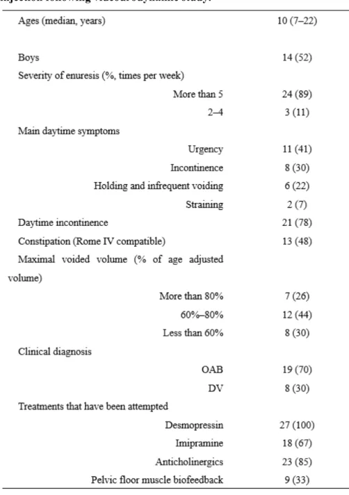
Results
Among the 282 patients with enuresis who received treatment at our hospital during the study period, 27 (10.4%; 14 boys and 13 girls) ultimately received endoscopic treatment (Table 1). All patients were considered to have NME and exhibited at least one kind of storage (urgency and frequency) or emptying (straining and intermittency) symptom. Concomitant daytime incontinence was found in 21 (77.8%) patients. A history of constipation was noted in 13 (48%) patients who received polyethylene glycol treatment for a median of 7 (4–31) months. Based on their clinical features, 19 and 8 patients were diagnosed with OAB and DV, respectively. Prior to endoscopic treatment, patients received a median of 16 (6–31) months of treatments for enuresis or daytime symptoms. Such treatment included standard urotherapy, such as constipation treatment, biofeedback for pelvic floor muscle exercise, anticholinergics, and desmopressin and imipramine, either alone or in combination. Overall, 7 (36.8%) and 3 (37.5%) patients with OAB and DV exhibited improvement in their daytime incontinence or daytime symptoms, though no patient indicated any improvement of their enuresis. With regard to voiding diary, none of the patients with OAB and only two patients with DV
showed normalized maximal and average voided volumes. Thus, both were found to be treatment resistant.
Figure 2 presents the VUDS results for the included patients. The two major abnormalities observed during the filling phase were DO and RC found in 14 (52%) and 22 (82%) patients, respectively. During the voiding phase, 15 (56%) patients had SH. Additionally, 9 (33%) patients showed gradual WBN during the filling phase. Among the 19 patients clinically presumed to have OAB, 11 (58%) and 16 (84%) had DO and RC, respectively. Four (21%) patients showed WBN during the filling phase. During the voiding phase, 9 (47%) patients showed synergic voiding. Among the 8 patients presumed to have DV, 3 (38%) and 6 (75%) had DO and RC, respectively. During the filling phase, 5 (63%) showed WBN, while during the voiding phase, same number of patients showed SH. Four patients presumed to have OAB failed to void despite being catheterized. They were subsequently diagnosed with SH and grouped with the six patients who showed RC but not DO. Nondilating vesicoureteral reflux was observed in two patients.
Consistency between clinical and urodynamic diagnoses was unsatisfactory with an overall concordance rate of 52%. The sensitivity and specificity of clinical versus urodynamic diagnosis were 75% and 33%, respectively.
Given our determination to treat patients according to their VUDS findings, intravesical and intrasphincteric BTX injections were attempted for urodynamic DO/RC and SH, respectively. Initially, only sphincteric BTX was attempted for the first four patients with SH, two of whom exhibited mild DO.
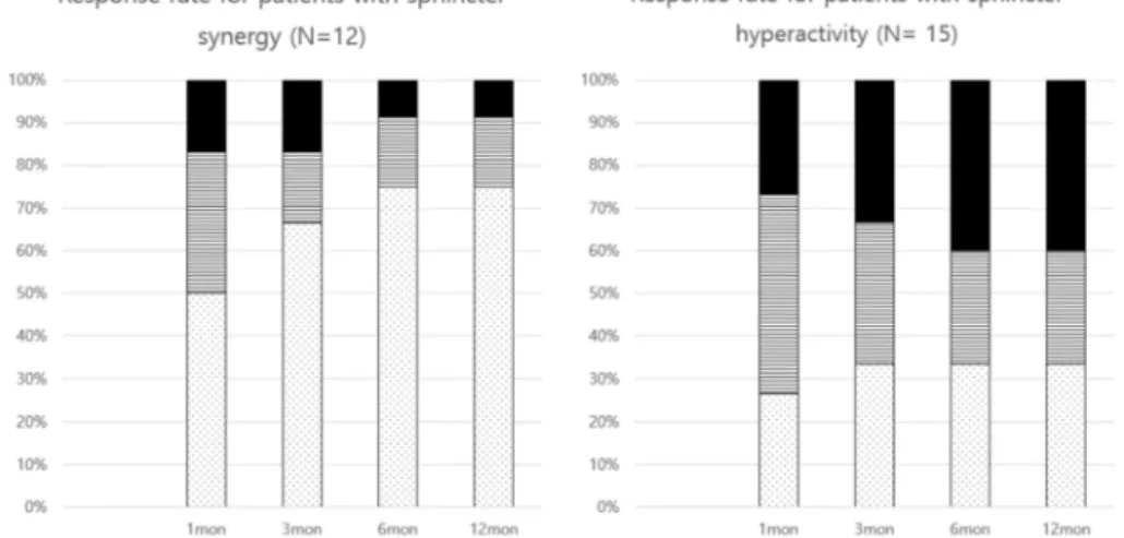
Assuming that SH was the main pathophysiology, we believed that addressing only SH would sufficiently relieve enuresis. However, after recognizing unsatisfactory results among patients who received only sphincteric BTX injection, the remaining nine patients with DV who had SH also received intravesical BTX. Figure 3 summarizes the treatments provided to patients and enuresis outcomes at the 1-year follow-up.
Figure 4 reveals that time courses for responses can differ according to the type of urethral movement during voiding. Accordingly, patients showing synergic urethral movement showed a better treatment course than those showing SH. All but one synergic patient showed at least improvement of enuresis, while 75% experienced CR. Patients showed a transition from PR or NR to CR after 6 months of treatment. On the other hand, initial responses worsened in the SH group, with 6 (40%) patients eventually showing NR.
Although 11 (73%) patients showed CR or PR after 1 month of treatment, two patients with PR experienced a loss in efficacy, leading to NR. On the other
hand, only one patient with PR transitioned to CR at 1 year. Responses became stabilized after 6 months of injection.
Apart from patients with CR who no longer required any treatment, four patients showing PR were able to control their enuresis with desmopressin or imipramine treatment.
Seven patients with NR, one with synergic voiding and six with SH, failed to show any meaningful improvement. Most of them did not receive intravesical BTX injection. Moreover, four patients, two of whom were girls, exhibited WBN in the filling phase and suffered persistent daytime incontinence and enuresis. One was found to have DO and synergic voiding, while the others showed SH.
In aspect of day time symptoms, there were only ten patients who submitted survey before and after surgery. Average of ‘before surgery’ was 10.0, and average of ‘after surgery’ was 5.5. Nine patients (90%) showed improvement in this questionnaire.
Neither significant anesthesia nor acute treatment related complication was noted around the procedure. No postoperative complication was noted during follow-up.
Discussion
The current study presented several urodynamic findings and combinations thereof that could explain treatment refractoriness. Our results showed that determining the injection site based on urodynamic findings promoted a response rate of more than 70%. Furthermore, improved responses were maintained for up to 12 months. As such, performing endoscopic treatment with BTX based on urodynamic findings within a single a day can be considered an effective salvage treatment strategy.
All patients included herein had been diagnosed with NME, which has been considered hard to treat and requires prolonged treatment durations.18 This is understandable given that the treatment of NME requires the treatment of both LUTD and enuresis. One caveat is that LUTD consists of heterogenous urodynamic abnormalities. Thus, individual LUTD should be clarified and treated accordingly. However, current management schemes are mainly nonspecific and noninvasive. As revealed by the European Bladder Dysfunction Study the clinical diagnosis of OAB or DV was inconsistent with urodynamic results, while treatment aimed at either condition showed cross- efficacy to the other.19, 20 This
redundancy in diagnosis and treatment supports current management systems, which we acknowledge as helpful in most cases. However, establishing the correct diagnosis and tailored treatment may be needed especially among refractory cases, such as those included herein.
Our data confirmed that noninvasive test results could not predict features observed during VUDS. Accordingly, only about half of the patients showed consistency between preoperative assumption and urodynamic diagnosis. We believe that the diagnostic discrepancy between clinical features and VUDS could partly explain treatment failures among patients. Moreover, five patients showed urodynamic features comprising both DO and SH, which shared features of OAB and DV. Such results cannot be determined from noninvasive diagnostic methods.
Unlike previous negative assertions for urodynamic study in refractory enuresis,9, 21all patients included herein showed at least one abnormal finding, highlighting the usefulness of VUDS. The most notable finding was that approximately half of those with clinically OAB turned out to have SH. This finding is consistent with previous reports of increased numbers of DV among refractory cases.22, 23Although the application of fluoroscopy facilitated better identification of SH, the lack of DO during UDS did not seem to justify withholding treatment for DO as reflected by the higher prevalence of NR
among those receiving only intrasphincteric BTX for urodynamic DV compared to those receiving both intravesical and intrasphincteric BTX.
Glassberg et al.24 indicated, that the patients with DV should receive anticholinergics to alleviate storage symptoms despite the absence of DO during VUDS.
Gradual WBN during the filling phase had been observed in 9 (35%) patients and was associated with daytime incontinence. Our result suggested that WBN may play a role in the development of treatment resistance.
Accordingly, one patient showed synergic voiding, while the three showed SH.
Considering that the other three patients exhibiting WBN reported CR after treatment, WBN may not be the sole risk factor for treatment resistance.
However, the presence of WBN as the only risk factor for nonresponsiveness in four patients may provide insight into its inhibitory role against continence.
We have no knowledge whether WBN is related to pelvic floor laxity25 or intrinsic sphincter problem.26 However, one previous study reported that colposuspension successfully treated girls with congenital bladder neck insufficiency.26
Unlike adults, most of the children who responded to treatment required just a single BTX shot. This is interesting considering that the effects of intravesical BTX are unlikely to last for more than a year. We speculated that
the endoscopic injection was helpful in correcting DO and DV, which may be evidence of immaturity in bladder control, and facilitated the achievement of normal bladder or urethral control to such a degree that further correction by injection was not necessary.
We must consider about limitations of BTX injection in children. Firstly, procedure needs general anesthesia in children. Because the risk for general anesthesia always exists, we must consider about that. Secondly, it is generally known that effect of BTX is only 6-12 months. That means, the patient may have to undergo repeated surgery. Regardless of one day scheduled simple procedure, repeated surgery can be a burden to the patient.
Nevertheless, BTX injection show gain to patient with refractory enuresis. So we propose not to hesitate BTX injection in patients with refractory enuresis.
Conclusion
The present study suggests that utilizing VUDS for those with refractory enuresis with LUTD can help reveal the underlying pathophysiology and determine how to address the problem. VUDS was especially helpful in revealing sphincter pathology. Moreover, BTX injection was quite effective in controlling enuresis when administered in accordance with VUDS results.
Those showing DO benefited from intravesical BTX injection, whereas those with only SH benefited from both intravesical and intrasphincteric injections.
Treatment resistance to BTX seemed to have been attributed to BNW. The beneficial effects of BTX was maintained once symptoms stabilized after 6 months of treatment.
References
1. Yeung, C. K., Sreedhar, B., Sihoe, J. D. et al.: Differences in characteristics of nocturnal enuresis between children and adolescents: a critical appraisal from a large epidemiological study. BJU Int, 97:1069, 2006
2. Theunis, M., Van Hoecke, E., Paesbrugge, S. et al.: Self-image and performance in children with nocturnal enuresis. Eur Urol, 41:660, 2002
3. Vande Walle, J., Rittig, S., Bauer, S. et al.: Practical consensus guidelines for the management of enuresis. Eur J Pediatr, 171:971, 2012
4. Franco, I., von Gontard, A., De Gennaro, M.: Evaluation and treatment of nonmonosymptomatic nocturnal enuresis: a standardization document from the International Children's Continence Society. J Pediatr Urol, 9:234, 2013
5. Glassberg, K. I., Combs, A. J., Horowitz, M.: Nonneurogenic voiding disorders in children and adolescents: clinical and videourodynamic findings in 4 specific conditions. J Urol, 184:2123, 2010
6. Kaufman, M. R., DeMarco, R. T., Pope, J. C. t. et al.: High yield of urodynamics performed for refractory nonneurogenic dysfunctional voiding in the pediatric population. J Urol, 176:1835, 2006
7. Anding, R., Smith, P., de Jong, T. et al.: When should video and EMG be added to urodynamics in children with lower urinary tract dysfunction and is this justified by
the evidence? ICI-RS 2014. Neurourol Urodyn, 35:331, 2016
8. Hjalmas, K.: Urinary incontinence in children: suggestions for definitions and terminology. Scand J Urol Nephrol Suppl, 141:1, 1992
9. Parekh, D. J., Pope, J. C. t., Adams, M. C. et al.: The use of radiography, urodynamic studies and cystoscopy in the evaluation of voiding dysfunction. J Urol, 165:215, 2001
10. Ballek, N. K., McKenna, P. H.: Lower urinary tract dysfunction in childhood. Urol Clin North Am, 37:215, 2010
11. Hoebeke, P., De Caestecker, K., Vande Walle, J. et al.: The effect of botulinum-A toxin in incontinent children with therapy resistant overactive detrusor. J Urol, 176:
328, 2006
12. Mokhless, I., Gaafar, S., Fouda, K. et al.: Botulinum A toxin urethral sphincter injection in children with nonneurogenic neurogenic bladder. J Urol, 176:1767, 2006 13. Bauer, S. B., Nijman, R. J., Drzewiecki, B. A. et al.: International Children's Continence Society standardization report on urodynamic studies of the lower urinary tract in children. Neurourol Urodyn, 34:640, 2015
14. Wen, J. G., Djurhuus, J. C., Rosier, P. et al.: ICS educational module: Cystometry in children. Neurourol Urodyn, 37:2306, 2018
15. t Hoen, L. A., van den Hoek, J., Wolffenbuttel, K. P. et al.: Breaking the vicious circle: Onabotulinum toxin A in children with therapy-refractory dysfunctional
voiding. J Pediatr Urol, 11:119.e1, 2015
16. Vricella, G. J., Campigotto, M., Coplen, D. E. et al.: Long-term efficacy and durability of botulinum-A toxin for refractory dysfunctional voiding in children. J Urol, 191:1586, 2014
17. Austin, P. F., Bauer, S. B., Bower, W. et al.: The standardization of terminology of lower urinary tract function in children and adolescents: Update report from the standardization committee of the International Children's Continence Society.
Neurourol Urodyn, 35:471, 2016
18. Rittig, N., Hagstroem, S., Mahler, B. et al.: Outcome of a standardized approach to childhood urinary symptoms-long-term follow-up of 720 patients. Neurourol Urodyn, 33:475, 2014
19. Bael, A., Lax, H., de Jong, T. P. et al.: The relevance of urodynamic studies for Urge syndrome and dysfunctional voiding: a multicenter controlled trial in children. J Urol, 180:1486, 2008
20. van Gool, J. D., de Jong, T. P., Winkler-Seinstra, P. et al.: Multi-center randomized controlled trial of cognitive treatment, placebo, oxybutynin, bladder training, and pelvic floor training in children with functional urinary incontinence. Neurourol Urodyn, 33:482, 2014
21. Soygur, T., Arikan, N., Tokatli, Z. et al.: The role of video-urodynamic studies in managing non-neurogenic voiding dysfunction in children. BJU Int, 93:841, 2004
22. Elmissiry, M., Abdelkarim, A., Badawy, H. et al.: Refractory enuresis in children and adolescents: how can urodynamics affect management and what is the optimum test? J Pediatr Urol, 9:348, 2013
23. Yeung, C. K., Chiu, H. N., Sit, F. K.: Bladder dysfunction in children with refractory monosymptomatic primary nocturnal enuresis. J Urol, 162:1049, 1999 24. Glassberg, K. I., Van Batavia, J. P., Combs, A. J.: Can children with either overactive bladder or dysfunctional voiding transition from one into the other: Are both part of a single entity? J Pediatr Urol, 12:217.e1, 2016
25. Bauer, S. B., Vasquez, E., Cendron, M. et al.: Pelvic floor laxity: A not so rare but unrecognized form of daytime urinary incontinence in peripubertal and adolescent girls. J Pediatr Urol, 14:544.e1, 2018
26. Dobrowolska-Glazar, B. A., Groen, L. A., Nieuwhof-Leppink, A. J. et al.: Open and Laparoscopic Colposuspension in Girls with Refractory Urinary Incontinence.
Front Pediatr, 5:284, 2017
Table 1. Clinical features of the 27 patients who underwent endoscopic injection following videourodynamic study.
Fig 1. Brief schematic diagram of this study.
Fig 2. VUDS results for the included patients
Fig 3. Treatments provided to patients and enuresis outcomes at the 1- year follow-up
Fig 4. Time courses for responses can differ according to the type of urethral movement during voiding
국 문 초 록
정규환 학번: 2015-23221 서울대학교 의학과 비뇨의학교실
서론: 이 연구는 난치성 야뇨증의 요역학적 특성을 결정하고 보툴리눔 독소(BTX)를 이용한 내시경 주사를 통해 관리 될 수 있는지 여부를 조사하는 데 목적이 있다.
대상 및 방법:
12 개월 이상 동안 보존적 치료 후 반응을 보이지 않은
비단일증상 야뇨증 (NME)을 갖는 총 27 명의 서울대병원 어린이병원 환자가 포함되었다. 환자들은 비디오 요역동학검사
(VUDS)를 받았고 같은 날에 BTX 의 내시경 주사를 받았다.
방광용적 감소 (RC), 배뇨근 과다활동(DO) 및 방광목 확대 (BNW)는 충만기에서 평가 된 3 가지 주요 비정상 결과였으며, 괄약근
과다활동 (SH)은 배뇨기 단계에서 유일하게 평가 된 이상이었다.
VUDS 결과에 따라 BTX 의 정맥 내 또는 괄약근 주사가
시도되었다. 치료 후 1, 3, 6 및 12 개월에 추적 관찰을 수행 하였다.
결과: 평균 연령은 10 세 (7-31)였다. 19 명과 8 명의 환자가 각각 수술 전에 과민성 방광 또는 기능 장애가 있었지만, 절반 이상이
VUDS 에 따라 다른 진단을 받았다. DO 를 나타내는 사람들은 방광내 BTX 주사에만 효과를 보인 반면, SH 만을 가진 사람들은 방광내 및 괄약근 주사에 효과를 보였다. BTX 에 대한 치료 내성은 BNW 에 기인 한 것으로 보인다. 주사 후 6 개월 동안 지속되는 시간은 효능에 명백한 영향을 미치지 않았다. 환자의 80 % 이상이 1 년 후에도 주사의 이점을 유지했다.
결론: VUDS는 요로 기능 장애를 특성화하고 NME 환자의 적절한 치료를 결정하는 데 유용했다. 괄약근 기능 장애는 기존 치료법에 대한 난치성에 중요한 역할을 한다.
--- 주요어: 야뇨증, 난치성, 비단일증상, 요역학, 보툴리눔 독소