저작자표시-비영리-변경금지 2.0 대한민국 이용자는 아래의 조건을 따르는 경우에 한하여 자유롭게
l 이 저작물을 복제, 배포, 전송, 전시, 공연 및 방송할 수 있습니다. 다음과 같은 조건을 따라야 합니다:
l 귀하는, 이 저작물의 재이용이나 배포의 경우, 이 저작물에 적용된 이용허락조건 을 명확하게 나타내어야 합니다.
l 저작권자로부터 별도의 허가를 받으면 이러한 조건들은 적용되지 않습니다.
저작권법에 따른 이용자의 권리는 위의 내용에 의하여 영향을 받지 않습니다. 이것은 이용허락규약(Legal Code)을 이해하기 쉽게 요약한 것입니다.
Disclaimer
저작자표시. 귀하는 원저작자를 표시하여야 합니다.
비영리. 귀하는 이 저작물을 영리 목적으로 이용할 수 없습니다.
변경금지. 귀하는 이 저작물을 개작, 변형 또는 가공할 수 없습니다.
이학석사 학위논문
Direct immersion in-tube microextraction
coupled with capillary electrophoresis
액체상 관 내 미세추출법과 모세관 전기영동의 연동
2014년 8월
서울대학교 대학원 화학부 분석화학전공
고 영 진
Abstract
Direct immersion in-tube microextraction coupled with capillary electrophoresis
Young Jin Koh Analytical Chemistry, School of Chemistry The Graduate School Seoul National University
Solid phase microextraction (SPME) and liquid phase microextraction (LPME) are widely used for sample enrichment and cleanup prior to an instrumental analysis. Compared to simply applicable SPME, LPME usually requires more manual maneuvers. In order to overcome such shortcomings of LPME, we developed in-tube microextraction (ITME). ITME uses a liquid plug inside a capillary as an acceptor phase and thus can be in-line coupled with capillary electrophoresis (CE) with ease. Since the acceptor phase is well protected inside the capillary, ITME can be carried out under severe extraction conditions.
Moreover, given that the extraction and injection processes take place
simultaneously, the extraction kinetics and efficiency are very high. Using a
commercial CE instrument, direct immersion (DI)-ITME was demonstrated for
neutral forms in an acidic aqueous donor solution were extracted into a pentanol acceptor plug in the capillary. In three-phase DI-ITME, a small amount of octanol was injected into a capillary containing a basic aqueous acceptor phase and acidic analytes in an acidic aqueous donor solution were enriched into the basic acceptor phase through the octanol layer by the driving force of a pH difference. Due to the lack of a hanging drop, DI-ITME is a quite simple and robust extraction method.
Keywords:
Liquid phase microextraction, Single drop microextraction, In-tube microextraction, Capillary electrophoresis
Student Number: 2012-20264
CONTENTS
ABSTRACT i
CONTENTS iii
LIST OF FIGURES AND TABLES
ⅳ1 INTRODUCTION 1
2 EXPERIMENTAL 2
2.1 Chemicals and materials 2
2.2 Apparatus 3
2.3 DI-ITME 4
2.3.1 Two-phase DI-ITME 4
2.3.2 Three-phase DI-ITME 5
3 RESULTS AND DISCUSSION 6
3.1 Two-phase DI-ITME-CE 6
3.2 Three-phase DI-ITME-CE 12
3.3 Comparison of two- and three-phase DI-ITME-CE 17
4 CONCLUSIONS 20
REFERENCES 21
ABSTRACT IN KOREAN 25
LIST OF FIGURES AND TABLES
Figure 1. LVSEP of samples in pentanol.
Figure 2. Optimization of two-phase DI-ITME;
(a) Pentanol volume, (b) Extraction time Figure 3. Electropherograms of two-phase DI-ITME.
Figure 4. Optimization of three-phase DI-ITME;
(a) Octanol thickness, (b) Organic solvent, (c) Extraction time Figure 5. Electropherograms of three-phase DI-ITME.
Table 1. Two-phase DI-ITME-CE performance
Table 2. Three-phase DI-ITME-CE performance
1 INTRODUCTION
In order to analyze a sample of low concentration or one in a complex matrix, pretreatment steps for sample enrichment and cleanup are often required [1, 2]. Solid phase extraction (SPE) and liquid phase extraction (LPE) are commonly utilized as sample pretreatment methods [3, 4]. While a cartridge or disk filled with sorbent is used for SPE, a sorbent-coated fiber is used for solid phase microextraction (SPME) [5-9]. Analytes collected on the SPME fiber can be directly injected into GC or LC [10, 11]. SPME is thus simple and fast and usually requires no solvent. Although LPE does not use a special cartridge as SPE does, LPE uses a large amount of solvents. Various schemes of liquid phase microextraction (LPME) including hollow fiber LPME [12- 15], dispersive liquid-liquid microextraction [16-19], and single drop microextraction (SDME) have thus been introduced to minimize the use of solvents and to improve the extraction performance [20-25]. Most LPME methods are usually operated in a manual manner and off-line coupled with an instrumental analysis [26, 27]. SDME using an acceptor drop hanging at the tip of a separation capillary, however, can easily be coupled in-line with capillary electrophoresis (CE) [28, 29]. By using a drop at a sub- microliter volume, high levels of sample enrichments can be obtained from SDME with a short extraction time. The sample cleanup and enrichment powers of SDME for CE have been demonstrated in a variety of applications [30-34]. To advance SDME further, we recently demonstrated more efficient and fully automated in-tube microextraction (ITME) for headspace extraction coupled with CE [35]. Unlike SDME and other LPME modes, ITME uses a liquid plug inside a syringe or capillary as an
acceptor phase. Consequently, ITME is much simpler in operation compared to SDME.
Moreover, since the analytes extracted to the acceptor phase of a very small volume are simultaneously injected into the separation capillary, the extraction kinetics and efficiency are very high. Here, we present direct immersion ITME coupled with CE (DI-ITME-CE). From two-phase DI-ITME of 30 min extracting 2,4-dinitrophenol (DNP), 3-bromobenzoic acid (BBA), and 4-iodobenzoic acid (IBA) in an acidic aqueous sample into a pentanol plug inside the capillary, enrichment factors (EFs) of 80, 52, and 24 were obtained, respectively. In comparison, much higher corresponding EFs of 510, 1600, and 600 were obtained from three-phase DI-ITME of 30 min for the three analytes in an acidic aqueous donor to a basic aqueous acceptor through a thin octanol layer at the entrance of a capillary.
2 EXPERIMENTAL
2.1 Chemicals and materials
DNP was purchased from TCI (Tokyo, Japan). BBA, IBA, sodium tetraborate decahydrate, HPLC-grade HCl, octadecyl trimethoxysilane (ODTS), 1-heptanol, 1- octanol, 1-nonanol, 1-decanol, 1-pentanol, and tris(hydroxymethyl)-aminomethane (Tris) were from Sigma-Aldrich (St. Louis, MO, USA). Acetic acid and HPLC-grade methanol were from Merck (Darmstadt, Germany). Deionized water was prepared by a Nanopure II system (Barnstead, Dubuque, IA, USA). 10 mM stock solutions of BBA, IBA, and DNP were prepared in methanol. Sample donor solutions were prepared by diluting the stock solutions with an aqueous HCl solution.
2.1 Apparatus
ITME and CE were performed using bare fused silica capillaries with various inner diameters (IDs) and lengths, but with a fixed outer diameter of 360 μm (Polymicro Technologies, Phoenix, AZ, USA) with a P/ACE MDQ CE system (Beckman, Fullerton, CA, USA). For electrophoresis, a reverse potential of –20 kV was applied across the capillary in two-phase DI-ITME-CE and a normal potential of +25 kV was applied in three-phase DI-ITME-CE. Analytes were monitored using a UV detector at 214 nm. During the electrophoresis process, the capillary temperature was set to 25°C.
2.3 DI-ITME
2.3.1 Two-phase DI-ITME
For two-phase DI-ITME of the three analytes in an acidic donor solution of pH 3.0, pentanol was used as an organic acceptor phase, as it is water-immiscible and of low viscosity for easy electrophoresis [36]. To separate analytes in a pentanol plug, nonaqueous CE (NACE) was carried out using a methanol run buffer prepared by titrating a 25 mM Tris solution in methanol to pH 8.0 with a 25 mM acetic acid solution in methanol [37-39]. A bare fused capillary with an ID of 50 μm and an effective/total length of 30/40 cm was conditioned with 0.5 M NaOH, water, and the run buffer for 5 min each at 70 psi. A plug of pentanol was then injected into the capillary filled with the methanol run buffer. After immersing the capillary inlet in a sample donor solution, two-phase DI-ITME was carried out. Both ends of the capillary were placed in run buffer vials and electrophoresis was then performed. The peak heights from NACE of a 100 µM sample in a methanol run buffer injected for 3 s at 0.3 psi were used as reference values to calculate EFs of two-phase DI-ITME.
The distribution coefficients Doa of the three analytes between the organic phase of pentanol and the aqueous donor phase were measured. A mixture of 1 mL of 1 mM HCl and 1 mL of 10 mM analytes in pentanol was vortexed overnight at room temperature, after which the two phases were separated by centrifugation for 10 min.
The Doa value was calculated from the analyte concentration in the aqueous phase measured by CE [40].
2.3.2 Three-phase DI-ITME
For three-phase DI-ITME of the three analytes in an acidic donor solution of pH 2.5, octanol was used as an organic phase. A 25 mM sodium tetraborate buffer of pH 9.2 was used as an acceptor phase and was also used as a run buffer for the subsequent CE.
A bare fused silica capillary having an ID of 25, 50, or 75 μm and an effective/total length of 20/30, 30/40, or 50/60 cm was used. Each day, the capillary inlet tip surface was hydrophobically coated by silanization. After cleansing the capillary tip with ethanol, the tip was dipped 1 mm into a solution of 5 vol% ODTS and 0.1 vol% acetic acid in ethanol for 20 s and dried in open air for 5 min [41].
Before the extraction process, the capillary was treated by sequentially rinsing it with 0.5 M NaOH, water, and the acceptor phase (run buffer), each for 5 min at 70 psi.
Then, octanol was hydrodynamically injected into the capillary containing the acceptor phase, the capillary inlet was immersed in the sample donor vial, and extraction was carried out. After extraction, the octanol layer was removed by dipping the capillary inlet tip into a methanol vial for 1.6 s. Both ends of the capillary were placed in run buffer vials and electrophoresis was then performed. The peak heights from CE of a 500 µM sample in a run buffer injected for 5 s at 0.5 psi were used as reference values to calculate EFs of three-phase DI-ITME.
3 RESULTS AND DISCUSSION
3.1 Two-phase DI-ITME-CE
When acidic analytes in a pentanol plug undergo electrophoresis using a methanol run buffer under a reverse potential, the analytes are stacked at the boundary between the pentanol and methanol matrix zones due to the large difference in the conductivities of the two zones, while the pentanol matrix is removed to the inlet side by the electroosmotic flow (EOF) [30]. This sample enrichment scheme is called large volume sample stacking using an EOF pump (LVSEP) [42]. Fig. 1 shows that higher peaks with longer migration times were obtained with longer injection of a sample in pentanol. Thus, a greater degree of sample enrichment can be obtained with a larger volume of sample up to full injection into the capillary. However, in our two-phase DI- ITME, analytes were extracted into the pentanol plug through the capillary opening and a pentanol plug with an excessive length led to sample dilution and longer migration times.
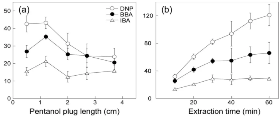
To optimize the pentanol plug volume, the EF values of the analytes extracted for 10 min from 500 nM sample solutions in HCl of pH 3.0 were compared as the pentanol plug length was varied from 1.25% (0.5 cm) to 9% (3.6 cm) of the capillary length. From the results shown in Fig. 2a, 3% (1.2 cm) of the capillary length was chosen as the optimal pentanol plug length. The volume of the pentanol plug was 24 nL. Fig. 2b shows EF with respect to the extraction time. As the extraction time was increased from 10 to 60 min, the EF values increased from 30 to 120 for DNP. BBA
the EF values and reproducibility, 30 min was chosen. Under the optimum extraction conditions, the EFs of DNP, BBA, and IBA were 80, 52, and 24 respectively. The RSDs of the migration times and peak heights (n = 4) were 2% and 4-18%, respectively. The limits of detection (LODs) (S/N = 3) were 39, 16, and 67 nM for DNP, BBA, and IBA, respectively. Analytical performance data of two-phase DI- ITME-CE are listed in Table 1.
Figure 1. LVSEP of samples in pentanol. Electropherograms of 10 μM samples in pentanol injected for (a) 30 s at 0.3 psi, (b) 15 s at 3 psi, (c) 40 s at 3 psi, and (d) 80 s at 3 psi. Fused silica capillary; 50 μm ID, 30/40 cm. Run buffer; 25 mM Tris/acetate (pH 8.0) in methanol, –20 kV, 214 nm, 25°C. Peak identification; 1) DNP, 2) BBA, and 3) IBA.
Figure 2. Optimization of 2-phase DI-ITME. (a) EF from 10 min extraction vs. the pentanol volume. (b) EF vs. the extraction time. Donor phase; 500 nM samples in HCl of pH 3.0. Acceptor phase; pentanol injected for 18 s at 0.3 psi. Other conditions as in Fig. 1.
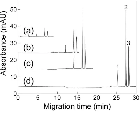
Figure 3. Electropherograms of (a) a 100 μM sample in a methanol run buffer injected for 3 s at 0.3 psi and (b) a 500 nM sample in HCl of pH 3.0 enriched by 30 min 2- phase DI-ITME to a pentanol acceptor plug. Other conditions as in Fig. 2. Peak identification: 1) DNP, 2) BBA, and 3) IBA.
0 2 6
4
4 2
0 6 8 10
Migration time (min)
3 2
1
1
2 3
A b so rb a n ce ( m A U )
(a)
(b)
Table 1. Two-phase DI-ITME-CE performance
Analyte EF
RSD (n = 4) LOD
(S/N = 3)
Linear range (nM)
Linearity (r
2) Migration
time
Peak height
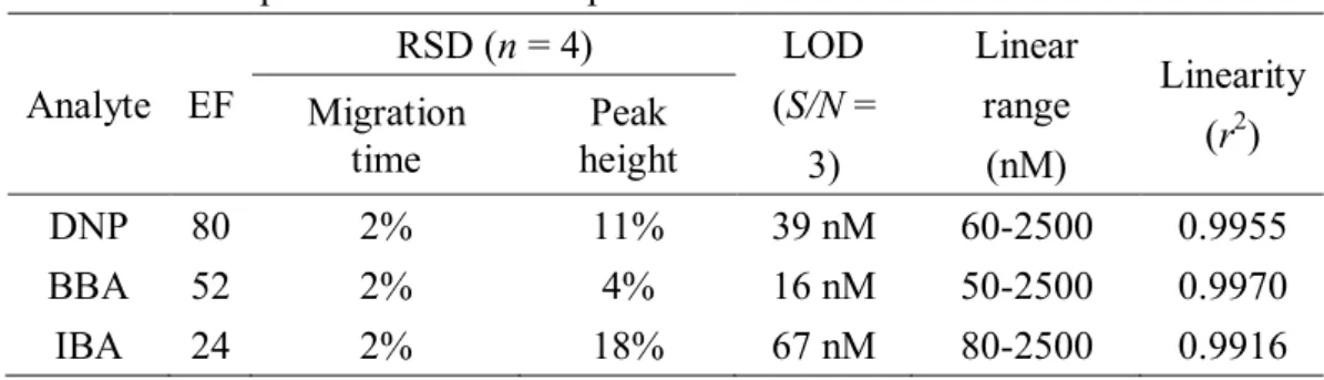
DNP 80 2% 11% 39 nM 60-2500 0.9955
BBA 52 2% 4% 16 nM 50-2500 0.9970
IBA 24 2% 18% 67 nM 80-2500 0.9916
3.2 Three-phase DI-ITME-CE
In three-phase LPE, an analyte is extracted from an aqueous donor phase to an organic (octanol) phase and back-extracted to an aqueous acceptor phase. For a finite extraction time, higher EFs are expected with a thinner organic phase. In order to place a thin octanol layer at the capillary entrance stably, the capillary tip surface was treated with a hydrophobic coating to improve its affinity with the organic phase. Without this coating, the success rate was about one in ten. With the coating, it was greater than nine in ten.
For a capillary of length L, the hydrodynamic injection volume v at pressure P for time t can be expressed by the Poiseuille equation [43]
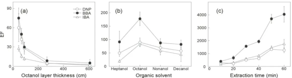
v = πID4tP/128hL, (1) with being the viscosity of the solution inside the capillary. Thus the octanol layer thickness is proportional to ID2 and inversely proportional to L. The minimum thickness of the octanol layer was determined by the minimum hydrodynamic injection condition P = 0.1 psi for 2 s, as given by our commercial CE instrument. Thus the layer thickness was varied instead by changing the capillary ID and L but with the minimum hydrodynamic injection at 0.1 psi for 2 s. When the ID of a 40-cm long capillary was changed from 75 to 25 μm, the octanol layer thickness was reduced from 610 to 67 μm. Then the EFs from 5 min extraction increased from 2 to 42 for DNP, from 5.4 to 50 for BBA, and from 3 to 16 for IBA. These 5- to 20-fold increases in the EF obtained with a 9-fold thinner octanol layer were greater than the 3-fold reduction in the absorbance due to the smaller ID. Therefore, a capillary ID of 25 μm was chosen.
When the length L was changed from 30 to 60 cm, the octanol layer thickness was reduced from 90 to 45 μm and the EFs from 5 min extraction increased from 28 to 60 for DNP, from 30 to 75 for BBA, and from 12 to 28 for IBA. Fig. 4a shows the increase in EF as the octanol layer became thinner. Therefore, we chose a capillary of 25 μm ID and 60 cm length.
Organic solvents other than octanol were tested as an organic phase. When heptanol, octanol, nonanol, and decanol were compared, the EFs obtained with octanol were highest as shown in Fig. 4b. The extraction time was also optimized. Fig. 4c shows the increase in EF over time. Considering the EFs and RSDs, 30 min was chosen.
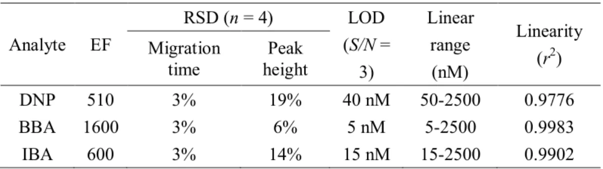
Under the optimum extraction conditions, i.e., injecting octanol at 0.1 psi for 2 s into a 60-cm capillary with an ID of 25 μm and extracting for 30 min, the EFs of DNP, BBA, and IBA were 510, 1600, and 600, respectively. The RSDs of the migration times and the peak heights (n = 4) were 3% and 6-19%, respectively. The LODs were 40, 5, and 15 nM for DNP, BBA, and IBA, respectively. Analytical performance data of three-phase DI-ITME-CE are listed in Table 2.
Figure 4. Optimization of 3-phase DI-ITME. (a) EFs from 5 min extraction of 10 μM samples in HCl of pH 2.5 vs. the octanol layer thickness. Fused silica capillary; 25, 50, or 75 μm ID, 20/30, 30/40, or 50/60 cm. (b) EFs from 5 min extraction of 5 μM samples in HCl of pH 3.5 vs. organic solvent. (c) EF for 500 nM sample in pH 2.5 HCl vs. the extraction time. Fused silica capillary; 25 μm ID, 50/60 cm. Organic solvent injection at 0.1 psi for 2 s. Run buffer; 25 mM sodium tetraborate buffer (pH 9.2). +25 kV, 214 nm, 25°C.
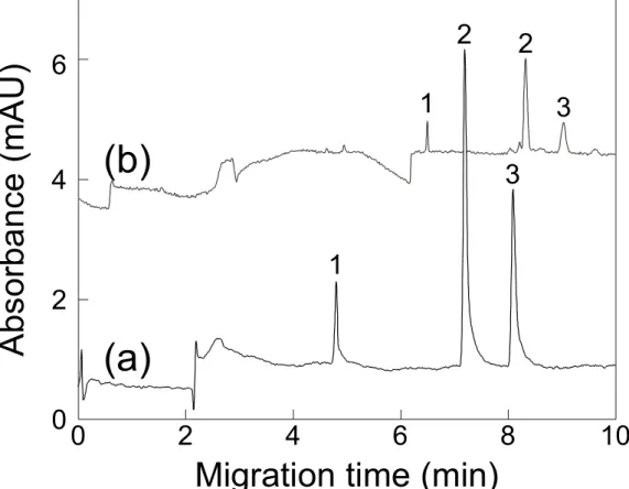
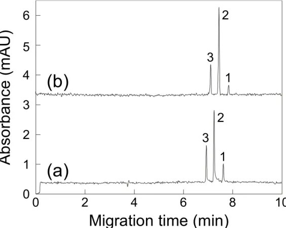
Figure 5. Electropherograms of (a) a 500 μM sample in HCl of pH 2.5 and (b) a 500 nM sample in pH 2.5 HCl enriched by 30 min 3-phase DI-ITME through a 45 μm thick octanol layer to a run buffer used as an acceptor. Fused silica capillary; 25 μm ID, 50/60 cm. Other conditions as in Fig. 4. Peak identification: 1) DNP, 2) BBA, and 3) IBA.
4 2
0 6 8 10
0 3 2 1 6 5 4
3 2
1
(a) (b)
Migration time (min)
A b so rb an ce ( m A U )
3 2
1
Table 2. Three-phase DI-ITME-CE performance
Analyte EF
RSD (n = 4) LOD
(S/N = 3)
Linear range (nM)
Linearity (r
2) Migration
time
Peak height
DNP 510 3% 19% 40 nM 50-2500 0.9776
BBA 1600 3% 6% 5 nM 5-2500 0.9983
IBA 600 3% 14% 15 nM 15-2500 0.9902
3.3 Comparison of two- and three-phase DI-ITME-CE
In two-phase LPE, an analyte is extracted from an aqueous donor phase (a) to an organic acceptor phase (o); the EF at equilibrium is given by [44, 45]
EFeq ≡ Co,eq
Ca,i
= 1
1
Doa+Vo Va, (2)where C and V are the concentration and volume of the phase denoted by the subscript, respectively. The subscripts i and eq denote the initial and equilibrium values, respectively. The theoretical maximum of EFeq is given by the distribution coefficient, Doa. For an acidic analyte, Doa can be increased up to the neutral form’s partition coefficient by lowering the donor pH below the analyte pKa value. Since the driving force is the preference towards the organic phase, two-phase LPE is suitable for hydrophobic compounds of large Doa values. Additionally, NACE is needed to separate analytes in the organic acceptor phase. Depending on the nature of analysis, NACE can be advantageous, but with some limitations [33]. In three-phase LPE, an analyte is extracted from an aqueous donor phase (a1) to an organic phase (o) and back-extracted an aqueous acceptor phase (a2); the EF at equilibrium is given by [46]
EFeq ≡ Ca2,eq
Ca1,i
= 1
D2 D1
( )
+D2(
Vo Va2)
+(
Va2 Va1)
, (3)where Dn is the distribution coefficient between the organic phase and the aqueous phase denoted by the subscript n. The driving force in three-phase LPE is the pH difference between the donor and acceptor phases. By increasing the difference in pH
and reducing the organic phase volume, EFeq can be maximized to the volume ratio Va1/Va2 [44]. Therefore, the EFeq in three-phase LPE can be much higher than the theoretical maximum Doa in two-phase LPE. Note that three-phase LPE is suitable for acidic or basic compounds not for neutral compounds and the liquid handling capability of a CE instrument limits the lower bound of the organic phase volume in three-phase DI-ITME. In practice, however, most analytical extractions are performed only for a finite time before reaching equilibrium to obtain EFeq of Eq. (2) or (3).
When the extraction time t is short, EF(t) can be approximated as [32], EF(t) ∝
A
V t
(4)where A and V are the surface area and volume of the acceptor phase, respectively.
Therefore, an acceptor phase with a small volume is preferred for high EF values.
The analytes used in this report were hydrophobic acids with a benzene ring, and thus both two-phase and three-phase DI-ITME were applicable. The experimentally measured Doa values of DNP, BBA, and IBA between pentanol and the aqueous donor phase were 850, 1300, and 200, respectively. When Va = 1800 μL and Vo ≈ 24 nL, Vo/Va ≈ 1.3 × 10⁻⁵ << 1/Doa. The actual EF value of 80 obtained with two- phase DI-ITME of 30 min was much smaller than this theoretical value at equilibrium.
In three-phase DI-ITME, Va1 = 1800 μL and Vo ≈ 22 pL for the injection at 0.1 psi for 2 s into a capillary with an ID of 25 μm and a length of 60 cm. The acceptor phase in a capillary can be approximated as a cylinder with length l from the longitudinal diffusion in one dimension [47]:
where D is the diffusion coefficient. Using the representative value of DNP, D = 9 × 10⁻⁶ cm2/s [48], the diffusion length l for 30 min of extraction was about 0.2 cm and Va2 ≈ 1 nL. When pHa1 = 2.5 and pHa2 = 9.2, using pKa = 4.09 and the partition coefficient of 81 between octanol and water for DNP [49], D2 = 6.3 × 10⁻⁴ and D2/D1 = 8 × 10⁻⁶. Then EFeq ≈ 4.5 × 10⁴ according Eq. (3). The actual EF value of 510 obtained with three-phase DI-ITME of 30 min was also much smaller than the theoretical value at equilibrium. Note that, however, the two EF values 1600 and 600 for BBA and IBA were larger than the theoretical maximum in two-phase DI-ITME, Doa, 1300 and 200, respectively.
From the same extraction time of 30 min, the EFs of 510 to 1600 obtained with three-phase DI-ITME were 6 to 30 times higher than those of 24 to 80 obtained with two-phase DI-ITME. The LODs for BBA and IBA from three-phase DI-ITME-CE were about 4 times lower than those from two-phase DI-ITME-CE while the LODs for DNP were similar. The discrepancies between the EF increases and LOD decreases were due to the effects: 1) The ID of the capillary for three-phase DI-ITME was 25 μm whereas that for two-phase DI-ITME was 50 μm, reducing the absorbance signal to less than half. 2) The baseline noise in NACE using a methanol run buffer (see Fig. 3) was smaller than that in aqueous CE (see Fig. 5). As a result, for an analyte of a given concentration, the signal to noise ratio from three-phase DI-ITME-CE was more than 3 times lower than that from two-phase DI-ITME-CE. In summary, three-phase DI- ITME, suitable for acidic and basic compounds, offered higher EF values and lower LODs than two-phase DI-ITME, suitable for hydrophobic compounds, although an additional step of hydrophobic coating of the capillary end surface was necessary.
4. CONCLUSIONS
Using a commercial CE instrument, two-phase DI-ITME from an aqueous sample solution to a pentanol acceptor plug and three-phase DI-ITME from an aqueous sample to an aqueous acceptor through a thin octanol layer were demonstrated.
The driving forces for the two-phase and three-phase extractions were the distribution coefficient and the pH difference, respectively. Due to the lack of a hanging acceptor drop, ITME was much simpler and more robust than SDME. Since the analytes extracted were already injected into the separation capillary, DI-ITME was easily and efficiently coupled in-line with CE. As well as the first demonstration of ITME for headspace extraction, DI-ITME offers a quite powerful but extremely easy method readily usable by anyone without special equipment or training.
REFERENCES
[1] J.R. Veraart, H. Lingeman, U.A.T. Brinkman, Coupling of biological sample handling and capillary electrophoresis, J. Chromatogr. A 856 (1999) 483-514.
[2] A.C. Mehta, Sample pretreatment in the trace determination of drugs in biological fluids, Talanta 33 (1986) 67-73.
[3] T. Hyotylainen, Critical evaluation of sample pretreatment techniques, Anal. Bioanal. Chem. 394 (2009) 743-758.
[4] L. Ramos, Critical overview of selected contemporary sample preparation techniques, J. Chromatogr. A 1221 (2012) 84-98.
[5] S. Ulrich, Solid-phase microextraction in biomedical analysis, J.
Chromatogr. A 902 (2000) 167-194.
[6] P. Puig, F. Borrull, M. Calull, C. Aguilar, Sorbent preconcentration procedures coupled to capillary electrophoresis for environmental and biological applications, Anal. Chim. Acta 616 (2008) 1-18.
[7] J.S. Fritz, M. Macka, Solid-phase trapping of solutes for further chromatographic or electrophoretic analysis, J. Chromatogr. A 902 (2000) 137-166.
[8] C. Dietz, J. Sanz, C. Camara, Recent developments in solid-phase microextraction coatings and related techniques, J. Chromatogr. A 1103 (2006) 183-192.
[9] I. Rodriguez, H.K. Lee, S.F.Y. Li, Ion-pair solid-phase extraction of biogenic amines before micellar electrokinetic chromatography with laser-induced fluorescence detection of their fluorescein thiocarbamyl derivatives, Electrophoresis 20 (1999) 1862-1868.
[10] C.L. Arthur, J. Pawliszyn, Solid-phase microextraction with thermal desorption using fused silica optical fibers, Anal. Chem. 62 (1990) 2145-2148.
[11] S. Risticevic, V.H. Niri, D. Vuckovic, J. Pawliszyn, Recent developments in solid-phase microextraction, Anal. Bioanal. Chem. 393 (2009) 781-795.
[12] L. Chimuka, M. Michel, E. Cukrowska, B. Buszewski, Advances in sample preparation using membrane-based liquid-phase microextraction techniques, Trac-Trends Anal. Chem. 30 (2011) 1781-1792.
[13] L.J. Lozano, C. Godinez, A.P. de los Rios, F.J. Hernandez-Fernandez, S.
Sanchez-Segado, F.J. Alguacil, Recent advances in supported ionic liquid membrane technology, J. Membrane Sci. 376 (2011) 1-14.
[14] B. Lindegard, H. Bjork, J.A. Jonsson, L. Mathiasson, A.M. Olsson, Automated column liquid-chromatographic determination of a basic drug in blood plasma using the supported liquid membrane technique for sample pretreatment, Anal. Chem. 66 (1994) 4490-4497.
[15] J.A. Jonsson, L. Mathiasson, Membrane-based techniques for sample
enrichment, J. Chromatogr. A 902 (2000) 205-225.
Berijani, Determination of organic compounds in water using dispersive liquid-liquid microextraction, J. Chromatogr. A 1116 (2006) 1-9.
[17] M.C. Breadmore, Ionic liquid-based liquid phase microextraction with direct injection for capillary electrophoresis, J. Chromatogr. A 1218 (2011) 1347-1352.
[18] T.G. Halvorsen, S. Pedersen-Bjergaard, K.E. Rasmussen, Liquid-phase microextraction and capillary electrophoresis of citalopram, an antidepressant drug, J. Chromatogr. A 909 (2001) 87-93.
[19] B. Horstkotte, M. Alexovic, F. Maya, C.M. Duarte, V. Andruch, V.
Cerda, Automatic determination of copper by in-syringe dispersive liquid-liquid microextraction of its bathocuproine-complex using long path-length spectrophotometric detection, Talanta 99 (2012) 349-356.
[20] L. Xu, C. Basheer, H.K. Lee, Developments in single-drop microextraction, J. Chromatogr. A 1152 (2007) 184-192.
[21] P.K. Dasgupta, S. Kar, Measurement of gases by a suppressed conductometric capillary electrophoresis separation system, Anal. Chem.
67 (1995) 3853-3860.
[22] H.H. Liu, P.K. Dasgupta, Analytical chemistry in a drop. Solvent extraction in a microdrop, Anal. Chem. 68 (1996) 1817-1821.
[23] W.H. Gao, G.P. Chen, Y.W. Chen, X.S. Zhang, Y.G. Yin, Z.D. Hu, Application of single drop liquid-liquid-liquid microextraction for the determination of fluoroquinolones in human urine by capillary electrophoresis, J. Chromatogr. B 879 (2011) 291-295.
[24] Z.F. Zhu, X.M. Zhou, N. Yan, L. Zhou, X.G. Chen, On-line combination of single-drop liquid-liquid-liquid microextraction with capillary electrophoresis for sample cleanup and preconcentration: A simple and efficient approach to determining trace analyte in real matrices, J. Chromatogr. A 1217 (2010) 1856-1861.
[25] W.H. Gao, G.P. Chen, T.F. Chen, X.S. Zhang, Y.W. Chen, Z.D. Hu, Directly suspended droplet microextraction combined with single drop back-extraction as a new approach for sample preparation compatible with capillary electrophoresis, Talanta 83 (2011) 1673-1679.
[26] A. Sarafraz-Yazdi, A. Amiri, Liquid-phase microextraction, Trac- Trends Anal. Chem. 29 (2010) 1-14.
[27] D. Han, K.H. Row, Trends in liquid-phase microextraction, and its application to environmental and biological samples, Microchim. Acta 176 (2012) 1-22.
[28] K. Choi, S.J. Kim, Y.G. Jin, Y.O. Jang, J.S. Kim, D.S. Chung, Single drop microextraction using commercial capillary electrophoresis instruments, Anal. Chem. 81 (2009) 225-230.
[29] K.W. Choi, Y. Kim, D.S. Chung, Liquid-phase microextraction as an on-line preconcentration method in capillary electrophoresis, Anal.
Chem. 76 (2004) 855-858.
[30] K. Choi, Y.G. Jin, D.S. Chung, In-line coupling of two-phase single
drop microextraction and large volume stacking using an electroosmotic
flow pump in nonaqueous capillary electrophoresis, J. Chromatogr. A
1216 (2009) 6466-6470.
[31] J. Choi, K. Choi, J. Kim, A.Y.B.H. Ahmed, Z.A. Al-Othman, D.S.
Chung, Sensitive analysis of amino acids with carrier-mediated single drop microextraction in-line coupled with capillary electrophoresis, J.
Chromatogr. A 1218 (2011) 7227-7233.
[32] S.T. Park, J. Kim, K. Choi, H.R. Lee, D.S. Chung, Headspace-single drop microextraction with a commercial capillary electrophoresis instrument, Electrophoresis 33 (2012) 2961-2968.
[33] Z.A. Al-Othman, M. Dawod, J. Kim, D.S. Chung, Single-drop microextraction as a powerful pretreatment tool for capillary electrophoresis: A review, Anal. Chim. Acta 739 (2012) 14-24.
[34] K. Cheng, K. Choi, J. Kim, I.H. Sung, D.S. Chung, Sensitive arsenic analysis by carrier-mediated counter-transport single drop microextraction coupled with capillary electrophoresis, Microchem. J 106 (2013) 220-225.
[35] H.R. Lee, S.M.Cho, J.Kim, D.S.Chung, Novel and simple headspace in- tube microextraction coupled with capillary electrophoresis, J.
Chromatogr. A http://dx.doi.org/doi:10.1016/j.chroma.2014.04.052 [36] S.P. Porras, M. Jussila, K. Sinervo, M.L. Riekkola, Alcohols and wide-
bore capillaries in nonaqueous capillary electrophoresis, Electrophoresis 20 (1999) 2510-2518.
[37] B. Kim, M.S. Chun, S. Shin, D.S. Chung, Nonaqueous capillary electrophoresis of chlorinated phenols, Bull. Korean Chem. Soc. 20 (1999) 1483-1486.
[38] E. Bosch, P. Bou, H. Allemann, M. Roses, Retention of ionizable compounds on HPLC. pH scale in methanol-water and the pK and pH values of buffers, Anal. Chem. 68 (1996) 3651-3657.
[39] I. Canals, J.A. Portal, E. Bosch, M. Roses, Retention of ionizable compounds on HPLC. 4. mobile-phase pH measurement in methanol/water, Anal. Chem. 72 (2000) 1802-1809.
[40] T.S. Ho, S. Pedersen-Bjergaard, K.E. Rasmussen, Recovery, enrichment and selectivity in liquid-phase microextraction comparison with conventional liquid-liquid extraction, J. Chromatogr. A 963 (2002) 3-17.
[41] A. Pallandre, B. de Lambert, R. Attia, A.M. Jonas, J.L. Viovy, Surface treatment and characterization: perspectives to electrophoresis and lab- on-chips, Electrophoresis 27 (2006) 584-610.
[42] D.S. Burgi, Large volume stacking of anions in capillary electrophoresis using an electroosmotic flow modifier as a pump, Anal. Chem. 65 (1993) 3726-3729.
[43] R.Skalak, S.Sutera, The history of poseuille's Law, Annu. Rev. Fluid Mech., Anuual Reviews Ins., California, 25 (1993) 1-19.
[44] K. Choi, J. Kim, D.S. Chung, Single-drop microextraction in bioanalysis, Bioanalysis 3 (2011) 799-815.
[45] M.A. Jeannot, F.F. Cantwell, Solvent microextraction into a single drop, Anal. Chem. 68 (1996) 2236-2240.
[46] M.H. Ma, F.F. Cantwell, Solvent microextraction with simultaneous
back-extraction for sample cleanup and preconcentration: Quantitative extraction, Anal. Chem. 70 (1998) 3912-3919.
[47] J.J. Vandeemter, F.J. Zuiderweg, A. Klinkenberg, Longitudinal diffusion and resistance to mass transfer as causes of nonideality in chromatography, Chem. Eng. Sci. 5 (1956) 271-289.
[48]
http://www.epa.gov/superfund/health/conmedia/soil/pdfs/part_5.pdf.[49]
http://www.epa.gov/ttn/atw/hlthef/dinitrop.html, 19.10.2013국문 초록
고체상 미세추출법(SPME)와 액체상 미세추출법(LPME)은 널리 쓰이는 샘플 전처리 방법이다. SPME와 달리, LPME는 더 복잡한 수동의 작동과정을 거친다. 이러한 LPME의 단점을 극복하기 위해 관 내 미세추출법 (ITME)을 개발하였다. ITME는 받개층을 모세관 안의 용액으로 사용하여 모세관 전기영동과 보다 쉽게 연동된다. 받개층이 모세관 안에서 보호되기 때문에, 추출에 영향을 미치는 다양한 조건을 적용하기에 용이하다. 또한 농축과 주입이 동시에 이루어지기 때문에, 추출 속도와 효율이 매우 높다. 상용화된 모세관 전기영동 기기를 이용하여 액체상 ITME가 2-, 3-상 추출이 가능함을 보였다. 2-상 액체상 ITME에서는, 산성 수용성 주개층에서 중성상태인 산성 분석물질이 모세관 안의 pentanol 받개층으로 추출된다. 3-상 액체상 ITME에서는, 염기성의 수용성 받개층이 들어있는 모세관 안으로 적은 양의 octanol을 주입하고 산성 수용성 주개층의 산성 분석물질이 pH 차이에 의해 octanol 층을 지나 염기성의 받개층으로 추출된다. 모세관 끝에 매달린 방울이 없기 때문에, 액체상 ITME는 매우 간편하고 튼튼한 추출법이다.
주요어: 액체상 미세추출법, 미세방울 추출법, 액체상 관 내 미세추출법, 모세관 전기영동
학번: 2012-20264