저작자표시-비영리-변경금지 2.0 대한민국 이용자는 아래의 조건을 따르는 경우에 한하여 자유롭게
l 이 저작물을 복제, 배포, 전송, 전시, 공연 및 방송할 수 있습니다. 다음과 같은 조건을 따라야 합니다:
l 귀하는, 이 저작물의 재이용이나 배포의 경우, 이 저작물에 적용된 이용허락조건 을 명확하게 나타내어야 합니다.
l 저작권자로부터 별도의 허가를 받으면 이러한 조건들은 적용되지 않습니다.
저작권법에 따른 이용자의 권리는 위의 내용에 의하여 영향을 받지 않습니다. 이것은 이용허락규약(Legal Code)을 이해하기 쉽게 요약한 것입니다.
Disclaimer
저작자표시. 귀하는 원저작자를 표시하여야 합니다.
비영리. 귀하는 이 저작물을 영리 목적으로 이용할 수 없습니다.
변경금지. 귀하는 이 저작물을 개작, 변형 또는 가공할 수 없습니다.
Doctor of Philosophy
Prognostic Significance of CD200 Expression and Correlation with COX-2 Expression in
Cutaneous Melanoma
The Graduate School of the University of Ulsan
Department of Medicine Lee, Mi-hye
[UCI]I804:48009-200000176968 [UCI]I804:48009-200000176968
Prognostic Significance of CD200 Expression and Correlation with COX-2 Expression in
Cutaneous Melanoma
Supervisor: Jee Ho Choi
A Dissertation
Submitted to
the Graduate School of the University of Ulsan In Partial Fulfillment of the Requirements
for the Degree of
Doctor of Philosophy
by
Lee, Mi-hye
Prognostic Significance of CD200 Expression and Correlation with COX-2 Expression in
Cutaneous Melanoma
This certifies that the dissertation of Mi-hye Lee is approved.
Committee Vice-chair Dr. Mi Woo Lee
Committee Member Dr. Sung Eun Chang
Committee Member Dr. Chong Hyun Won
Committee Member Dr. Woo Jin Lee
Committee Member Dr. Chun Wook Park
Department of Medicine
Ulsan, Korea
Febuary 2019
감사의 글
이번 연구에 관심과 격려를 아끼지 않고 좋은 박사학위 논문이 나올 수 있도 록 이끌어 주신 최지호 지도교수님, 언제나 좋은 연구를 진행할 수 있게 믿고 지 지해 주시는 따뜻한 이미우 교수님, 연구가 진행되는 내내 전반적인 내용을 아우 르며 실험 과정과 결과들을 하나하나 챙기고 도와 주신 장성은 교수님, 틈틈이 관심과 애정으로 소중한 충고와 조언을 해주시며 이끌어주신 원종현 교수님, 또 한 부족한 저에게 논문의 기틀을 잡아 주시고 실질적인 연구 진행을 챙겨주시며 학위 논문의 밥상을 차려주신 이우진 교수님, 바쁘신 와중에도 논문을 심사해 주 시고 더 나은 논문이 될 수 있도록 조언을 해 주신 박천욱 교수님께 깊은 감사 의 인사를 올립니다. 연구실 실험과정과 내용이 미숙한 저의 개인적인 질문에 대 해 귀찮아하는 내색 없이 많은 가르침을 주신 백승환 선생님, 문익준 선생님, 서 현이 선생님께 감사드립니다. 박사 학위 동안 훌륭한 동료 임상강사 선생님들과 함께 재직할 수 있던 것도 너무 다행스럽고 감사한 일이었습니다. 신청한 슬라이 드를 찾아와 달라는 부탁을 수도 없이 했던 것 같은데, 매번 도와준 우리 서울아 산병원 피부과 의국 전공의 영재, 현지, 호정이, 학태, 재원이, 명은이, 창진이, 건 희에게 모두 감사합니다. 멀리 있어도 영원한 내 소중한 동기인 예진이에게도 감 사의 말을 전합니다. 매주 서로의 실험 진행에 대해서 함께 토론하고 배울 수 있 는 랩미팅 시간을 함께한 방승현 선생님 이하 연구실 선생님들께 깊은 감사 말 씀 올리며, 무엇보다 종종 늦은 밤이나 주말까지도 저의 사소한 궁금증에도 진지 하게 상담해 주시고 실험에 대해 함께 고민해주신 윤경아 선생님께 특별히 깊은 미안함과 그보다 더진한 감사함을 전합니다.
항상 저를 믿고 격려와 응원을 아끼지 않는 어머니와 아버지, 귀여운 동생 지 현이와 미현이, 사랑하는 가족들에게 이 논문을 바칩니다.
Abstract
Prognostic significance of CD200 expression and correlation with COX-2 expression in cutaneous melanoma
Background: Immune escape by tumors can occur by various mechanisms. As immunotherapy targeting immune checkpoints such as Cytotoxic T-lymphocyte-associated protein 4 and anti-programmed cell death protein 1 is considered as a good alternative to conventional chemotherapy in melanoma patients, more research on new immune checkpoints, which allows better therapeutic efficacy with less toxicity is needed.
Objectives: We evaluated CD200 and COX-2 expression in melanoma and their correlation, and determined their effects on clinicopathological characteristics including survival data.
Materials and Methods: Tumor samples obtained from primary melanoma lesions were analyzed for both CD200 and COX-2 expression by IHC analysis. Clinidopathological features including survival data were analyzed according to the expression of CD200 and COX-2. Co-localization of PD-L1 and COX-2 expression was analyzed by double fluorescence staining. Lastly the BRAFV600E A375 melanoma cell lines were used to evaluate the effect of COX-2 inhibition by celecoxib on expression of CD200 in vitro.
Results: CD200 and COX-2 expressions showed a tendency to be associated with aggressive clinicopathologic features. CD200 expression positively correlated with COX-2 expression in human melanoma cells. CD200 and COX-2 expression in melanoma were independent prognostic markers for worse overall survival. Lastly, inhibition of COX-2 activity by celecoxib down-regulated the expression of CD200 in BRAFV600E melanoma cell lines.
Conclusion: Our study suggests that CD200 expression by melanoma cells is associated with more aggressive pathologic features and worse survival data. Anti-CD200 treatment might be therapeutically beneficial for melanoma treatment.
Key words: CD200, Tumor microenvironment, immune checkpoint, Melanoma
Contents
Abstract...ⅰ Contents...ⅲ List of tables...ⅳ List of figures...ⅴ
Introduction...1
Materials and Methods...4
Chemical reagents and antibodies…...4
Cell culture experiments………...4
Cell viability…...5
Cell proliferation…...6
Evaluation of CD200 and COX-2 expression in cutaneous melanoma.…………...6
Immunofluorescence (IF) staining………9
Western blot analysis...5
Statistical analysis...7
Results...8
Discussion...26
Conclusion...32
References...33
국문요약...39
List of Tables
Table 1. Clinico-histopathological characteristics of the 118 cutaneous melanomas according to CD200 and COX-2 expression...19
Table 2. Survival outcomes of the 118 cutaneous melanoma patients according to CD200 and COX-2...19
Table 3. Univariate and multivariate analyses for OS and PFS...19
List of Figures
Figure 1. Immunohistochemical staining for CD200 and COX-2 in cutaneous melanoma.
(A) CD200 negative melanoma, (B) CD200 positive melanoma and (C) COX-2 positive melanoma. Figure 1B and 1C were from same patient specimen (x100)...11 Figure 2. Clinicopathologic differences according to CD200 and COX-2 expressions.
Pathologically, high expression of CD200(A) and COX-2(B) were associated with deeper breslow thickness respectively. Clinically, high expression of CD200(C) and COX-2(D) were associated with the advanced AJCC stage……...12 Figure 3. Immunofluorescence double staining for (A) melan-A and CD200, (B) melan-A
and COX-2, (C) CD200 and COX-2. Melan-A positive melanoma cells expressed both CD200(A) and COX-2(B). CD200 positive cell also expressed COX- 2(C)...13 Figure 4. Correlation of CD200 and COX-2 expression in melanoma. It shows strong
positive correlation between CD200 expression and COX-2 expression in melanoma………...15 Figure 5. (A) COX-2 inhibitor (celecoxib) inhibited melanoma cell growth, (B,C) Down- regulation of CD200 by COX-2 inhibitor (celecoxib) in melanoma cell lines...
………...17 Figure 6. Overall survival of the entire population according to (A) CD200 and (B) COX-2
expression and progression free survival according to (C) CD200 and (B) COX-2 expression in melanoma patients………...25
Introduction
Tumor-associated inflammation and immune responses are known to be key components in the tumor microenvironment (TME) that regulates tumor growth, progression and metastasis.1,2 There have been many reports on the tumor microenvironment as a target for anticancer treatment, which could reverse tumor immune escape by suppression of immune checkpoints in multiple types of cancers3-5 including melanoma.6,7The approval of anti-cytotoxic T-lymphocyte-associated protein 4 (CTLA-4) and anti-programmed cell death protein 1 (PD-1) antibodies for human use has already resulted in significant improvements in disease outcomes for various cancers, especially melanoma.8-10 Although such immunotherapy is considered as an good alternative to conventional chemotoxic agents, its response rate has been reported to range from 15.2% to 57.6%.21 That is, there is a need for further research on new immune checkpoints, which allows better therapeutic efficacy with less toxicity.
It has been generally reported that expression of CD200 on cancer cells could suppress antitumor response and have a pro-tumor effect.11-16 In hematologic malignancies CD200 expression was first reported in chronic lymphocytic leukemia11, followed by acute myeloid leukemia14 and multiple myeloma.13Recently, it is being reported in solid tumors including breast cancer,16bladder cancer,17 and has recently been reported in skin squamous cell carcinoma18and melanoma.19
There have been only a few reports of CD200 expression in melanoma.19-22Some have reported that melanoma can express CD200, thereby potentially suppressing anti-tumor immune responses.19-21 However, they were all limitied mouse experiments or in vitro experiments using cell lines, and the role of CD200 expression in tumor growth and immunity are not well established.21,22There was no reports on clinical significance of CD200 expression in melanoma patients.
Cyclooxygenase-2(COX-2) is also currently investigated as a major player of tumor progression in several type of malignancies including melanoma.3 COX-2 is frequently expressed in malignant melanomas23 and its inhibition may prevent melanoma progression24. It has been reported that COX‑2 expression positively correlates with PD‑L1 expression in human melanoma,7but there has been no study on correlation between COX-2 and CD200 expression in melanoma.
In this study we investigated CD200 and COX-2 expression in melanoma patients and their
clinicopathological characteristics including survival data. We also tested whether CD200 expression correlates with COX-2 expression in human melanoma.
Materials and Methods
This study was approved by the Institutional Review Board of our institution. The database of Asan Medical Center was searched for cases of cutaneous melanoma that were confirmed by skin biopsy between January 1998 and December 2016. All histological and immunophenotypic data pertaining the 118 cases included were reviewed. The biopsy slides were reviewed, and the following parameters were analyzed: Breslow thickness, ulceration, and vertical growth phase.
Chemical reagents and antibodies
The following chemicals or reagents are used in this study. Dulbecco’s Modified Eagle’s Medium (DMEM), Minimum Essential Medium Eagle (MEM), HEPES Buffer Solution (1 M), and fetal bovine serum (FBS) were purchased from WelGENE Inc. (Daegu, Korea).
Antibiotic-Antimycotic solution 100 X (AA) (Thermo Fisher Scientific, Gibco).
Dimethylsulfoxide (DMSO, Duchefa Biochemie, the Netherlands). Bio-Rad protein assay dye reagent concentrate (Bio-Rad Laboratories, Richmond, CA) Celecoxib (Selleckchem, Huston, TX, USA). 1 % SDS lysis buffer for cell lysis in western blot: 1 % SDS, 100 mM Tris-HCl pH 8.0.
The following are the primary antibodies used in this study. Dilution rates used in western blot analysis and immunofluorescence staining are stated: Anti-COX2 antibody was from abcam (cat. no. ab15191, Cambridge, UK). For western blot analysis, dilution rate was 1:200.
For immunofluorescence staining, dilution rate was 1:50. CD200 antibody was from R&D systems (cat. no. AF2724, Minneapolis, MN USA). For western blot analysis, dilution rate was 1:1000. For immunofluorescence staining, dilution rate was 1:10. Melan-A (A 103) antibody was from Cell Marque (cat. no. 281M-85, 1:50 dilution; Rocklin, CA, USA). COX- 2(12C4F12) antibody was purchased from Thermo Scientific (cat. no. A-6404, 1:50 dilution;
Rockford IL, USA).
Secondary antibodies for Immunofluorescence staining used were as follows: Goat Anti- Rabbit IgG(H+L)-FITC was from Southern biotech (cat. no. 4055-02, 1:500 dilution;
Birmingham, AL). Rabbit Anti-Goat IgG(H+L)-FITC was from Southern biotech (cat. no.
6160-02, 1:500 dilution; Birmingham, AL). Goat anti-Mouse IgG (H+L) Cross-Adsorbed,
Alexa Fluor 546 was from Thermo scientific (cat. no. A-11003, 1:500 dilution; Rockford IL, USA).
Cell culture experiments
The A375, A2058, SK-MEL-5 human melanoma cells were cultured in DMEM supplemented with 10 % FBS, 1 % AA solution. The MNT1 human melanoma cell was cultured in MEM containing 10 % DMEM, 20 % FBS, 0.9 % AA solution and 20 mM HEPES. All cell lines were cultured at 37 °C in a humidified atmosphere containing 5% CO2.
For western blot analysis, cells were plated onto 60-mm tissue culture plates at the density of 55x10^4. After 48 h of cultivation, cells were treated with Celecoxib and incubated another 24 hours.
Cell viability
A 375 cells was seeded into 24-well plate at 2 x10^4 per well. After 72 h of cultivation at 37 °C in a 5% CO2, indicated doses of celecoxib was added to the cells. In control cells, DMSO (vehicle of the drug) concentration was maintained at 0.03 %. Following 24 h incubation, Cell proliferation rates were determined by an EZ-Cytox cell viability assay kit (Daeil Labservice, Seoul, Korea) according to the manufacturer’s instructions.
Cell proliferation
A 375 cell were seeded into 6 well plate at the density of 1 x 10^5 per well. After 72 h of cultivation at 37 °C in 5% CO2, celecoxib was treated with the indicated doses. DMSO (vehicle of the drug) concentration was maintained at 0.03 % in control cells. Following 24 h incubation, cell viability was analyzed by the MuseTM Count and Viability Kit (MuseTM Cell Analyzer, Millipore) according to the manufacturer’s protocol.
using primary antibody anti-CD200 (1:5000, catalogue number 10886-R333, Sino Biological, Beiing, China) and anti-COX-2 (1:100, ABCAM, Cambridge, UK). Two dermatologists (LWJ, LMH) agreed on CD200 expression in 103 of 112 (88%, k=0.653) tumors, and on COX-2 expression in 101 of 118 (86%, k=0.639) tumors. Cutoff values for CD200 and COX-2 expression that showed the most significant differences in overall survival (OS) were selected. A sample was considered CD200 positive if positive cells made up 35% or more of the overall melanoma cells. A sample was considered COX-2 positive if 40% or more of melanoma cells showed reactivity to the COX-2 antibody, whereas a sample was considered COX-2 negative if COX-2 expression was detected in <40% of the melanoma cells.
Immunofluorescence (IF) staining
A375, A2058, SK-MEL-5 and MNT1 cells were cultured on sterile glass coverslips (Deckglaser Cover Glasses), using 12 well. The density of cells was 2 x 10^5 or 4 x 10^5 per well. When the confluency of the cells was reached about 70-80 % of the well, cells were washed with Dulbecco's phosphate buffered saline (DPBS) and fixed for 10-20 min with IC fixation buffer(eBioscience). Coverslips were washed with DPBS and permeabilized using PBST (PBS buffer containing 0.1 % tween 20) about 10 min and incubated with blocking solution (5 % normal serum and 0.3 % triton x-100) for 1 h. Followed by an incubation with primary antibodies overnight at 4 °C (Anti-COX2 antibody) and RT for 1h (Melan-A (A 103) antibody). After that, they were detected using secondary antibodies Goat Anti-Rabbit IgG(H+L)-FITC for COX-2 and Goat anti-Mouse IgG (H+L) Cross-Adsorbed, Alexa Fluor 546 for Melan-A. Followed by washing with PBST, Coverslips were counterstained the nuclei with 4′, 6-diamidino-2-phenylindole (DAPI) at a final concentration of 0.5 ug/ml.
After washing 3 times, slides were mounted onto micro slides using Dako Fluorescence Mounting Medium. After drying at room temperature for 1-2 h, slides were examined using a fluorescent microscope (Zeiss ZEN LSM 710, Zeiss, Inc., Thornwood, NY).
For CD200 and Melan A double IF staining, formalin-fixed paraffin embedded (FFPE) melanoma tissues were deparaffinized and rehydrated with an ethanol. Slide were subjected to antigen-retrieval using antigen unmasking solution (Vector Laboratories) in a microwave for 15 min at 110 °C. After cooling for 20 min and washing with PBST, slides were
incubated with blocking solution for 20 min. Prepared slides were then incubated with primary antibodies at 4 °C for 2-3 days (CD200 antibody) and overnight at 4 °C (Melan-A (A 103) antibody). The samples were then detected using secondary antibodies Rabbit Anti- Goat IgG(H+L)-FITC for CD200 and Goat anti-Mouse IgG (H+L) Cross-Adsorbed, Alexa Fluor 546 for Melan-A.
For COX-2 and CD200 double IF staining, FFPE melanoma tissues were deparaffinized and rehydrated with an ethanol. Slides were then treated with antigen unmasking solution, cooling and washing with PBST, slides were incubated with blocking solution for 20 min.
Prepared slides were incubated with primary antibody at 4 °C for 2-3 days (COX-2(12C4F12) antibody and CD200 antibody). The samples were then washed, incubated with secondary antibody Goat anti-Mouse IgG (H+L) Cross-Adsorbed, Alexa Fluor 546 for COX-2 and Rabbit Anti-Goat IgG(H+L)-FITC for CD200. Followed by washing steps, each double stained slide for CD200 and Melan A / COX-2 and CD200 were then counterstained with DAPI and mounted with mounting medium. Stored at 4 °C, slides were examined using a fluorescent microscope.
Western blot analysis
A375 cells were lysed using 1 % SDS lysis buffer and boiling for 10 min at 95 °C. Proteins were quantified using a standard commercial kit (Bio-Rad Lab. Ltd) with bovine serum albumin as a standard. 25-30 ug of protein per lane was resolved on 8 % SDS- polyacrylamide gel electrophoresis. Gel was transferred to nitrocellulose membranes, which were then blocked with 5% nonfat milk (Bioworld, Dublin, OH, USA) or BSA (Bioworld, Dublin, OH, USA) in Tris-buffered saline containing 0.1 % tween 20. Blots were then incubated with primary antibodies, followed by incubation with horseradish peroxidase- conjugated secondary antibodies. Immunodetection was conducted using an enhanced
SPSS Inc., Chicago, IL). A value of P less than 0.05 was considered statistically significant.
Survival was analyzed using the Kaplan-Meier method, and comparisons were made using the log-rank test. Prognostic factors independently associated with overall survival (OS) and progression-free survival (PFS) at the time of diagnosis were identified by multivariate analysis using Cox proportional hazards regression modelling. Comparisons between subgroups of patients were performed using a chi-square test for categorical variables, and a t-test for continuous variables. Person’s correlations coefficient was used to evaluate associations for continuous variables.
Results
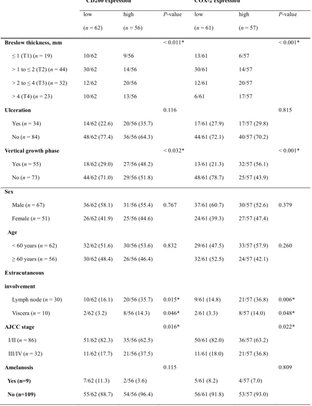
A total of 118 cases of cutaneous melanoma were included in the study. The demographic data and clinical features of the patients are summarized in Table 1.
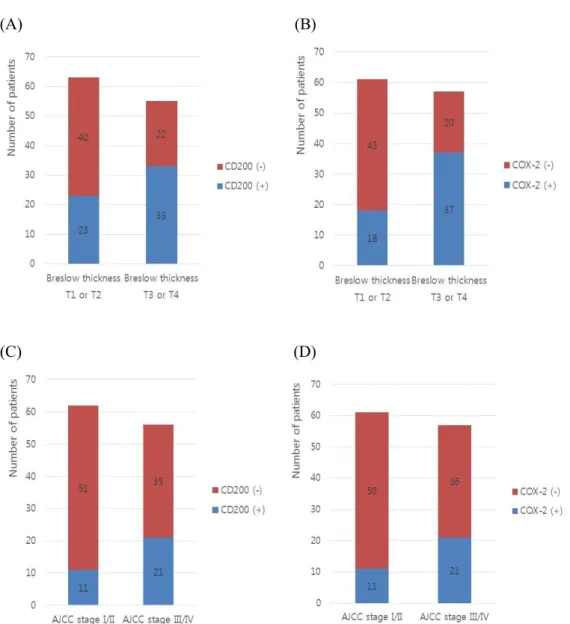
Expression of CD200 and COX-2 and their association with clinicopathological features Of 118 patients, CD200 and COX-2 (Fig. 1) were positive in 56 (47.4%) and 57 (48.3%) patients, respectively. Clinicopathological variables were stratified depending on expression of these proteins in tumor tissues to assess their associations (Table 1, Fig. 2).
There were significant correlations between the high expression of CD200 and pathological findings such as a deeper Breslow thickness (P=0.011) and more vertical growth (P=0.032).
High CD200 expression was also associated with higher frequency of lymph node (LN) involvement (P=0.015), visceral involvement (P=0.031) and a higher frequency of advanced American Joint Committee on Cancer (AJCC) stage (P=0.016; Table 1, Fig. 2).
High COX-2 expression was associated with pathological variables such as deeper Breslow thickness (P < 0.001) and more vertical growth phase (P < 0.001). High expression of COX- 2 was associated with higher frequency of LN involvement (P = 0.006), visceral involvement (P=0.036) and advanced AJCC stage (P = 0.022) (Table 1, Fig. 2).
Correlation of CD200 and COX-2 expression
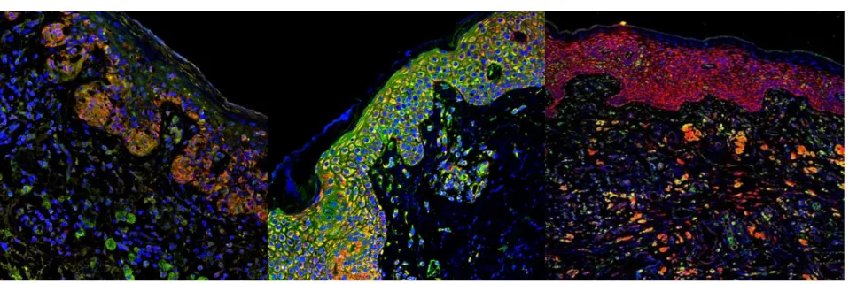
Double IF staining of melanoma tissue showed that melan-A-positive melanoma cells expressed CD200 (Fig. 3A) and COX-2 (Fig. 3B). CD200 positive cell also expressed COX- 2 (Fig. 2C). Of 56 cases with high expression of CD200, 44 (78.6%) cases showed the high expression of COX-2, and there was significant association between CD200 and COX-2 expression (P < 0.001). When expression values were evaluated as continuous variables,
melanoma cell lines were used as an in vitro model. A375 was treated with the selective COX-2 inhibitor celecoxib at different doses. As shown in Fig. 5, following a 24h incubation, celecoxib inhibited the growth of A375 melanoma cell lines. The growth of melanoma cells reduced by around 50% after the dose of 90 μM. Western blot analyses demonstrated that treatment with celecoxib significantly down-regulated the total protein level and surface expression of COX-2 in A375 melanoma cells as compared to untreated cells. (Fig. 5)
Prognostic significance of CD200 and COX-2 expression
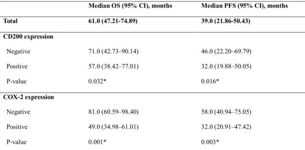
When all patients were combined into a single cohort, the 5-year OS rate was 46% and the median OS period was 61.0 months (95% confidence interval [CI]: 50.79–81.74 months).
Survival data in relation to CD200 and COX-2 expression are summarized in Table 2.
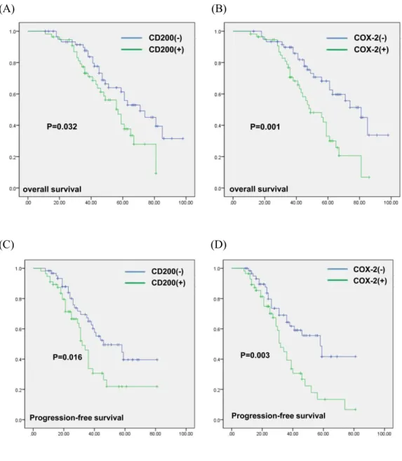
OS was significantly shorter in patients with high expression of CD200 compared to those with low expression of CD200 (P = 0.032, Fig. 6A). PFS was also significantly better in patients with low expression of CD200 (P = 0.016, Fig. 6C). Patients with high expression of COX-2 showed inferior OS than patients with low expression of COX-2 (P = 0.001, Fig. 6B).
PFS was also affected by expression of COX-2 (P = 0.003, Fig. 6D).
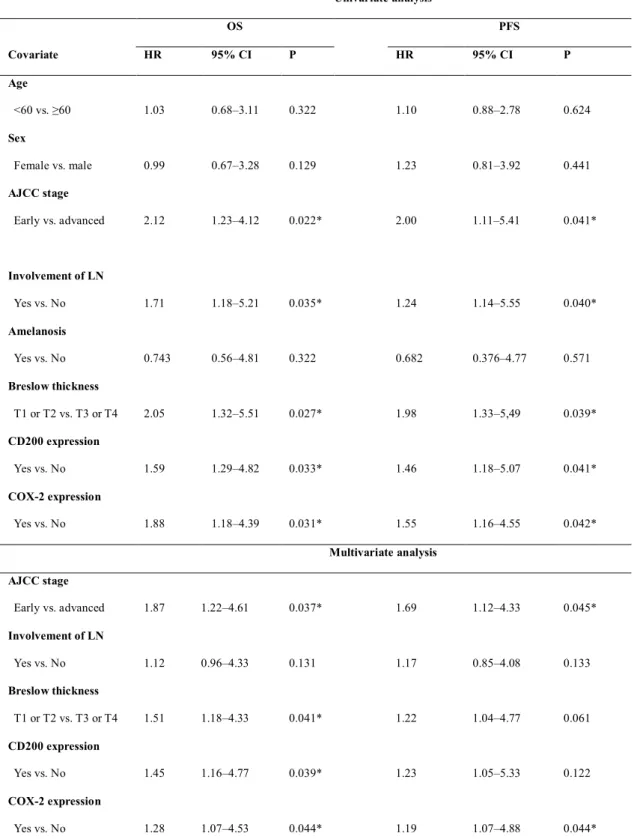
The results of univariate and multivariate analysis using clinicopathological variables were summarized in Table 3. By univariate analysis, CD200 expression (hazard ratio (HR) = 1.59, 95% CI = 1.29-4.82, P = 0.033), and COX-2 expression (HR = 1.88, 95% CI = 1.18-4.39, P
= 0.031) indices were associated with worse OS. By multivariate analysis, CD200 expression (HR = 1.45, 95% CI = 1.16-4.77, P = 0.039), and COX-2 expression (HR = 1.28, 95% CI = 1.07-4.53, P = 0.044) were independent prognostic markers for worse OS (Table 3).
Figure 1. Immunohistochemical staining for CD200 and COX-2 in cutaneous melanoma. (A) CD200 negative melanoma, (B) CD200 positive melanoma and (C) COX-2 positive melanoma. Figure 1B and 1C were from same patient specimen (x100)
(A) (B)
(C) (D)
Figure 2. Clinicopathologic differences according to CD200 and COX-2 expressions.
Pathologically, high expression of CD200(A) and COX-2(B) were associated with deeper breslow thickness respectively. Clinically, high expression of CD200(C) and COX-2(D) were associated with the advanced AJCC stage.
Table 1. Clinico-histopathological characteristics of the 118 cutaneous melanomas according to CD200 and COX-2 expression
CD200 expression COX-2 expression
low (n = 62)
high (n = 56)
P-value low (n= 61)
high (n= 57)
P-value
Breslow thickness, mm
≤ 1 (T1) (n= 19)
> 1 to ≤ 2 (T2) (n= 44)
> 2 to ≤ 4 (T3) (n= 32)
> 4 (T4) (n= 23) Ulceration
Yes (n= 34) No (n= 84) Vertical growth phase
Yes (n= 55) No (n= 73)
10/62 30/62 12/62 10/62
14/62 (22.6) 48/62 (77.4)
18/62 (29.0) 44/62 (71.0)
9/56 14/56 20/56 13/56
20/56 (35.7) 36/56 (64.3)
27/56 (48.2) 29/56 (51.8)
< 0.011*
0.116
< 0.032*
13/61 30/61 12/61 6/61
17/61 (27.9) 44/61 (72.1)
13/61 (21.3) 48/61 (78.7)
6/57 14/57 20/57 17/57
17/57 (29.8) 40/57 (70.2)
32/57 (56.1) 25/57 (43.9)
< 0.001*
0.815
< 0.001*
Sex
Male (n= 67) Female (n= 51) Age
< 60 years (n= 62)
≥ 60 years (n= 56) Extracutaneous involvement
Lymph node (n= 30) Viscera (n = 10) AJCC stage
I/II (n= 86) III/IV (n= 32)
36/62 (58.1) 26/62 (41.9)
32/62 (51.6) 30/62 (48.4)
10/62 (16.1) 2/62 (3.2)
51/62 (82.3) 11/62 (17.7)
31/56 (55.4) 25/56 (44.6)
30/56 (53.6) 26/56 (46.4)
20/56 (35.7) 8/56 (14.3)
35/56 (62.5) 21/56 (37.5)
0.767
0.832
0.015*
0.046*
0.016*
37/61 (60.7) 24/61 (39.3)
29/61 (47.5) 32/61 (52.5)
9/61 (14.8) 2/61 (3.3)
50/61 (82.0) 11/61 (18.0)
30/57 (52.6) 27/57 (47.4)
33/57 (57.9) 24/57 (42.1)
21/57 (36.8) 8/57 (14.0)
36/57 (63.2) 21/57 (36.8)
0.379
0.260
0.006*
0.048*
0.022*
(A) (B) (C)
Figure 3. Double immunofluorescence staining of (A) melan-A and CD200 (melan-A, red; CD200, green), (B) melan-A and COX-2 (melanA, red;
COX-2, green), (C) CD200 and COX-2 (CD200, green; COX-2, red). Melan-A positive melanoma cells expressed both CD200(A) and COX-2(B).
CD200 positive cell also expressed COX-2(C)
(A) (B) (C)
Figure 5. (A) COX-2 inhibitor (celecoxib) inhibited melanoma cell growth, (B,C) Down-regulation of CD200 by COX-2 inhibitor (celecoxib) in melanoma cell lines.
Figure 5. Histologic appearance of human skin explants cultured at 37˚C for 0 days, 1 day, 3 days and 7 days after no treatment or various fractional laser resurfacing (Hematoxylin and eosin,×1
00
). YAG, yttrium aluminum garnett.Table 2. Survival outcomes of the 118 cutaneous melanoma patients according to CD200 and COX-2 expression
Median OS (95% CI), months Median PFS (95% CI), months
Total 61.0 (47.21-74.89) 39.0 (21.86-50.43)
CD200 expression Negative Positive P-value
71.0 (42.73–90.14) 57.0 (38.42–77.01) 0.032*
46.0 (22.20–69.79) 32.0 (19.88–50.05) 0.016*
COX-2 expression Negative Positive P-value
81.0 (60.59–98.40) 49.0 (34.98–61.01) 0.001*
58.0 (40.94–75.05) 32.0 (20.91–47.42) 0.003*
COX-2, cyclooxygenase-2; CI, confidence interval; OS, overall survival; PFS, progression-free survival.
(A) (B)
(C) (D)
Figure 6. Overall survival of the entire population according to (A) CD200 and (B) COX-2 expression and progression free survival according to (C) CD200 and (B) COX-2 expression
Table 3. Univariate and multivariate analyses for OS and PFS
Univariate analysis
OS PFS
Covariate HR 95% CI P HR 95% CI P
Age
<60 vs. ≥60 1.03 0.68–3.11 0.322 1.10 0.88–2.78 0.624
Sex
Female vs. male 0.99 0.67–3.28 0.129 1.23 0.81–3.92 0.441
AJCC stage
Early vs. advanced 2.12 1.23–4.12 0.022* 2.00 1.11–5.41 0.041*
Involvement of LN
Yes vs. No 1.71 1.18–5.21 0.035* 1.24 1.14–5.55 0.040*
Amelanosis
Yes vs. No 0.743 0.56–4.81 0.322 0.682 0.376–4.77 0.571
Breslow thickness
T1 or T2 vs. T3 or T4 2.05 1.32–5.51 0.027* 1.98 1.33–5,49 0.039*
CD200 expression
Yes vs. No 1.59 1.29–4.82 0.033* 1.46 1.18–5.07 0.041*
COX-2 expression
Yes vs. No 1.88 1.18–4.39 0.031* 1.55 1.16–4.55 0.042*
Multivariate analysis AJCC stage
Early vs. advanced 1.87 1.22–4.61 0.037* 1.69 1.12–4.33 0.045*
Involvement of LN
Yes vs. No 1.12 0.96–4.33 0.131 1.17 0.85–4.08 0.133
Breslow thickness
T1 or T2 vs. T3 or T4 1.51 1.18–4.33 0.041* 1.22 1.04–4.77 0.061
CD200 expression
Yes vs. No 1.45 1.16–4.77 0.039* 1.23 1.05–5.33 0.122
COX-2 expression
Yes vs. No 1.28 1.07–4.53 0.044* 1.19 1.07–4.88 0.044*
AJCC , American Joint Committee on Cancer; COX-2, cyclooxygenase-2; LN, lymph node; VEGF, vascular endothelial growth factor; CI, confidence interval; OS, overall survival; PFS, progression-free survival.
Discussion
CD200 (also known as OX-2) is a type-1 membrane glycoprotein containing two immunoglobulin domains, normally expressed in a broad range of cells including B cells, activated T cells, certain vascular endothelia, kidney, placenta and neurons.11,24 CD200 has been well characterized as an immunosuppressive protein that inhibits immune responses through its receptor.11,12 CD200R is mainly expressed on myeloid cells, T cell and B cells, inactivating leukocyte through negative immune signals.11,25 In melanoma, CD200 has been shown to be targeted after RAS/RAF/MEK/ERK activation.19 Its binding with CD200 receptor (CD200R) is able to induce an immunosuppressive signal and, in animal models, favors the tumor growth.14,26The positivity of CD200 and COX-2 expression in melanoma patients were 47% and 48% in our study, simillar with the result of previous reports.7,19 CD200R signaling plays a role in inhibiting melanoma growth and metastasis. It has been reported that CD200R is not only a receptor having the subsequent effect of CD200 but also has an independent function. CD200R controls tumor outgrowth independently of CD200 expression by the tumor cells themselves in mice.26 In a recent study, it was reported that CD200R-deficient mice exhibited accelerated growth of CD200-positive melanoma.21
Expression of CD200 is an independent prognostic factor for various forms of leukemia predicting worse overall survival of the patients.26However, there has been no report on the clinical significance of CD200 expression in melanoma patients. We demonstrated for the first time that CD200 expression of melanoma was associated with some clinicopathological variables, which are indexes of disease progression and poor prognosis.
indicate CD200 and COX-2 could be prognostic biomarkers and targets for immunotherapy in melanoma patients.
Some previous studies have reported that CD200 expression does not have a protumor effect, challenging the current paradigm that tumor expression of CD200 promotes tumor progression and metastasis. Fatemeh Talebian et al. reported that subcutaneous injection of CD200-positive B16 melanoma cells rather inhibit tumor formation and growth in mice.
According to them, the expression of CD200 inhibits tumor formation and metastasis via inhibiting the functions of CD200R+ myeloid cells.22 Nuray Erin et al. demonstrated that CD200 analogues (CD200fc) might have therapeutic potential in the treatment of aggressive breast carcinoma.16Although CD200fc mimic the effect of CD200,11,16it acts as a CD200R1 agonist, unlike CD200 expression having an inhibitory signal in CD200R in human.14,26 Although these murine models could provide important information with regard to the role of CD200, various aspects of the mouse immune system differ from the human immune system, making inferences from these models unpredictable at times. In contrast to humans where only a single CD200 receptor has been identified, mice have multiple CD200 receptors, including stimulatory and inhibitory receptors.27,28 Considering these points, we should pay attention to the interpretation of these conflicting results.
It has been reported that COX-2 expression positively correlates with PD-L1 expression in human melanoma cells.7 To the best of our knowledge, This is the first report to reveal the positive correlation between CD200 and COX-2. Recently, there are various clinical trials examining the comination of immune checkpoint inhibitors. In this regard more research is needed to develop better treatment using new combination of immune checkpoint inhibitors and small molecule inhibitors.9,10
In conclusion, Our study suggests that CD200 could be both a prognostic factor and a potential target of immunotherapy of melanoma. More research aiming to reverse the “do not eat me” signal of CD200 or to manipulate the suppressive immune microenvironment
induced by CD200 binding to its receptor.
Conclusion
Our study suggests that CD200 expression by melanoma cells is associated with more aggressive pathologic features and worse survival data. Anti-CD200 treatment might be therapeutically beneficial for melanoma treatment.
References
1. Mantovani A, Allavena P, Sica A, et al. Cancer-related inflammation. Nature.
2008;454(7203):436-444.
2. Zarour HM. Reversing T-cell dysfunction and exhaustion in cancer. Clin Cancer Res.
2016;22(8):1856-1864.
3. Coles SJ, Gilmour MN, Reid R, et al. The immunosuppressive ligands PD-L1 and CD200 are linked in AML T-cell immunosuppression: identification of a new immunotherapeutic synapse. Leukemia. 2015;29(9):1952-1954.
4. Wein F, Weniger MA, Hoing B, et al. Complex immune evasion strategies in classical hodgkin lymphoma. Cancer Immunol Res. 2017;5(12):1122-1132.
5. Xie J, Wang J, Cheng S, et al. Expression of immune checkpoints in T cells of esophageal cancer patients. Oncotarget. 2016;7(39):63669-63678.
6. Amaria RN, Reddy SM, Tawbi HA, et al. Neoadjuvant immune checkpoint blockade in high-risk resectable melanoma. Nat Med. 2018;24(11):1649-1654.
7. Botti G, Fratangelo F, Cerrone M, et al. COX-2 expression positively correlates with PD-L1 expression in human melanoma cells. J Transl Med. 2017;15(1):46.
8. Seidel JA, Otsuka A, Kabashima K. Anti-PD-1 and Anti-CTLA-4 therapies in cancer:
Mechanisms of action, efficacy, and limitations. Front Oncol. 2018;8:86.
9. Wolchok JD, Kluger H, Callahan MK, et al. Nivolumab plus ipilimumab in advanced melanoma. N Engl J Med. 2013;369(2):122-133.
10. Larkin J, Hodi FS, Wolchok JD. Combined nivolumab and ipilimumab or monotherapy in untreated melanoma. N Engl J Med. 2015;373(13):1270-1271.
11. Xiong Z, Ampudia-Mesias E, Shaver R, et al. Tumor-derived vaccines containing CD200 inhibit immune activation: implications for immunotherapy. Immunotherapy.
Multiple Myeloma: another piece of the puzzle. Leuk Res. 2013;37(12):1616-1621.
14. Damiani D, Tiribelli M, Raspadori D, et al. Clinical impact of CD200 expression in patients with acute myeloid leukemia and correlation with other molecular prognostic factors. Oncotarget. 2015;6(30):30212-30221.
15. Palumbo GA, Parrinello N, Fargione G, et al. CD200 expression may help in differential diagnosis between mantle cell lymphoma and B-cell chronic lymphocytic leukemia.
Leuk Res. 2009;33(9):1212-1216.
16. Erin N, Tanriover G, Curry A, et al. CD200fc enhances anti-tumoral immune response and inhibits visceral metastasis of breast carcinoma. Oncotarget. 2018;9(27):19147- 19158.
17. Rexin P, Tauchert A, Hanze J, et al. The immune checkpoint molecule CD200 is associated with tumor grading and metastasis in bladder cancer. Anticancer Res.
2018;38(5):2749-2754.
18. Stumpfova M, Ratner D, Desciak EB, et al. The immunosuppressive surface ligand CD200 augments the metastatic capacity of squamous cell carcinoma. Cancer Res.
2010;70(7):2962-2972.
19. Petermann KB, Rozenberg GI, Zedek D, et al. CD200 is induced by ERK and is a potential therapeutic target in melanoma. J Clin Invest. 2007;117(12):3922-3929.
20. Siva A, Xin H, Qin F, et al. Immune modulation by melanoma and ovarian tumor cells through expression of the immunosuppressive molecule CD200. Cancer Immunol Immunother. 2008;57(7):987-996.
21. Liu JQ, Talebian F, Wu L, et al. A critical role for CD200R signaling in limiting the growth and metastasis of CD200+ melanoma. J Immunol. 2016;197(4):1489-1497.
22. Talebian F, Liu JQ, Liu Z, et al. Melanoma cell expression of CD200 inhibits tumor formation and lung metastasis via inhibition of myeloid cell functions. PLoS One.
2012;7(2):e31442.
23. Lee C, Ramirez JA, Guitart J, et al. Expression of cyclooxygenase-2 and peroxisome proliferator-activated receptor gamma during malignant melanoma progression. J Cutan Pathol. 2008;35(11):989-994.
24. Barclay AN, Clark MJ, McCaughan GW. Neuronal/lymphoid membrane glycoprotein
MRC OX-2 is a member of the immunoglobulin superfamily with a light-chain-like structure. Biochem Soc Symp. 1986;51:149-157.
25. Harshman LC, Choueiri TK, Drake C, et al. Subverting the B7-H1/PD-1 pathway in advanced melanoma and kidney cancer. Cancer J. 2014;20(4):272-280.
26. Rygiel TP, Karnam G, Goverse G, et al. CD200-CD200R signaling suppresses anti- tumor responses independently of CD200 expression on the tumor. Oncogene.
2012;31(24):2979-2988.
27. Gorczynski R, Chen Z, Kai Y, et al. CD200 is a ligand for all members of the CD200R family of immunoregulatory molecules. J Immunol. 2004;172(12):7744-7749.
28. Wright GJ, Cherwinski H, Foster-Cuevas M, et al. Characterization of the CD200 receptor family in mice and humans and their interactions with CD200. J Immunol.
2003;171(6):3034-3046.
29. Gowda R, SubbaRao V, Dhimant Desai et al. Simultaneous Targeting of COX-2 and AKT Using Selenocoxib-1-GSH to Inhibit Melanoma. Mol Cancer Ther. 2013;12(1):3- 15.
30. Rigel DS, Carucci JA. Malignant melanoma: prevention, early detection, and treatment in the 21st century. CA Cancer J Clin. 2000; 50:215-36.
국문 요약
피부 흑색종에서 CD200 발현이 예후에 미치는 임상병리학적 의미와 COX- 2 발현과의 연관성
배경: 면역관문을 타겟으로 한 면역 치료는 다양한 항암치료에 적용되어 오고 있 다. 흑색종 환자에게 Cytotoxic T-lymphocyte-associated protein 4 와 programmed cell
death protein 1 을 타겟으로 한 치료는 기존 항암제의 독성을 최소화하고 더 좋은
효과를 보이는 새로운 대안으로 널리 사용되고 있다. 새로운 면역 관문에 작용하 는 물질을 연구하는 것은 기존 치료에 불응하거나 부작용을 보이는 환자에게 더 나은 치료를 제공해 줄 수 있다.
목적: 흑색종 세포의 CD200, COX-2 발현 여부 그들간의 연관성을 연구한다. 또한
CD200 발현이 갖는 임상병리학적 의미 및 예후인자로서의 연관성을 분석하고자
한다.
방법: 흑색종 환자의 조직에 CD200, COX-2에 대한 면역화학염색을 시행하고, 그 발현에 따른 임상병리학적 특징 및 예후에 미치는 영향을 분석한다. BRAFV600E
A375 흑색종 세포를 사용하여 COX-2 억제제가 CD200 발현에 미치는 영향을 조
사한다.
결과: 흑색종의 CD200, COX-2 발현은 나쁜 임상병리학적 특징와 연관되었으며각
각 독립적인 나쁜 예후인자로 작용하였다. CD200과 COX-2 발현에는 양의 상관관 계가 관찰되었다. COX-2 억제제 처리시 CD200 발현이 감소하였다.
결론: 흑색종에서 CD200 발현은 COX-2 발현과 연관되며, 임상병리학적으로 나쁜 예후와 연관된다. 흑색종 세포에서 CD200을 타겟으로 한 치료제가 흑색종 치료 의 새로운 대안이 될 가능성이 있다.
중심단어: CD200, 종양미세환경, 면역관문, 흑색종