Ⅰ. INTRODUCTION
After the pulp exposure, pulpal tissue responds by laying down a matrix of tertiary dentin which may be reactionary or reparative, beneath the site of injury
1). Survived odontoblasts secrete reactionary dentin in
response to environmental stimuli. The ability to in- duce dentinogenesis in a controlled and timely man- ner around the remaining walls of intact dentin would be therapeutically useful
1-3). This concept of using biologic agents to control pulpal response across the existing layer of dentin or the transdenti- nal approach is a relatively new approach to a long recognized clinical problem
4,5). Stimulation of a specif- ic cellular response in the dentin pulp complex at the site of injury would allow a biologically directed ap- proach to tissue repair rather than a simple mechan- ical approach. New strategies based on these ap-
Gene Expression of Exposure to Mineral Trioxide Aggregate(MTA) on Dental Pulp Cells
Yu-Seok Choi, Nan-Young Lee, Sang-Ho Lee
Department of Pediatric Dentistry, College of Dentistry, Chosun University
Dental pulp cells are assumed to possess the capacity to elaborate both bone and dentin matrix under the pathological conditions following tooth injury. The purpose of this study is to examine the effects of mineral trioxide aggregate (MTA) on various gene expression regarding dentinogenesis and cell viability assay in cultured primary human dental pulp cells. The author also examined the effects of this material on cellular alkaline phosphatase activity as a potential indicator of dentinogenesis. For gene expression on MTA, reverse transcriptase polymerase chain reaction was performed using primer sets of glyceralde- hyde-3-phosphate dehydrogenase, type I collagen, alkaline phosphatase(ALP), osteonectin, and dentin sialoprotein after 2 and 4 days. Cell viability assay showed that the proportion of MTA-treated pulp cells which had been exposed for 5 days to MTA was higher than that of the control cells. Among the genes in- vestigated in this study, ALP and osteonectin(SPARC) were increased in MTA treated group than in con- trol. These findings suggest that this dental pulp culture system may be useful in the future as a model for studying the mechanisms underlying dentin regeneration after the treatment with MTA. Exposure to MTA material would not induce cytotoxic response in the dental pulp cells. In addition, MTA could influ- ence the behavior of human pulp cells by increasing the ALP activity and SPARC synthesis.
Key words
: Mineral trioxide aggregate(MTA), Dentinogenesis, Alkaline phosphatase activity, Osteonectin(SPARC)
Abstract
교신저자 : 이 상 호
광주광역시 동구 서석동 375번지 조선대학교 치과대학 소아치과학교실 Tel: 062-220-3860
E-mail: [email protected]
proaches will have to address the problems of deliv- ery and control of the bioactive molecules
2,3,5).
Calcium hydroxide has been the “gold standard”for inducing pulpal repair
6). Its effectiveness at promot- ing dentinal bridge formation over small pulpal expo- sure sites is believed to be related to a combination of antimicrobial activity(attributed to high pH) and its ability to stimulate tertiary dentin formation(at- tributed to the release of calcium ions). Recently, mineral trioxide aggregate(MTA) has been proposed as an alternative to calcium hydroxide for pulp re- generation. In vitro and in vivo studies suggest that MTA may be more effective at inducing dental hard tissue formation than calcium hydroxide, possibly via a physicochemical reaction in which released calcium ions which react with tissue phosphates to form hy- droxyapatite
7,8).
Mineral trioxide aggregate(MTA) is being widely used for fillings the end of root, pulp capping, repairs of root perforation, and other endodontic procedures.
Because of its high pH value during its pre-harden- ing stage, MTA caused lysis of adjacent cell and de- naturation of medium protein. After the hardening, MTA shows favorable biocompatibility, with no effect on cell morphology and limited impact on cell growth.
MTA has a similar effect to calcium hydroxide when implanted in rat subcutaneous connective tissue
9). When MTA was used as a root-end filling material, fibrous connective tissue and thin layers of hard tis- sue were formed in direct contact to it
10,11). The for- mation of cementum and periodontal ligament fibers was also observed on its surface
12,13). MTA stimulates a biologic response in osteoblasts
14,15)and also pro- vides a biocompatible surface for cell adhesion
14,16).
MTA plays the same role with calcium hydroxide on pulp capping
3,17)and apexification
13). MTA has also been successfully used for repairing external root re- sorption
18)and root perforations
6,19).
Although, MTA has clinically shown to induce pulp tissue regeneration, the mechanisms that MTA influ- ences on cell function are not known. The purpose of present study is to assess the effects of MTA materi- als on the survival of osteogenic or dentinogenic gene expression in human dental pulp cells. We also ex- amined the effects of these materials on the cellular alkaline phosphatase activity as a potential indicator of dentinogenesis.
Ⅱ. MATERIALS AND METHODS 1. MTA specimen
A commercial brand of white ProRoot MTA (Dentsply Maillefer, Ballaigues, Switzerland) was used for this study. The cement was prepared ac- cording to the manufacturer’s specifications, and their manipulation was carried out in a laminar flow hood under aseptic conditions. The pellets of cement were allowed to set for 24 hours at 37℃ in a humidi- fied CO
2incubator for complete hardening.
Subsequently, the pellets were sterilized using EO gas. The diameter of each pellet was approximately 1 cm. In one set of the specimens, the fresh pellet was immediately placed over a transwell insert measuring 24 mm in diameter(Corning, NY, USA) that fitted one well of a six-well cell culture plate, as depicted(Fig. 1). The transwell insert contained a permeable membrane(0.4 um-pore size) and was
Fig. 1. Photograph and schematic drawing of transwell insert used in the six-well plates to prevent the direct physical contact between the pulp cells and the MTA.
Transwell insert Upper compartament Cell culture media Permeable membrane Pulp cell
MTA
used to prevent direct physical interaction between the cells and the specimens while allowing for soluble compounds from the specimens to reach the cells(Fig.
1). Untreated controls were cells which were cul- tured in a six-well plate, with transwell inserts, but without the MTA specimen.
2. Cultures of dental pulp cells
All teeth used in this study were immature third molars extracted during normal treatment of 16- year-old patients. The teeth were all normal, freshly extracted, and used with the patient’s informed con- sents. Immediately after the extraction, the teeth were swabbed with 70%(v/v) alcohol and then washed with sterile phosphate-buffered saline(PBS, 0.01 M, pH 7.4). The teeth were then transferred in- to a laminar flow tissue culture hood in order to per- form the rest of the procedures under sterile condi- tions. The apical part of the teeth was removed with sterile scalpels and the dental pulps were gently re- moved with forceps. Dental pulps were minced with scalpels and rinsed with PBS. The explants were cul- tured in 100 mm diameter culture dishes(Becton Dickinson Labware, Lincoln Park, NJ) in modified Eagle’s medium(MEM)(GIBCO) containing 10%
heat inactivated fetal bovine serum(FBS), 100 ug/ml of streptomycin, and 100 U/ml of penicillin. The cul- tures were maintained at 37℃ in a humidified at- mosphere of 5% CO
2, 95% air. The culture medium was changed every other day. When the dishes get confluent, cell were collected by trypsinization(0.2%
trypsin and 0.02% EDTA) and subcultured.
3. Cell Viability Assay (MTT assay)
Cells were cultured in 24-well plates at a density of 3×10
4cells/well. Twenty-four hours later, cells were treated with MTA for 12 hrs or 24 hrs. Cell via- bility was determined by using the 3-(4,5-Dimethyl- 2-thiazolyl)-2,5-diphenyl-2H-tetrazolium bromide (MTT) cell proliferation assay(measured at 570 nm).
4. Von Kossa staining
After dental pulp cells were fixed with 70% ethanol for 1 hr, the culture dishes were washed with dis-
tilled water and then they were treated with 1%
AgNO
3for 20 min. After they were washed with dis- tilled water, they were treated with 2.5% sodium thiosulfate for 5 min. The samples were then exam- ined without counterstaining or after Mayer’s hema- toxylin counterstaining.
5. Alkaline phosphatase activity
Cells were seeded in 6-well plates at a density of 1
×10
5cells per well and cultured in complete medium containing MTA or transwell only. After 2 or 4 days in culture, the cell layers were rinsed with PBS, scraped into 1 ml buffer(10 mM TRIS. HCl, 5 mM MgSO
4, 0.1% Triton X-100, 0.1% NaNO
3), frozen(- 20℃) and thawed three times, sonicated for 5 min to disrupt cell membranes, and centrifuged(4,000 g) at 4 C for 15 min. ALP activity was determined by the hydrolysis of p-nitrophenyl phosphate in 2-amino-2- methyl-1-propanol buffer(pH 10.4) at 37℃ for 30 min. Absorbance at 405 nm was measured with a spectrophotometer(Bio-Rad 3550 microplate Reader).
ALP activity was corrected for the total protein con- tent(determined by the Bradford method with bovine serum albumin as a standard) and was expressed as nano moles of para nitrophenol per milligram of total protein.
6. Isolation of total RNA and RT-PCR analysis
The total RNA was isolated using TRIzol reagent(Invitrogen, Carlsbad, CA) according to the manufacturer’s instructions. The integrity of RNA was verified following electrophoresis on denaturing agarose gel.
Semi-quantitative RT-PCR analysis was used to
confirm the expression of several genes. Total RNA
was isolated using TRIzol reagent(Invitrogen,
Carlsbad, CA) and RT-PCR was performed using
SuperScript One-Step RT-PCR Systems(Invitrogen,
Carlsbad, CA) according to the manufacturer’s in-
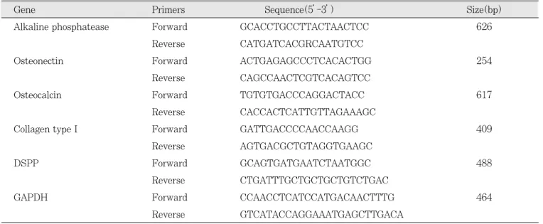
structions. Specific primers used for RT-PCR are
shown in Table 1. RT-PCR was performed by using
the following conditions: One cycle of 30 min at 50℃,
1 cycle of 2 min at 94℃, and 30 cycles of 30 sec at
94℃, 30 sec at 60℃, 30 sec at 72℃, with a final ex-
tension of 10 min at 72℃. For caspase 7 and
GAPDH, PCR reactions were performed for 25 cycles.
The PCR products were then run on 1.5% agarose gels and visualized by ethidium bromide staining.
Ⅲ. RESULTS 1. Cell Viability Assay (MTT assay)
The results of cell viability are shown in Fig. 2.
The incubation time did not influence the cell growth after 5 days of culture. The proportion of MTA treat- ed pulp cells exposed for 5 days to MTA was higher than the control cells. However, there was no differ- ence in the percentages of live cells between the two groups. According to this result, exposure to MTA material would not induce cytotoxic response in the dental pulp cells.
2. Von Kossa staining
Von Kossa staining did not reveal formation of cal- cium-positive mineralization nodule on both groups(Data was not shown).
3. Alkaline phosphatase activity
Alkaline phosphatase activity of each group was in- creased gradually, but MTA induced slight levels of alkaline phosphatase activity, and did the same to control pulp cells(Fig. 3). Maximal enzyme activity was detected after 4 days in culture for both cell populations, while this expression was delayed for 2 days with MTA.
4. RT-PCR analysis
MTA had no effect on alkaline phosphatase expres- sion after 2-day incubation. However, the expression of alkaline phosphatase was enhanced after 4-day in- cubation(Fig. 4). This result corresponds to the alka- line phosphatase activity assay. Like the effect on expression of alkaline phosphatase, MTA also in- creased DSPP and collagen type I expression in all the tested periods. However, there was no difference in the expression levels between the two groups(P<0.05). Interestingly, the expression of os- teonectin of MTA treated pulp cells exposed for 4 days to MTA was statistically higher than that of the control cells(P<0.05).
Table 1. Primers used for RT-PCR
Gene Primers Sequence(5’-3’) Size(bp)
Alkaline phosphatease Forward GCACCTGCCTTACTAACTCC 626 Reverse CATGATCACGRCAATGTCC
Osteonectin Forward ACTGAGAGCCCTCACACTGG 254
Reverse CAGCCAACTCGTCACAGTCC
Osteocalcin Forward TGTGTGACCCAGGACTACC 617
Reverse CACCACTCATTGTTAGAAAGC
Collagen type I Forward GATTGACCCCAACCAAGG 409
Reverse AGTGACGCTGTAGGTGAAGC
DSPP Forward GCAGTGATGAATCTAATGGC 488
Reverse CTGATTTGCTGCTGCTGTCTGAC
GAPDH Forward CCAACCTCATCCATGACAACTTTG 464
Reverse GTCATACCAGGAAATGAGCTTGACA
Fig. 2. Survival and proliferation of dental pulp cells on MTA.
Fig. 4. A. Transcript alteration by MTA. B. Intensities of bands mRNA shown in A were normalized with respect to those for GAPDH.
Fig. 3. Effects of MTA on alkaline phosphatase activity.
Alkaline phosphatase DSPP
A
B
Collagen type I Osteonectin
%
Con
0 2 4 0 2 4 days
Alkaline phosphatase Osteonectin
Collagen type I DSPP
GAPDH
MTA
Ⅳ. DISCUSSION
Since its introduction to clinical dental practice in 1993, mineral trioxide aggregate(MTA) has gained acceptance as the material of choice for several clini- cal procedures. It has been shown to be an effective pulp capping materia
l6,17). In addition, MTA has been used to seal and repair root perforations
18,19), and to create an apical barrier in teeth with open apices
10,11). Success as a root-end filling material has also been reported
12,13). MTA seems to be an adequate material for these procedures because of its sealing ability when compared with other materials such as amal- gam, IRM or SuperEBA
12,20). Several investigators have demonstrated that MTA is biocompatible with the surrounding tissues
13,14). It has been shown to promote osteoblastic activity
15)and to be less cytotox- ic than amalgam, IRM or SuperEBA
12,20). Furthe- rmore, MTA has been found to possess antibacterial activity
21).
The author hypothesized that MTA would induce dental pulp cells to express genes associated with dentin formation. This is based on studies in both human and dogs in which dentin deposition was his- tologically observed on MTA
12,17). Our results indicat- ed that dental pulp cells are capable of expressing genes which indicate mineralization, especially alka- line phosphatase and osteonectin. Alkaline phos- phatase(ALP) is a phosphate-releasing protein, and its activity is considered as an important indicator of bone formation and a phenotypic marker of os- teoblasts
22). The observation that alkaline phos- phatase transcript levels peak prior to any detectable increase in alkaline phosphatase activity following exposure to MTA was expected; however, the finding that enzyme activity levels increased after a decline in transcript levels was unexpected. Further studies will be needed to be performed for a more careful ex- amination of the correlation of transcript and protein stability with the respect to the regulation of func- tional levels of phosphatase activity
23).
A major collagenous protein found in dentin, DSP and DPP, plays a regulatory role in the mineraliza- tion of dental hard tissue. The production of DSP and DPP is restricted to cells that function in a min- eralizing capacity, including osteoblasts, odonto- blasts, and cementoblasts
23). The strong expression of
DSPP gene expression by dental pulp cells on both MTA and control is suggestive that the process of dentin formation can be proceeded under experimen- tal conditions. These results support MTA as being dentin-conductive by allowing the expression of genes and proteins which are consistent with the dentinogenesis process. The results of this investiga- tion compare positively with the results of other studies which indicate a favorable biologic response by MTA comparing with other endodontic materi- als
9,13,17).
Changes in expression patterns of several tran- scripts associated with hard-tissue formation were also detected. Among the genes investigated in this study, ALP and osteonectin are the most strongly as- sociated with mineralization. ALP activity is essen- tial for normal osteoblast function
15), and it is gener- ally considered as an early marker of mineralization.
In this study ALP expression by dental pulp cells at days 2 and 4 appeared on MTA and control sub- strates. These results suggest again that MTA is permissive for dentinogenesis gene expression.
Osteonectin(SPARC) is a calcium binding matricel- lular glycoprotein expressed by odontoblasts but not by the pulp cells
24,25). Furthermore, an increased level of SPARC was detected during the healing process of pulp tissue
26). ALP is an enzyme usually detected in the mineralized tissue forming cells
22,27). Thus, an in- crease in SPARC and ALP synthesis might be con- sidered as a marker of pulp cell differentiation.
SPARC is a kind of phosphorylated glycoprotein,
which is associated with tissue development, tissue
remodeling, and repair
28). It has been recently shown
that odontoblasts and unique dental pulp cells ex-
pressing SPARC, demonstrated increased expression
of SPARC in the initial stage of tertiary dentin for-
mation
24-26). Although the function of SPARC in
dentin formation is still unknown, it is suggested
that odontoblasts may release SPARC to stimulate
proliferation of some fraction of pulp cells to replace
the injured tissue due to dental caries or cavity
preparation
24). SPARC may also regulate the produc-
tion of extracellular matrix and matrix metallopro-
teinases
29,30)which might be involved in the modula-
tion of matrix for dentinogenesis. Moreover, SPARC
has been shown to play a role in differentiation of
some cell types
31). Thus, upregulation of SPARC and
ALP activity in dental pulp cells by the treatment of MTA, as demonstrated in this study, might also re- flect the changes in their function or differentiation of these cells.
In conclusion, this dental pulp culture system may be useful in the future as a model for studying the mechanisms underlying dentin regeneration after the treatment of MTA. Exposure to MTA material would not induce cytotoxic response in the dental pulp cells. MTA could influence the behavior of human pulp cells by increasing the ALP activity and SPARC synthesis.
REFERENCES
1. Yamamura T : Differentiation of pulpal cells and inductive and influences of various matrices with reference to pulpal wound healing. J Dent Res, 64:530-540, 1985.
2. Tziafas D : Basic mechanisms of cytodifferentia- tion and dentinogenesis during dental pulp re- pair. Int J Dev Biol, 39:281-290, 1995.
3. Tziafas D, Smith AJ, Lesot H : Designing new treatment strategies in vital pulp therapy. J Dent, 28:77-92, 2000.
4. Smith AJ, Cassidy M, Perry H, et al. : Reactionary dentinogenesis. Int J Dev Biol, 39:273-280, 1995.
5. Smith AJ, Lesot H : Induction and regulation of crown dentinogenesis: embryonic events as a template for dental tissue repair?. Crit Rev Oral Biol Med, 12:425-37 2001.
6. Murray PE, Windsor LJ, Smyth TW, et al. : Analysis of pulpal reactions to restorative proce- dures, materials, pulp capping, and future thera- pies. Crit Rev Oral Biol Med, 13:509-520, 2002.
7. Ham KA, Witherspoon DE, Gutmann JL, et al. : Preliminary evaluation of BMP-2 expression and histologic characteristics during apexification with calcium hydroxide and mineral trioxide ag- gregate. J Endod, 31:275-279, 2005.
8. Sarkar NK, Caicedo R, Ritwik P, et al. : Physicochemical basis of the biologic properties of mineral trioxide aggregate. J Endod, 31:97-100, 2005.
9. Holland R, Souza V, Nery MJ, et al . : Reaction of rat connective tissue to implanted dentin tubes
filled with mineral trioxide aggregate or calcium hydroxide. J Endod, 25:161-166, 1999.
10. Regan JD, Gutmann JL, Witherspoon DE : Comparison of Diaket and MTA when used as root-end filling materials to support regeneration of the periradicular tissues. Int Endod J, 35:840- 847, 2002.
11. Economides N, Pantelidou O, Kokkas A, et al. : Short-term periradicular tissue response to min- eral trioxide aggregate(MTA) as root-end filling material. Int Endod J, 36:44-48, 2003.
12. Torabinejad M, Hong CU, Lee SJ, et al. : Investigation of mineral trioxide aggregate for root end filling in dogs. J Endod, 21:603-608, 1995.
13. Torabinejad M, Pitt Ford TR, McKendry DJ, et al. : Histologic assessment of MTA as root end filling in monkeys. J Endod, 23:225-228, 1997.
14. Koh ET, McDonald F, Pitt Ford TR, et al. : Cellular response to Mineral Trioxide Aggregate.
J Endod, 24:543-547, 1998.
15. Koh ET, Torabinejad M, Pitt Ford TR, et al. : Mineral trioxide aggregate stimulates a biological response in human osteoblasts. J Biomed Mater Res, 37:432-439, 1997.
16. Zhu Q, Haglund R, Safavi KE, et al. : Adhesion of human osteoblasts on root-end filling materi- als. J Endod, 26:404-406, 2000.
17. Aeinehchi M, Eslami B, Ghanbariha M, et al. : Mineral trioxide aggregate(MTA) and calcium hy- droxide as pulp-capping agents in human teeth:
a preliminary report. Int Endod J, 36:225-231, 2003.
18. White C Jr, Bryant N : Combined therapy of mineral trioxide aggregate and guided tissue re- generation in the treatment of external root re- sorption and an associated osseous defect. J Periodontol, 73:1517-1521, 2002.
19. Holland R, Filho JA, de Souza V, et al. : Mineral trioxide aggregate repair of lateral root perfora- tions. J Endod, 27:281-284, 2001.
20. Keiser K, Johnson CC, Tipton DA : Cytotoxicity of mineral trioxide aggregated using human peri- odontal ligament fibroblasts. J Endod, 26:288- 291, 2000.
21. Estrela C, Bammann LL, Estrela CR, et al. :
Antimicrobial and chemical study of MTA,
Portland cement, calcium hydroxide paste, Sealapex and Dycal. Braz Dent J, 11: 3-9, 2000.
22. Kuru L, Griffiths GS, Petrie A, et al. : Alkaline phosphatase activity is upregulated in regenerat- ing human periodontal cells. J Periodontal Res, 34:123-127, 1999.
23. Bonson S, Jeansonne BG, Lallier TE : Root-end filling materials alter fibroblast differentiation. J Dent Res, 83:408-413, 2004.
24. Reichert T, Storkel S, Becker K, et al. : The role of osteonectin in human tooth development: an immunohistological study. Calcif Tissue Int, 50:468-472, 1992.
25. Begue-Kirn C, Smith AJ, Loriot M, et al. : Comparative analysis of TGF beta s, BMPs, IGF1, msxs, fibronectin, osteonectin, and bone sialoprotein gene expression during normal and in vitro-induced odontoblast differentiation. Int J Dev Biol, 38:405-420, 1994.
26. Itota T, Nishitani Y, Sogawa N, et al. : Alteration of odontoblast osteonectin expression following dental cavity preparation. Arch Oral Biol, 46:829-834, 2001.
27. Wlodarski KH, Reddi AH : Alkaline phosphatase as a marker of osteoinductive cells. Calcif Tissue Int, 39:382-385, 1986.
28. Yan Q, Sage EH : SPARC, a multicellular glyco- protein with important biological functions. J Histochem Cytochem, 47:1495-1506, 1999.
29. Tremble PM, Lane TF, Sage EH, et al. : SPARC, a secreted protein associated with morphogenesis and tissue remodeling, induces expression of metalloproteinases in fibroblasts through a novel extracellular matrix-dependent pathway. J Cell Biol, 121:1433-1444, 1993.
30. Francki A, Bradshaw AD, Bassuk JA, et al. : SPARC regulates the expression of collagen type I and transforming growth factor-beta1 in mesangial cells. J Biol Chem, 274:32145-32152, 1999.
31. Bassuk JA, Birkebak T, Rothmier JD, et al. : Disruption of the Sparc locus in mice alters the differentiation of lenticular epithelial cells and leads to cataract formation. Exp Eye Res, 68:
321-331, 1999.
국문초록
Mineral Trioxide Aggregate(MTA)에 의한 치수세포의 유전자 발현변화