511 http://dx.doi.org/10.4196/kjpp.2013.17.6.511
ABBREVIATIONS: K2P, two-pore domain K+ channel; TWIK, Tandem P domain weak inwardly rectifying K+ channel; TASK, TWIK-related acid-sensing K+ channel; TREK, TWIK-related channel; CDK, cyclin-dependent kinase; qRT-PCR, quantitative real-time reverse transcription-polymerase chain reaction; siRNA, small interfering RNA; Kv, voltage-dependent K; KCa, Ca2+-activated K+.
Received October 8, 2013, Revised November 11, 2013, Accepted November 19, 2013
Corresponding to: Yangmi Kim, Department of Physiology, College of Medicine, Chungbuk National University, 52 Naesudong-ro, Heung- deok-gu, Cheongju 361-763, Korea. (Tel) 82-43-261-2855, (Fax) 82- 43-272-1603, (E-mail) [email protected]
*These two authors contributed equally to this work.
This is an Open Access article distributed under the terms of the Creative Commons Attribution Non-Commercial License (http://
creativecommons.org/licenses/by-nc/3.0) which permits unrestricted non-commercial use, distribution, and reproduction in any medium, provided the original work is properly cited.
The TREK2 Channel Is Involved in the Proliferation of 253J Cell, a Human Bladder Carcinoma Cell
Kyung-Sun Park1,*, Min Ho Han2,6,*, Hee Kyung Jang3,6, Kyung-A Kim4,6, Eun-Jong Cha4,6, Wun-Jae Kim5,6, Yung Hyun Choi2,6, and Yangmi Kim3,6
1Division of Integrative Biosciences and Biotechnology, Pohang University of Science and Technology, Pohang 790-784, 2Department of Biochemistry, Dongeui University College of Oriental Medicine, Busan 614-714, Departments of 3Physiology, 4Biomedical Engineering,
5Urology, 6Personalized Tumor Engineering Research Center, College of Medicine, Chungbuk National University, Cheongju 361-763, Korea
Bladder cancer is the seventh most common cancer in men that smoke, and the incidence of disease increases with age. The mechanism of occurrence has not yet been established. Potassium channels have been linked with cell proliferation. Some two-pore domain K+ channels (K2P), such as TASK3 and TREK1, have recently been shown to be overexpressed in cancer cells. Here we focused on the relationship between cell growth and the mechanosensitive K2P channel, TREK2, in the human bladder cancer cell line, 253J. We confirmed that TREK2 was expressed in bladder cancer cell lines by Western blot and quantitative real-time PCR. Using the patch-clamp technique, the mechanosensitive TREK2 channel was recorded in the presence of symmetrical 150 mM KCl solutions. In 253J cells, the TREK2 channel was activated by polyunsaturated fatty acids, intracellular acidosis at - 60 mV and mechanical stretch at −40 mV or 40 mV. Furthermore, small interfering RNA (siRNA)-mediated TREK2 knockdown resulted in a slight depolarization from − 19.9 mV±0.8 (n=116) to − 8.5 mV±1.4 (n=74) and decreased proliferation of 253J cells, compared to negative control siRNA. 253J cells treated with TREK2 siRNA showed a significant increase in the expression of cell cycle boundary proteins p21 and p53 and also a remarkable decrease in protein expression of cyclins D1 and D3. Taken together, the TREK2 channel is present in bladder cancer cell lines and may, at least in part, contribute to cell cycle-dependent growth.
Key Words: Bladder cancer, Cell cycle, Proliferation, Small interfering RNA, TREK2
INTRODUCTION
K+ channels regulate membrane potential and may affect the oncogenic state of cells through cell cycle regulation, migration, invasiveness and Ca2+ signaling [1]. Nevertheless, the involvement of changes in membrane potential in cell growth mechanisms remains controversial [1]. Several K+ channels are expressed in a variety of malignant cancer cells [1-4]. The type of K channels related to proliferation include voltage-gated channels, such as KV1.3, KV10.1 (Eag1), KV1.5, KV3.1, KV3.4, and KV11.1 (HERG), Ca2+-dep- endent K+ channels, such as KCa1.1 and KCa3.1 (also called IKCa), ATP-dependent K+ channels and inward rectifiers
[1,4].
In addition, some two-pore domain K+ channels (K2P), which are divided into TWIK, TREK, TASK, TALK, TRESK and THIK, are also expressed in various tissues [5]. K2P channels have been known to regulate resting membrane potential and are modulated by antidepressants, anti- psychotics, volatile anesthetics, flavonoids, hypoxia, temper- ature, hydrogen, polyunsaturated fatty acids, cell volume (membrane stretch), and lysophospholipids [5,6]. Therefore, similar to other K+ channels, K2P channels might play an essential role in cancer cells [1,4,5]. Supporting data show that TASK3 was revealed as an oncogene that is frequently overexpressed in breast, lung, colon, and metastatic pros- tate cancers [7]. TREK1 was also reported as a novel tumor marker in prostate cancer cells [8]. More recently, the pres- ence of the TREK2 channel was reported in normal human ovaries and ovarian cancer [9]. Nonetheless, sufficient data on TREK2 in cancer cells is limited.
More than 90% of bladder cancers are transitional cell carcinomas (TCC), and one third of superficial tumors prog-
ress to muscle invasive cancer [10,11]. In addition, the re- currence rate of bladder cancer is relatively high compared to other cancers, and diagnosis is difficult due to low sensi- tivity biomarkers [10,11]. Furthermore, the mechanism of bladder cancer development and progression, especially in terms of ion channels, is not well established. There are only a few reports showing that the growth modulation mechanism involves ion channels in bladder cancer cells, including BKCa in 253J cells [12], induction of apoptosis by TRPV2 in T24 cells [13], and cell cycle arrest by capsaicin in RT4 cells [14].
In the present study, we investigated the involvement of the TREK2 channel in bladder cell growth. The primary objective of the study is to determine whether TREK2 is functionally expressed in bladder cancer cells, and if TREK2 is involved in cell growth. To achieve this aim, we used electrophysiological and molecular biological methods.
METHODS Cell lines and culture conditions
The human bladder cancer cell line 253J cells were main- tained in Dulbecco's modified Eagle's medium (DMEM) sup- plemented with 10% fetal bovine serum (FBS), 100 units per ml penicillin, and 100 μg per ml streptomycin in a hu- midified incubator at 37oC with 5% CO2. EJ, HT1376 and TCCSUP cells were maintained in DMEM, and T24, 5637 and J82 cells were maintained in RPMI-1640 supplemented with 10% FBS.
Electrophysiological recording and chemicals
Electrophysiological recording was performed in whole cell configurations and single channel recordings [15] using a patch clamp amplifier Axopatch 200B (Axon Instruments, Foster City, USA). Whole cell patch pipettes (Harvard Apparatus, Edenbridge, UK) were prepared at a resistance of 2∼3 megaohms. To measure resting membrane poten- tial, the current was clamped (I=0) in a whole cell patch configuration. The solution for the High-K+ pipette (internal solution) was composed of 150 mM KCl, 1 mM MgCl2, 5 mM Mg-ATP, and 2 mM EGTA (pH 7.2), and the normal Tyrode’s (NT) bath solution (external solution) was prepared as 143 mM NaCl, 5.4 mM KCl, 0.5 mM NaH2PO4, 0.5 mM MgCl2, 1.8 mM CaCl2, 5 mM HEPES, and 10 mM glucose (pH 7.4). For single TREK2 channel recordings, the pipette and bath solutions were applied symmetrically with 150 mM KCl, 1 mM MgCl2, 10 mM HEPES, and 5 mM EGTA (pH 7.2). The recorded signal was filtered at 1 kHz using an 8-pole Bessel filter (−3 dB: Frequency Devices) and transferred to a computer using the Digidata 1322A interface (Axon Instruments, Union City, CA) at a sampling rate of 2 kHz. Single-channel currents were analyzed with the pClamp program version 9.02 (Axon Instruments, Union City, USA). Data were described as channel activity (NPo). N is the number of channels in the patch, and PO
is the probability of a channel being open. The current trac- ings shown in the figures were filtered at 1 kHz. All record- ings were performed at room temperature (22∼24oC).
Isolation of total RNA, reverse transcription-poly- merase chain reaction (RT-PCR) and quantitative real-time RT-PCR (qRT-PCR)
Total RNA isolation was carried out with a MN NucleoSpin RNAII Kit (Macherey-Nagel, Germany), fol- lowed by first-strand cDNA synthesis with an iScript cDNA synthesis kit (Bio-Rad, Hercules, USA), according to the manufacturers’ protocols. RT-PCR was performed using 2 μl of cDNA in a 30 μl reaction containing 0.4 mM of each primer. The cycling conditions were as follows: 40 cycles at 96oC for 15 s, 57oC for 30 s, and 72oC for 1 min. An aliquot (20 μl) of the RT-PCR product was analyzed on a 2% agarose gel prepared in Tris-acetic acid-EDTA (TAE).
The identity of each of the PCR products was confirmed by sequence analysis. The sequences for primers were as follows: TREK1 (Accession No; AF129399), sense 5’
GGATTTGGAAACATCTCACCACGCACA 3’, antisense 5’
GATCCACCTGCAACGTAGTC 3’ (355 base pair [bp]); TREK2 (Accession No; AF279890), sense 5’ AAGCATGGGCAGGG- TGCGTC3’, antisense 5’ TCCGGCTCCCGGTCTTTGGT 3’
(291 bp). Quantitative real-time RT-PCR (qRT-PCR) was performed using CFX 96 real-time thermal cycler (Bio-Rad, Hercules, USA), and the relative amount of TREK2 ‘‘target’’
in bladder cancer cells was normalized to the endogenous control “GAPDH”. The cycling conditions were as follows:
40 cycles at 95oC for 10 s, 60oC for 10 s and 72oC for 30 s with SYBR green (Bio-Rad, Hercules, USA). Oligonucleo- tide primers for qRT-PCR to detect human TREK2 were forward 5’ CACCTCCAGACTCACCAA 3’ and reverse 5’
TCCTCCTCTTTCTTCTCCTC’, and forward 5’ACCAGGTG- GTCTCCTCTGAC 3’ and reverse 5’ TGCTGTAGCCAAA- TTCGTTG 3’ were used to detect GAPDH. The relative gene expression was analyzed using CFX manger software sys- tem version 3.0 (Bio-Rad, Hercules, USA).
Downregulation of TREK2 expression by small inter- fering RNA
TREK2 small interfering RNA (siRNA), negative control siRNA and fluorescein isothiocyanate (FITC)-labeled siRNA were purchased from Bioneer (Daejon, Korea). The TREK2 siRNA sequences were as follows: sense 5’ CAUCUUU- GGGAAAGCAUU 3’ and antisense 5’ AAUGCUUUUCC- CAAAGAU G 3’. The negative control siRNA sequences were as follows: sense 5’ CCUACGCCACCAAUUUCG 3’
and antisense 5’ ACGAAAUUGGUGGCGUAGG 3’. Lipofec- tamine RNAiMax (Invitrogen, Carlsbad, USA) was used for siRNA transfection. Cells were incubated for 72 hours after transfection.
Proliferation assay
Cell proliferation was monitored using an XTT cell pro- liferation assay kit (Biological Industries Israel Beit-Hae- mek Ltd., Kibbutz Beit-Haemek, Israel) following the proto- cols described by the manufacturer. Briefly, 253J cells were prepared at a density of 10,000 cells/ml in a 96-well plate containing 2% FBS, 100 units per ml penicillin, and 100 μg per ml streptomycin. Sample absorbance was measured with a SpectraMax M5e spectrophotometer (Molecular Devices, Sunnyvale, USA) at a wavelength of 450∼500 nanometers. Reference absorbance to measure non-specific readings was measured at a wavelength of 630∼690
Fig. 1. TREK2 channel expression in human bladder cancer cells. (A) Messenger RNA (mRNA) of ion channels related to the TREK1 (Accession No; AF129399) and TREK2 (Accession No; AF279890) were amplified by reverse transcription-polymerase chain reaction (RT-PCR) analysis. TREK1 (355base pair [bp]) and TREK2 (291 bp) were detected. (B) Immunoblot showed presence of TREK2 channel protein in the human bladder cancer 253J cell line. TREK2 transfected CHO cells were used as a control. (C) Representative confocal micro- scopic analysis of TREK2 in bladder cancer cell line 253J. Cells were stained with an anti-TREK2 antibody and F-actin. Scale bar, 20 μm.
nanometers.
Western blot assay
Bladder cancer 253J cells and TREK2 transiently trans- fected CHO cells were homogenized in lysis buffer (50 mM HEPES, pH 7.5, 150 mM NaCl, 100 mM NaF, 10% glycerol, 1% Triton X-100, 200 μM Na3VO4, 1 mM phenylmethyl- sulfonyl fluoride, 1 μg/ml aprotinin, and 1 μg/ml leupeptin).
Samples containing 40 μg total protein were separated by 10% sodium dodecyl sulfate polyacrylamide (SDS-PAGE) gel, transferred onto Immobilon-P membranes (Millipore Corporation, Bedford, USA) and incubated with rabbit poly- clonal anti-hKCNK10 (TREK2) (concentration of 1:1,000, Alomone labs, Jerusalem, Israel) antibodies. Proteins were visualized using polyclonal goat anti-rabbit IgG conjugated to horseradish peroxidase (concentration of 1:5,000, Santa Cruz Biotechnology, Santa Cruz, USA) and enhanced chem- iluminescence (ECL) reagent (Amersham Biosciences, Pis- cataway, USA) on a Lumi-Imager F1 scanner (Roche Applied Science, Indianapolis, USA). Images were analyzed using Lumi-analyst software version 3.1 (Roche Applied Science, Indianapolis, USA). Antibodies used to confirm the expression of cell cycle boundary proteins, anti-cdk2, anti- cdk4, anti-cdk6, anti-cyclin D1, anti- cyclin D2, anti-cyclin D3, anti- cyclin E, anti-actin and anti-GAPDH, were pur- chased from Santa Cruz Biotechnology (Santa Cruz, USA).
Anti-p21 and anti-p53 were purchased from Oncogene Science (Cambridge, USA). Anti-p27 was purchased from Calbiochem (Dramstadt, Germany).
Cell-cycle analysis
To measure the cellular DNA content, TREK2 siRNA treated cells were fixed with ethanol in PBS, treated with 20 μg/ml RNase A (Sigma-Aldrich, St Luis, USA) and stained with 50 μg/ml of propidium iodide (Sigma-Aldrich, St Luis, USA). Samples were then analyzed by a fluo- rescence-activated cell sorting (FACS) on a Calibur flow cy- tometer (BD Bioscience, San Jose, USA). Modfit LT cell cy- cle analysis software version 3.0 (Verity software house,
Topsham, USA) was used to determine the relative DNA content based on the presence of red fluorescence.
Immunocytochemistry
The 253J cells were fixed with 3.7% paraformaldehyde in phosphate-buffered saline (PBS) for 20 min at room tem- perature and were permeabilized with 0.5% Triton X-100 in PBS for 3 min. The cells were blocked for 30 min at 25oC with 3% BSA in PBS. For the staining, the cells were in- cubated with rabbit polyclonal anti-hKCNK10 (TREK2) (concentration of 1:200, Alomone Labs, Jerusalem, Israel) for overnight at 4oC, followed by incubation with an Alexa Fluor 488 conjugated secondary antibody (concentration of 1:200, Santa Cruz Biotechnology, Santa Cruz, USA) for 1 h. To visualize F-actin, cells were stained with Alexa Fluor 594 conjugated phalloidin (concentration of 1:100, Life Technologies, Grand lsland, USA) for 30 min at 25oC.
The images were acquired confocal microscopy (TCS-SP2 AOBS, Leica Microsystems, Heidelberg, Germany).
Statistics
The data are shown as the means±standard error (S. E.), and n represents the number of cells tested. The sig- nificance of the differences between the means was estab- lished using Student’s t-test. p<0.05 was regarded as significant. All statistical analyses were conducted with the Origin program version 6.0 (Microcal, Northampton, USA).
RESULTS
To explore the functional role of TREK2 channels in blad- der cancer cells, we first examined their expression in blad- der cell lines using qRT-PCR and Western blot (Fig. 1A and Fig. 1B). TREK2 mRNA was expressed and its protein was detected in 253J cells. Figure 1C showed representative confocal microscopic image of TREK2 in 253J cells. TREK2 was observed in plasma membrane and cytosol.
We performed the patch clamp technique to confirm the
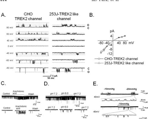
Fig. 2. The physiologic properties of TREK2 at a single channel level in bladder cancer 253J cells. (A) TREK2 in CHO cells transfected with DNA enco- ding TREK2 and GFP and in 253J cells was measured in the excised inside-out patch configuration at the holding po- tential values shown on the left. The current trace was obtained in sym- metrical 150 mM KCl solutions. The letters “c” and “o” represent the “closed”
and “open” states of the channels, re- spectively. (B) The I-V relationships showed inward rectification, and each point is the mean of 4 experiments with standard error (S.E) represented by the error bars. (C~E) Current tracing showed arachidonic acid, intracellular pH, and mechanosensitivity of the native TREK2- like channel in 253J cells at −60 mV,
+40 mV, and −40 mV. Negative pressure (−10 mmHg or −20 mmHg) was applied through the pipette. The panel below each of the figures shows the single channel trace on an expanded time scale.
functional expression of TREK2 using single channel re- cording in symmetrical 150 mM KCl solutions. We were able to record the TREK2 channel in the bladder cancer cell line 253J but not 5637 or EJ. Thus, we carried out the remainder of the study in 253J cells. The current-voltage (I-V) relation curve showed slightly inward rectification (Fig. 2A and 2B), and the single channel conductance was 123 pS (123.2±5.8 pS, n=7, Fig. 2A) at −60 mV. The chan- nel is similar in TREK2-transiently overexpressing CHO cell, as shown (145.0±8.7 pS, n=4, Fig. 2A). To observe the similarity with the electrophysiological properties of the TREK2 channel, we tested the effect of known physiological stimulants of TREK2 including mechanical stretch, arach- idonic acid (AA), and intracellular acid pH (pH 6.0) on the TREK2-like channel in 253J cells (Fig. 2C, 2D, and 2E) [16].
TREK2-like channel activity showed an 11-fold increase due to AA (relative NPo, 10.8±0.6, n=4) and an 8-fold in- crease due to intracellular acidic pH (relative NPo, 8.5±0.3, n=4) compared with the control (Fig. 2C and 2D). Mechani- cal stretch also activated the TREK2-like channel at +40 mV and −40 mV in 253J cells (Fig. 2E). These results re- vealed TREK2 is functionally expressed in bladder cancer 253J cell.
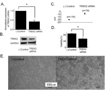
To confirm the effect on membrane potential and cell growth, knockdown was performed with siRNA specific to TREK2. We first measured the level of TREK2 expression on knockdown using qRT-PCR and Western blot 72 hours after transfection. TREK2 mRNA was decreased to ~43%
in cells transfected with TREK2 siRNA compared to neg- ative control siRNA (42.9±5.0%, n=8 with TREK2 siRNA, Fig. 3A). The protein level was also decreased by knock- down (Fig. 3B). To measure the membrane potential, we performed current clamp (I=0) with a whole cell patch configuration. The membrane potential was slightly depo- larized by knockdown of TREK2 with siRNA (−19.9±0.8 mV, n=116 with negative control siRNA vs. −8.5±1.4 mV, n=74 with TREK2 siRNA) (Fig. 3C). Next, we observed the effect of TREK2 knockdown on cell proliferation. Cell
growth decreased to ∼64% after TREK2 silencing com- pared to cells expressing the negative control (63.7±5.2%, n=38 with TREK2 siRNA, Fig. 3D). Figure 3E showed rep- resentative images of 253J cells transfected with TREK2 siRNA and negative control siRNA.
To elucidate the influence of TREK2 knockdown on cell-cycle progression, we checked the DNA content using flow cytometry. The silencing of TREK2 for 72 hours led to 253J cell-cycle arrest, with an increase of cells in the G0/G1 phases (51.8±2.4% in control vs. 56.3±1.9% in TREK2 siRNA, n=8) and a decrease in the percentage of cells in the S phase (42.7±3.3% in control vs. 36.6±3.0% in TREK2 siRNA, n=8) (Fig. 4A). These data prove that the inhibition of DNA synthesis on TREK2 knockdown resulted from cell cycle arrest at G0/G1. Because silencing of TREK2 channels affected G0/G1 phase progression in bladder can- cer cells, we examined the expression of central cell cycle regulatory proteins such as cyclin D1, cyclin D3, cdk2, cdk4, cdk6, p21, and p53. The protein levels of p53 and p21 were increased and cyclin D1, cyclin D3, cdk2, cdk4, and cdk6 were decreased (Fig. 4B). Therefore, these results clearly demonstrated that TREK2 was related to G0/G1 phase ar- rest in bladder cancer growth.
Taken together, TREK2 was functionally expressed in bladder cancer 253J cells and membrane depolarization by knockdown of TREK2 caused cell growth inhibition through cell cycle arrest at the G0/G1 phase.
DISCUSSION
In the present study, we demonstrated for the first time that TREK2 was functionally expressed in bladder cancer 253J cells. Furthermore, membrane depolarization induced by TREK2 knockdown inhibited growth of 253J cells via cell cycle arrest.
Changes in membrane potential by K+ channel regu- lation are generally accepted to affect the regulation of the
Fig. 4. Cell cycle arrest at G0/G1 after TREK2 siRNA transfection in bladder cancer 253J cells. (A) The bar graph shows that TREK2 siRNA treated cells resulted in an increased percentage of cells in G0/G1 and a decreased percentage of cells in the S phase of the cell cycle (t-test p<0.05) (B) Representative Western blot showing changes in the levels of associated proteins in cell cycle arrest of human bladder cancer 253J cells. Knockdown of TREK2 decreased the expression of cyclin D1, cyclin D3, cdk2, cdk4, and cdk6 and increased protein levels of p21 and p53.
Fig. 3. The effect of TREK2 knockdown on the growth of 253J bladder cancer cells. (A and B) TREK2 mRNA and protein levels in 253J cell were determined after knockdown of TREK2 by siRNA through qRT-PCR and Western blot. Expression of TREK2 mRNA after transfection of TREK2 siRNA or negative control siRNA was normalized. TREK2 protein was examined by Western blot analysis after transfection of the negative control or TREK2 siRNA in 253J cells. GAPDH was used as a control. (C) The membrane potential was measured at current clamp (I=0) in a whole cell patch configuration. The 253J cells were treated with FITC-labeled negative control siRNA and FITC-labeled TREK2 siRNA for 72 hours. The data were represented as the mean±S.E. (t-test, p value
<0.05). (D) The antiproliferative effect of TREK2 knockdown by siRNA in 253J cells. Cells were treated for three days with TREK2 siRNA or (−)control siRNA in 2% serum culture media. After treatment, proliferation was measured by XTT assay. Error bars represent the mean±S.E for 38 separate experiments. Asterisks indicate values that are different from the respective control (t-test, p<0.05). (E) Effect of TREK2 siRNA on 253J cell growth. Cells were captured 48 hours after transfection with TREK2 siRNA using a Nikon microscope at 10×10 magnification. Scale bar, 100 μm.
proliferative state [1]. Depolarization by K channel blockers leads to increased cell volume by water influx followed by an increase in solute, resulting in decreased proliferation [2,3,17]. Changes in membrane potential may also be in- volved in the control of cell cycle boundary proteins, such as cyclin-dependent kinase inhibitors and/or cyclin ex- pression [18,19]. Previous reports indicate that the in- hibition of KATP or hERG1 (KV11.1) caused cell cycle arrest at the G0/Gl phase [20,21], and cells in G0/G1 phase arrest were depolarized [22-24]. In addition, it has been reported that silencing hEag1 (KV10.1) in cells led to a decrease in cyclin expression and an increase of p21 [25]. These reports support our data that depolarization induced by knockdown of TREK2 with siRNA induced cell growth inhibition and cell-cycle arrest at the G0/G1 phase through an increase of p21 and p53 and a decrease of cyclin D1 and cyclin D3 (Fig. 3 and Fig. 4). Furthermore, the increase in p21 ex- pression inhibited the cyclin D/cdk complex and altered cel- lular proliferation by regulating cell cycle progression at G1 [26]. In this study, we found that TREK2 knockdown re- sulted in the downregulation of cyclin D1 and D3, leading us to expect a decrease of cyclin D/cdk complex formation necessary for the G1/S transition (Fig. 4) [26]. In addition,
we found an increase in the tumor suppressor gene p53, which delays the G1 phase of the cell cycle [26]. Therefore, these data clearly demonstrated that TREK2 participated in cell cycle arrest at the G0/G1 phase through the down-regulation of cdks kinase activity, via the selective induction of p21-mediated p53 expression.
During the experiments, we needed to distinguish be- tween TREK1 and TREK2 during single channel recording, as both have very similar electrophysiological properties [27-29]. In bladder cancer cells, we were able to distinguish these proteins using single channel conductance and I-V relationships. TREK2 had a higher single channel con- ductance than TREK1, and TREK2 showed a more inward rectifying I-V relationship than TREK1 in symmetrical 150 mM KCl solutions (Fig. 2). Thus, we could conclude that TREK2 was functionally present in bladder cancer 253J cells because our single channel data were similar to pre- vious results in other cells [28,29]. In present study, we could not record TREK2 channel in 5637 or other cell lines except 253J cell. The reason might be due to TREK2 trans- location deficiency into plasma membrane by unknown sig- nals or a low density of the channels in plasma membrane.
TREK2 generally has been known to be activated by is- chemic insults such as osmotic stress or change in pH to regulate resting membrane potential. Consequently, the TREK2 channel might act to protect the cancerous cells, similar to TASK3, which promotes tumor formation [7].
Therefore, the physiological relevance of the TREK2 chan- nel requires further study to provide a candidate for ad- juvant therapy along with anticancer drugs, as well as to provide the mechanism of disease progression, recurrence, and therapy in bladder cancer.
ACKNOWLEDGEMENTS
This work was supported by the National Research Foundation of Korea (NRF) grant funded by the Korea gov- ernment (MEST) (No. 2008-0062611) and Basic Science Research Program through the NRF funded by the Ministry of Education, Science and Technology (NRF-2010-0010719).
REFERENCES
1. Becchetti A. Ion channels and transporters in cancer. 1. Ion channels and cell proliferation in cancer. Am J Physiol Cell Physiol. 2011;301:C255-265.
2. Kunzelmann K. Ion channels and cancer. J Membr Biol. 2005;
205:159-173.
3. Ouadid-Ahidouch H, Ahidouch A. K+ channel expression in human breast cancer cells: involvement in cell cycle regulation and carcinogenesis. J Membr Biol. 2008;221:1-6.
4. Li M, Xiong ZG. Ion channels as targets for cancer therapy.
Int J Physiol Pathophysiol Pharmacol. 2011;3:156-166.
5. Es-Salah-Lamoureux Z, Steele DF, Fedida D. Research into the therapeutic roles of two-pore-domain potassium channels.
Trends Pharmacol Sci. 2010;31:587-595.
6. Kim KA, Kim Y. The effect of flavonoids on the TREK-1 channel. Journal of the Korea Academia-Industrial Cooperation Society. 2011;12:2660-2667.
7. Pei L, Wiser O, Slavin A, Mu D, Powers S, Jan LY, Hoey T.
Oncogenic potential of TASK3 (Kcnk9) depends on K+ channel function. Proc Natl Acad Sci U S A. 2003;100:7803-7807.
8. Voloshyna I, Besana A, Castillo M, Matos T, Weinstein IB, Mansukhani M, Robinson RB, Cordon-Cardo C, Feinmark SJ.
TREK-1 is a novel molecular target in prostate cancer. Cancer Res. 2008;68:1197-1203.
9. Innamaa A, Jackson L, Asher V, van Schalkwyk G, Warren A, Keightley A, Hay D, Bali A, Sowter H, Khan R. Expression and effects of modulation of the K2P potassium channels TREK-1 (KCNK2) and TREK-2 (KCNK10) in the normal human ovary and epithelial ovarian cancer. Clin Transl Oncol.
2013;15:910-918.
10. Kakehi Y, Hirao Y, Kim WJ, Ozono S, Masumori N, Miyanaga N, Nasu Y, Yokomizo A. Bladder Cancer Working Group report.
Jpn J Clin Oncol. 2010;40 Suppl 1:i57-64.
11. Ploeg M, Aben KK, Kiemeney LA. The present and future burden of urinary bladder cancer in the world. World J Urol.
2009;27:289-293.
12. Kim Y, Kim WJ, Cha EJ. Quercetin-induced growth inhibition in human bladder cancer cells is associated with an increase in Ca2+-activated K+ channels. Korean J Physiol Pharmacol.
2011;15:279-283.
13. Yamada T, Ueda T, Shibata Y, Ikegami Y, Saito M, Ishida Y, Ugawa S, Kohri K, Shimada S. TRPV2 activation induces apoptotic cell death in human T24 bladder cancer cells: a potential therapeutic target for bladder cancer. Urology. 2010;
76:509 e501-507.
14. Li Q, Wang XH, Yang ZH, Wang HP, Yang ZW, Li SW, Zheng XM. [Induction of cell cycle arrest in bladder cancer RT4 cells by capsaicin]. Zhonghua Yi Xue Za Zhi. 2010;90:1230-1233.
15. Hamill OP, Marty A, Neher E, Sakmann B, Sigworth FJ.
Improved patch-clamp techniques for high-resolution current recording from cells and cell-free membrane patches. Pflugers Arch. 1981;391:85-100.
16. Bang H, Kim Y, Kim D. TREK-2, a new member of the mechanosensitive tandem-pore K+ channel family. J Biol Chem.
2000;275:17412-17419.
17. Rouzaire-Dubois B, Dubois JM. K+ channel block-induced mammalian neuroblastoma cell swelling: a possible mechanism to influence proliferation. J Physiol. 1998;510 (Pt 1):93-102.
18. Wonderlin WF, Strobl JS. Potassium channels, proliferation and G1 progression. J Membr Biol. 1996;154:91-107.
19. Ghiani CA, Yuan X, Eisen AM, Knutson PL, DePinho RA, McBain CJ, Gallo V. Voltage-activated K+ channels and mem- brane depolarization regulate accumulation of the cyclin-depen- dent kinase inhibitors p27(Kip1) and p21(CIP1) in glial progenitor cells. J Neurosci. 1999;19:5380-5392.
20. Woodfork KA, Wonderlin WF, Peterson VA, Strobl JS.
Inhibition of ATP-sensitive potassium channels causes reversible cell-cycle arrest of human breast cancer cells in tissue culture.
J Cell Physiol. 1995;162:163-171.
21. Smith GA, Tsui HW, Newell EW, Jiang X, Zhu XP, Tsui FW, Schlichter LC. Functional up-regulation of HERG K+ channels in neoplastic hematopoietic cells. J Biol Chem. 2002;277:18528- 18534.
22. Wonderlin WF, Woodfork KA, Strobl JS. Changes in membrane potential during the progression of MCF-7 human mammary tumor cells through the cell cycle. J Cell Physiol. 1995;165:177- 185.
23. Ouadid-Ahidouch H, Le Bourhis X, Roudbaraki M, Toillon RA, Delcourt P, Prevarskaya N. Changes in the K+ current-density of MCF-7 cells during progression through the cell cycle:
possible involvement of a h-ether.a-gogo K+ channel. Receptors Channels. 2001;7:345-356.
24. Ouadid-Ahidouch H, Roudbaraki M, Delcourt P, Ahidouch A, Joury N, Prevarskaya N. Functional and molecular identifi- cation of intermediate-conductance Ca2+-activated K+ channels in breast cancer cells: association with cell cycle progression.
Am J Physiol Cell Physiol. 2004;287:C125-134.
25. Borowiec AS, Hague F, Gouilleux-Gruart V, Lassoued K, Ouadid-Ahidouch H. Regulation of IGF-1-dependent cyclin D1 and E expression by hEag1 channels in MCF-7 cells: the critical role of hEag1 channels in G1 phase progression. Biochim Biophys Acta. 2011;1813:723-730.
26. Vermeulen K, Van Bockstaele DR, Berneman ZN. The cell cycle: a review of regulation, deregulation and therapeutic targets in cancer. Cell Prolif. 2003;36:131-149.
27. Park KS, Kim Y. Functional expression of mechanosensitive two-pore domain potassium channel in human bladder carci- noma cells. J Biomed Res. 2013;14:71-76.
28. Kang D, Kim D. TREK-2 (K2P10.1) and TRESK (K2P18.1) are major background K+ channels in dorsal root ganglion neurons.
Am J Physiol Cell Physiol. 2006;291:C138-146.
29. Han J, Gnatenco C, Sladek CD, Kim D. Background and tandem-pore potassium channels in magnocellular neurosecre- tory cells of the rat supraoptic nucleus. J Physiol. 2003;546:
625-639.