비소세포성폐암에 대한 자연살해세포의 항암효능
박민경*·성혜란**·박지성·김지연·한상배·이종길·윤병규***·송석길#
충북대학교 약학대학, *식품의약품안전평가원, **Moffitt Cancer Center and Research Institute, ***(주)엔케이바이오 (Received June 9, 2011; Revised June 10, 2011; Accepted June 13, 2011)
Anticancer Effect of Activated Natural Killer Cells on Human Non-small Cell Lung Cancer
Min Gyeong Park*, Hyeran Sung**, Ji-sung Park, Jee Youn Kim, Sang-Bae Han, Chong-Kil Lee, Byung Kui Yun*** and Sukgil Song#
College of Pharmacy, Chungbuk National University, Cheongju 361-763, Korea
*Division of Pharmacological Research, KFDA, Osong Health Technology Administration Complex, Chungbuk 361-763, Korea
**Department of Comprehensive Melanoma Research Center (CMRC), H. Lee Moffitt Cancer Center & Research Institute, 12902 Magnolia Drive, Tampa, Florida 33612
***NKBIO Ltd., Seongnam 462-807, Korea
Abstract — Human NK cells, identified 30 years ago based on their ability to spontaneously kill tumor cells, constitute a subset of lymphocytes, which play an important role in the first line of immune defense and the effective function of these cells are enhanced by cytokines. Lung carcinoma has been one of the most commonly diagonosed cancer as well as the lead- ing cause of cancer death in male. Here we provide the evidence that human natural killer cells has inhibitory effects on tumor growth of human lung cancer cell NCI-H460 (non-small cell lung cancer). Enriched NK cell population was obtained by 2 weeks cultivation in interleukin-2(IL-2)-containing medium. The resulting population comprised 26% CD3+ cells, 9%
CD3+CD4+ cells, 16% CD3+CD8+ cells, 76% CD56+ cells, 6% CD3+CD56+ cells and 70% CD3-CD56+ cells. Activated NK cells at doese of 2.5, 5, and 10 million cells per mouse inhibited 2%, 12% and 45% of NCI-H460-induced tumor growth in nude mouse xenograft assays, repectively. This result suggests that NK cell-based immunotherapy may be used as an adoptive immunotherapy for lung cancer patients.
Keywords □ natural killer cells, immunotherapy, NCI-H460 lung cancer
전체 암 발생의 4위에 해당하는 폐암은 여성보다 남성의 발병 율이 더 높으며 세계적으로 점차 발생빈도가 증가하는 추세이다.1) 폐암은 병리학적으로 소세포성 폐암과 비소세포성 폐암으로 분 류되며, 병기에 따라 수술적 제거, 방사선 치료, 항암 화학 요법 의 세가지 방법으로 치료한다. 병기가 이르고 발병부위가 국소 적인 경우 수술과 방사선치료를 병행하며, 전신에 해당할 경우 항암약물치료요법으로 진행한다.2) 하지만, 현재의 치료법은 암세 포뿐 아니라 인근의 정상 세포까지 파괴하며 다른 질병에 쉽게 노출되는 등의 부작용을 야기하며 수술 및 보조요법에도 불구하 고 재발이나 전이 가능성이 높아 진단 5년 후 생존율이 14%로
나타난다. 환자의 생존율을 향상시키기 위해서는 보다 선택적이 며 효과적인 새로운 치료요법이 대한 개발이 필요하다. 면역세 포를 이용한 항암치료는 암세포에 특이적으로 작용하여 독성이 적다는 장점을 갖고 있다.3-9) 이러한 장점에서 특정 면역세포들 을 이용한 항암효능에 대한 연구가 활발하게 진행되어지고 있으 며, 자연살해세포가 새롭게 부각되어졌다. 자연살해세포는 T세 포, B세포와 더불어 림프구에 있는 하나의 군으로 외부항원이나 비정상적인 세포들에 가장 먼저 반응하는 선천성 면역반응의 중 추적인 역할을 하며, 세포 독성 림프구로서 바이러스 감염 세포 나 종양세포를 용해하거나 직접적으로 세포독성을 나타낸다. 자 연살해세포의 암세포 작용기전은 일반적으로 항체의존성 세포매 개형 세포독성(Antibody-dependent cell-mediated cutotoxicity, ADCC)이다. 자연살해세포는 lgG의 RC에 대한 수용체인 CD16 을 발현하고, 이를 통하여 다른 형태의 MHC 비 제한성 살해를
#본 논문에 관한 문의는 저자에게로 (전화) 043-261-2817 (팩스) 043-268-2732 (E-mail) [email protected]
종설
수행할 수 있다. ADCC는 표적세포를 인식하는 항체의 존재에 의존적이다. 즉, ADCC의 특이성은 항체의 특이성으로부터 유래 된 것이다. 자연살해세포는 이러한 기작을 통하여 tumor-specific T cell의 공격에서 벗어났거나 항원제시능에 결함이 있는 암세포 또는 MHC발현이 결핍된 암세포에 대해 항암효과를 나타낼 수 있다. 자연살해세포는 CD3-CD56+림프구로 정의되며, 전체 순 환하는 림프구 중 약 10% 내외를 차지하며 간, 복강 그리고 태 반을 포함하는 peripheral tissue에서 발견된다. IFN-α 나 IL-2 와 같은 사이토카인을 처리하여 활성화 시키거나, NKp30, NKp44, NKp46와 같은 자연살해세포 활성 수용체의 리간드를 자극함으로서 자연살해세포의 항암효과를 향상시킬 수 있다. 활 성화된 자연살해세포는 세포독성과립(perforin 및 granzyme)이 나 세포사멸 유도인자(TRAIL, FasL, cytokine및 chemokine) 를 발현하여 표적 세포에게 손상을 입힌다.10-12)이와 같이 활 성화된 자연살해세포를 이용한 항암치료법은 기존의 화학요법 제인 항암제가 가졌던 문제를 해결하고 다양한 암에 대한 암 특이 항원에 대하여 세포반응을 유발할 수 있어 암세포에 특이 적으로 작용하며, 전이된 암세포까지 치료가 가능하다는 장점 이 있다. 본연구진은 결장암에 대한 자연살해세포의 항암능력 을 검증하여 보고한바 있으며,13)기 정립된 최적화된 시험법을 이용하여 인간 폐암(비소세포성)에 대해 활성화된 자연살해세 포의 암세포 성장억제능력을 확인하였다. 인간말초혈액을 채취, IL-2와 함께 14일간 배양함으로써 활성화된 자연살해세포의 종 양 억제 효능을nude mouse xenograft model을 이용하여 평가 하였다.14)
실험방법
세포배양
NCI-H460(ATCC # HTB-177) 세포는 전체 폐암의 85%를 차 지하고 있는 non-small lung cancer(NSCLC)에서 유래한 세포주 의 하나로 wild type p35 유전자와 mutant K-mas 유전자를 가 지고 있다. 종양형성 유도를 위해 NCI-H460는 10% fetal bovin serum, 100 U/ml peniciilin, 100 U/ml streptomycin(Invitrogen, USA)를 함유한 RPMI-1640 medium에서 배양하였다. 자연살해 세포의 증폭은 건강한 지원자의 말초혈액 단핵세포(peripheral mononuclear cells. PBMC)로 만들어졌다. 지원자에게 사전의 충 분한 설명을 통한 자유의사에 의한 동의를 거친 후, heparin과 함께 40 ml의 혈액을 채취하였다. Ficoll-Hypaque density centrifugation을 통해 말초혈액 단핵세포가 포함하고 있는 buffy coats를 분리하고 PBS를 이용하여 세척하였다. 세척 후, 5%
human serum(Biowhitrraker-Cambrex, Malkersville, MD)가 함유된 Lymphomedia로 1×10⁶cells/ml로 현탁하여, 고정화 한 anti-CD3 antibody(OKT-3 10 ng/ml; BD Pharmingen, NJ,
USA)와 recombinant human IL-2(Proleukin 500 U/ml, Chiron, Emeryville, USA) 함께 배양하였다. 5일 배양 후 rhIL-2(500 U/
ml)와 5% human serum가 함유된 배지로 교체한 뒤, 매 배양주 기마다 1×10⁶cells/mm의 세포수를 유지하며 rhIL-2가 포함된 배지로 보충하며 배양하여준다. 이와 같은 세포 배양 방법을 통 하여 얻은 자연살해세포에서 87%의 생존율을 확인하였다.
세포표현형 분석
IL-2와 함께 배양하여 활성화된 자연살해세포를 약 1×10⁶cells 를 1% Bovine serum albumin(BSA)을 첨가한 PBS(BSA/PBS) 로 한번 세척한 후, 50µl의 PBS/BSA를 이용하여 세포를 현탁하 였다. 세포에 인간 항체 anti-CD3-FITC/CD16+CD56-PE와 anti-CD4-FITC/CD8-PE/CD3-PerCP(BD Biosciences, USA)를 첨가한 후 4oC에서 20분 동안 반응시킨 후, PBS로 두 번 세척하 고 400 µl의 PBS로 다시 현탁시켜 FACS Canto flow cytometer (BD Biosciences, USA)로 측정한 뒤, WinMDI statistical software(Scripps, La Jolla, USA)을 이용하여 분석하였다. 세포 의 생존율은 동일하게 PBS/BSA로 세척한 뒤, 50 µl의 PBS/BSA 를 이용하여 세포를 현탁하여 propidium iodide(1 µg/ml)를 세포에 10분간 염색하여 FACS Canto flow cytometer(BD Biosciences, USA)를 측정, PI positive 세포는 죽은 것으로 간주하였다.
Nude mouse xenograft 분석
In vivo에서 활성화된 자연살해세포의 항암능력을 판단하기 위 하여 SLC Japan, Inc.에서 생산된Specific pathogen-free female BALB/c-nu/nu mice(nude mice)를 이용하여 tumor xenograft 시험을 진행하였다. Mice는(6~8 weeks old) 충북대학교 실험동 물연구지원센터의 SPF시설에서 1주일 순화시킨 후 진행하였다.
시험 당일(0 day) 비소세포성 폐암 세포주인 NCI-H460을 2×
106cells/mouse의 농도로 nude mice에 피하 이식하였다. 활성화 된 자연살해세포은 1주일에 한번씩(day 0, 7, 14) 2.5, 5, 10×106 cells/mouse로 정맥 투여하였으며, 양성대조군인 Adriamycin (ADR; Sigma-Aldrich, St. Louis, USA)도 매주 1회(2 mg/kg)씩 총 3회 투여하였다. 최종일(21 day)에 종양을 분리한 후 종양의 무게를 측정하고 종양의 크기는 가로(mm)×세로(mm)×높이 (mm)/2로 측정하였다. 또한, 자가활성 자연살해세포의 독성을 확 인하기 위하여 매주 투여하기 전nude mouse의 체중변화를 측 정하였다.
자료분석 및 통계처리
In vivo 시험결과는 실험군 당 6마리 mice를 분석하였으며, in vitro 결과는 three samples의 mean values으로 나타냈다. 표준 편차(SD)와 p-values는 Student’s t-test 및 ANOVA(GraphPad Prism, GraphPad Software, USA)를 사용하여 산출하였다.
실험결과
Activated human NK cell의 표현형 분석
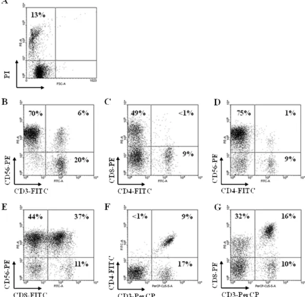
인간말초혈액에서 5~10%를 차지하는 자연살해세포를 IL-2 를 함유한 배지에서 2주간 배양하여, 약 7배 정도의 고동도로 배양됨을 확인했다. 인간항체를 이용하여 Fluorescence- activated cell sorting(FACS) 분석을 통해 활성화된 자연살해 세포의 표현형을 분석하였다. 분석결과, 배양한 세포는 87%의 생존율을 나타냈다(Fig. 1A). 또한, 26% CD3+ T cells, 9%
CD3+CD4+ T cells, 16% CD3+CD8+ T cells, 76% CD56+ cells, 6% CD3+CD56+ cells, 그리고 70% CD3-CD56+자연 살해세포가 포함되어 있음을 분석을 통하여 확인하였다(Fig.
1B~G).
In vivo antitumor effect of NK cells
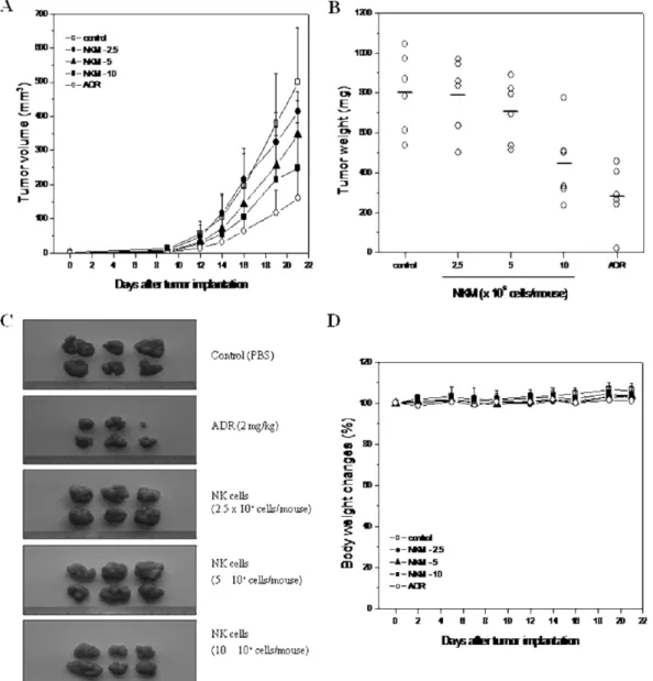
활성화된 자연살해세포의 암세포 성장 억제 효능을 확인하기 위하여 nude mouse xenograft 분석을 시행하였다. 예비시험에 서 3×10⁸cells(유효용량의 약 30배)의 활성자연살해세포를 nude mouse에 투여한 결과 사망, 탈모, 이상행동, 체중 감소의 유의한 독성은 관찰되지 않았다. 항암효과를 검증하기 위하여 Specific pathogen-free female BALB/c-nu/nu mice(nude mice)에 비소 세포성 폐암 세포주인 NCI-H460을 2×10⁶cells/mouse로 피하 이식하여 21일 후 500±160 mm3(n=6) 크기로 성장을 확인했다.
즉, 형성된 종양의 부피를 기준으로, 각 실험군에서 자연살해세 포를 2.5×10⁶cells/mouse로 저용량 투여한 경우에는 413±59 mm3종양크기를 나타내어 17%의 성장 억제를, 5×10⁶cells/
mouse 중용량 투여군에서는 347±98 mm3으로 31%의 성장 억
Fig. 1 − The phenotypic characterization of the autologous activated NK cells, which was used in injection of nude mouse xenograft assay.
Human PBMCs were cultured in the presence of IL-2 for 14 days and the resulting NK cell populations were stained with Propidium iodide (A) or human antibodies, such as anti-CD3-FITC/CD56-PE (B), anti-CD4-FITC/CD8-PE (C), anti-CD4-FITC/CD56-PE (D), anti-CD8-FITC/CD56-PE (E), anti-CD3-perCP/CD4-FITC (F) or anti-CD3-perCP/CD8-PE (G), followed with FACS analysis.
제를, 10×106cells/mouse 고용량 투여군에서는 249±132 mm3 (p<0.05)으로 50%의 종양 성장 억제 효과를 나타내었다. 양성 대조군으로 사용한 아드리아마이신 투여군에서는 159±80 mm3 (p<0.001)으로 68%의 강력한 종양 성장 억제 효과를 나타냄을 확인하였다(Fig. 2A). 최종일(21 day)에 nude mouse에서 종양을 분리하여 무게를 측청함으로써 NCI-H460에 대한 자연살해세포 의 종양억제능력을 증명하였다. 대조군의 평균 종양 무게는 802
±199 mg이었으며, 자연살해세포를 2.5×10⁶cells/mouse로 투여 한 경우에는 788±184 mg으로 2%의 종양 성장 억제를, 5×10⁶
cells/mouse 투여군에서는 706±156 mg으로 12%의 종양 성장 억제를, 10×106cells/mouse 투여군에서는 445±193 mg (p<
0.05)으로 45%의 종양 성장 억제 효과를 나타내었다. 양성대조 군으로 사용한 아드리아마이신 투여군에서는 279±152 mg (p<0.001)으로 65%의 종양 성장 억제 효과를 나타내었다(Fig.
2B). 이 같은 결과는 활성화된 자연살해세포가 농도의존적으로 비소세포성 폐암종양의 성장을 억제한다는 사실을 말해준다.
또, 자연살해세포에 의하여 유발되는 독성을 확인하기 위하여 nude mouse의 체중 변화를 21일 동안 측정하였으며, 육안으로 행
Fig. 2 − Antitumor effect of activated NK cells to NCI-H460-induced lung tumor. NCI-H460 cells were implanted subcutaneously to nude mice (n=6) on day 0. Activated NK cells at doses from 2.5 ro 10 million cells/mouse were injected intravenously once in a week (day 0, 7, and 14). Adriamycin (ADR) was injected intravenously at 2 mg/kg. Tumor volumes were estimated by the formula: length (mm)×width (mm)×height (mm)/2. Statistical significance was determined using the Student’s t-test versus PBS-treated control group (*p<0.05, ***p<0.001)(A). On day 21, the mice were sacrificed and the tumor weights were measured (B). Representative photographs are shown (C). Statistical significance was determined using the Student’s t-test versus PBS-treated control group (**p<0.01). The body weights of the tumor-bearing nude mice were measured to estimate toxicity (D).
동 변화를 관찰하였다. Day 0 의 체중(100%)과 비교하였을 때, day 21에 대조군의 경우 106%, 저, 중, 고용량의 투여군에서 각각 104%, 103%, 103%으로 정상적인 체중변화를 나타내었으며 이상 행동 역시 관찰되지 않았다. 양성대조군인 아드리아마이신 투여군 역시 101%로 정상적인 체중 변화를 나타내었다(Fig. 2C and D).
결론 및 고찰
본 연구에서는 인간 폐암(non-small cell lung cancer, NCI- H460)에 대한 ex vivo에서 활성화된 자연살해세포의 종양 억제 효과를 nude mouse xenograft model을 이용하여 in vivo상에서 검증하였다. 폐암은 전체 암 사망률 29%를 차지하며 기존의 수 술 및 보조적인 치료에도 불구하고 재발과 전이가 쉬워 2008년 미국 남성 암사망율 1위로 보고되었다. 이와 같은 폐암 진단 환 자의 생존율을 높이기 위하여 새로운 치료법의 도입이 필요하다.
암환자는 건강한 사람에 비해 자연살해세포의 양이 현저히 낮다 고 보고되었다.15,16)최근에는 이와 같은 면역세포를 이용한 항암 효능에 대한 연구가 활발히 진행되어지고 있다. 대식세포(dendritic cell), 자연살해세포, 림포카인활성 살해세포(LAK cell), 싸이토카 인 유도살해세포(CLK cell), 세포독성 T세포(cytotoxic T cell)와 같은 면역세포를 이용한 항암치료법은 ex vivo 상태에서 증식한 면역세포를 이식하는 방법이다.17-20) 이미 전이성 신장암이나 뇌 암에 대해서도 긍정적인 치료 영향과 함께 효능이 제한적이라고 도 보고되고 있다.21-24)
안전하고 재현성 있는 동물효능시험 및 임상결과를 얻기 위해 서 활성화된 자연살해세포의 증식방법에 대한 연구가 진행되어 왔다. 최근, GMP 조건하에서 IL-2와 함께 ex vivo에서 자연살해 세포의 증식이 가능하다는 것이 증명되었으며, 이를 통해 활성화 된 자연살해세포가 최근 대체요법으로 사용되어지고 있다.25-29)기 존의 연구보고에 의하면 동물시험에서 자연살해세포의 투여용량 은 1×10⁵cells/mouse에서 1×10⁸cells/mouse로 다양한데, 이는 암세포주의 종류, 이식되는 암세포의 수, 암세포의 이식부의, 면 역세포의 투여횟수, 사용되는 실험동물의 종류 및 실험기간에 따 라서 설정되는 것으로 판단된다. 자연살해세포의 항암능력에 대 한 차이는 있으나 통계적으로 유의한 항암효과를 나타내었다.30,31) 결장암(SW-620)에서의 종양억제효능을 검증하여 정립된 실험법 을 인용하여, In vivo에서의 항암효과를 확인하기 위하여 비소세 포성 폐암(NCI-H460)를 2×10⁶cells/mouse 피하이식하였다. 시 험기간인 21일 이내에 통계적으로 유의한 크기의 고형암을 형성 하였으며, 시험기간 동안 자연살해세포에 의한 체중변화 및 육 안적 이상행동은 관찰되지 않았다. 또한, 유효용량의 30배에 달 하는 3×10⁸cells/mouse를 이식하여 관찰해봄으로써 활성화된 자연살해세포의 독성이 없음을 검증하였다. 투여한 자연살해세 포의 cytokine 발현을 확인하기 위하여 semiquantitative RT-
PCR을 실시했던 결과, 성숙한 수지상세포의 활성화 및 antigen- specific T세포의 priming, macrophage에 있어 IL-12, IL-15와 같은 monokine들의 발현을 증가시키는 INF-α, 미성숙 수지상세 포를 자극하는 TNF-α와 같은 Th1-type cytokines은 주로 활성 화된 자연살해세포에서 생산하며, perforin, granzyme-B같은 cytolytic molecules을 발현함을 확인하였다(Data was not shown).13) Nude mice에 비소세포성 폐암 NCI-H460를 이식한 후 저, 중, 고용량(2.5~10×106cells/mouse)의 자연살해세포를 정맥주사하여 in vivo에서의 항암효능을 확인하였다. 자연살해세 포는 1주일 1회씩 투여하여 저, 중, 고용량하였을 때, 각 2%, 12%, 45%의 억제효과를 확인하였다. 동일한 양의 자연살해세포 가 투여되었을때, 결장암(SW-620)이 이식된 nude mice에서 나 타난 25%, 44%, 52%의 종양억제효과에 비해 상대적으로 낮은 효능을 나타냈으나, 자연살해세포의 투여양에 비례하며 항암효 능이 증대되어 것으로 미루어 유의한 효능으로 판단된다. 본 연 구를 통하여 활성화된 자연살해세포의 Human non-small lung cancer에 항암효능이 있음을 nude mouse xenograft model을 이 용하여 규명하였고, 향후 lung cancer 치료를 위한 cell immunotherapeutics로 사용 가능성을 알 수 있었다.
감사의 말씀
이 연구는 2009년 충북대학교 학술연구지원에 의하여 연구되 었음.
참고문헌
1) Ahmedim, J., Bray, F., Melissa, M., Jacques, F., Ward, E. and Forman, D. : Global cancer statistics. CA Cancer. J. Ciln. 69 (2011).
2) Gaijar, A. and Lichtman, S. M. : Treatment of advanced lung cancer in erderly. Hosp. Pract. 39, 107 (2011).
3) Albertsson, P. A., Basse, P. H., Hokland, M., Goldfarb, R. H., Nagelkerke, J. F., Nannmark, U. and Kuppen, P. J. : NK cells and the tumour microenvironment: implications for NK-cell function and anti-tumour activity. Trends. Immunol. 24, 603 (2003).
4) Gross, S. and Walden, P. : Immunosuppressive mechanisms in human tumors: Why we still cannot cure cancer. Immunol.
Lett. 116, 7 (2008).
5) Hallett, W. H. and Murphy, W. : Natural killer cells: biology and clinical use in cancer therapy. Cell. Mol. Immunol. 1, 12 (2004).
6) Herberman, R. B. : Cancer immunotherapy with natural killer cells. Semin Oncology 3, 27 (2002).
7) Klingemann, H. G. : Natural killer cell-based immunotherapeutic strategies. Cytotherapy 7, 16 (2005).
8) Ljunggren, H. G. and Malmberg, K. J. : Prospects for the use of NK cells in immunotherapy of human cancer. Nat. Rev.
Immunol. 7, 329 (2007).
9) Sentman, C. L., Barber, M. A., Barber, A. and Zhang, T. : NK cell receptors as tools in cancer immunotherapy. Adv. Cancer.
Res. 95, 249 (2006).
10) Zamai, L., Ponti, C., Mirandola, P., Gobbi, G., Papa, S., Galeotti, L., Cocco, L. and Vitale, M. : NK cells and cancer. J. Immunol.
178, 4011 (2007).
11) Bremers, A. J., Kuppen, P. J. and Parmiani, G. : Tumour immunotherapy: the adjuvant treatment of the 21st century.
Eur. J. Surg. Oncol. 26, 418 (2000).
12) Kalinski, P., Nakamura, Y., Watchmaker, P., Giermasz, A., Muthuswamy, R. and Mailliard, R. B. : Helper roles of NK and CD8+ T cells in the induction of tumor immunity. Polarized dendritic cells as cancer vaccines. Immunol. Res. 36, 137 (2006).
13) Sung, H., Kim, J. Y., Park, M. G., Kim, I., Lee, D. W., Han, S., Lee, C. and Song, S. : Anticancer effect of activated natural killer cells on human colorectal tumor. Yakhak Hoeji 54, 192 (2010).
14) Thorne, S. H., Negrin, R. S. and Contag, C. H. : Synergistic antitumor effects of immune cell-viral biotherapy. Science 311, 1780 (2006).
15) Whiteside, T. L. and Herberman, R. B. : The role of natural killer cells in immune surveillance of cancer. Curr. Opin.
Immunol. 7, 704 (1995).
16) Bobek, V., Boubelik, M., Fiserová, A., L'uptovcová, M., Vannucci, L., Kacprzak, G., Kolodzej, J., Majewski, A. M. and Hoffman, R. M. : Anticoagulant drugs increase natural killer cell activity in lung cancer. Lung Cancer. 47, 215 (2005).
17) Voskoglou-Nomikos, T., Pater, J. L. and Seymour, L. : Clinical predictive value of the invitro cell line, human xenograft, and mouse allograft preclinical cancer models. Clin. Can. Res. 9, 4227 (2003).
18) Le Maux Chansac, B., Moretta, A., Vergnon, I., Opolon, P., Lecluse, Y., Grunenwald, D., Kubin, M., Soria, J. C., Chouaib, S. and Mami-Chouaib, F. : NK cells infiltrating a MHC class I- deficient lung adenocarcinoma display impaired cytotoxic activity toward autologous tumor cells associated with altered NK cell-triggering receptors. J. Immunol. 175, 5790 (2005).
19) Sun, Z., Chi, L., Zhang, H., Shao, Y., Lin, Y., Li, X. and Bai, C.
Immune modulation and safety profile of adoptive immuno- therapy using expanded autologus activeated lymphocutes against advanced cancer. Clin. Immunol. 138, 23 (2011).
20) Salaginanni, M., Lekka, E., Moustaki, A., Lliopoulou, E. G., Baxevanis, C. N., Papamichail, M. and Perez, S. A. : Nk cell adoptive transfer combined with Ontak-mediated regulatory T cell elimination induces effective adaptive antitumor immune responses. J. Immunol. 186, 3327 (2011).
21) Kakimi, K., Nakajima, J. and Wada, H. : Active specific immunotherapy and cell-transfer therapy for treatment of non- small cell lung cancer. Lung. Cancer. 65, 1 (2009).
22) Kim, H. M., Kang, J. S., Lim, J., Park, S. K., Lee, K., Yoon, Y. D., Lee, C. W., Lee, K. H., Han, G., Yang, K. H., Kim, Y. J., Kim, Y.
and Han, S. B. : Inhibition of human ovarian tumor growth by cytokine-induced killer cells. Arch. Pharm. Res. 30, 1464 (2007).
23) Imai, K., Matsuyama, S., Miyake, S., Suga, K. and Nakachi, K. : Natural cytotoxic activity of peripheral-blood lymphocytes and cancer incidence: an 11-year follow-up study of a general population. Lancet. 356, 1795 (2000).
24) Whiteside, T. L. and Herberman, R. B. : Role of human natural killer cells in health and disease. Clin. Diagn. Lab. Immunol. 1, 125 (1994).
25) Escudier, B., Farace, F., Angevin, E., Charpentier, F., Nitenberg, G., Triebel, F. and Hercend, T. : Immunotherapy with interleukin-2 (IL2) and lymphokine-activated natural killer cells: improvement of clinical responses in metastatic renal cell carcinoma patients previously treated with IL2. Eur.
J. Cancer. 30A, 1078 (1994).
26) Carlens, S., Gilljam, M., Chambers, B. J., Aschan, J., Guven, H., Ljunggren, H. G., Christensson, B. and Dilber, M. S. : A new method for in vitro expansion of cytotoxic human CD3- CD56+ natural killer cells. Hum. Immunol. 62, 1092 (2001).
27) Klingemann, H. G. : Natural killer cell-based immunotherapeutic strategies. Cytotherapy 7, 16 (2005).
28) Alici, E., Sutlu, T., Björkstrand, B., Gilljam, M., Stellan, B., Nahi, H., Quezada, H. C., Gahrton, G., Ljunggren, H. G. and Dilber, M. S. : Autologous antitumor activity by NK cells expanded from myeloma patients using GMP-compliant components. Blood. 111, 3155 (2008).
29) Guven, H., Gilljam, M., Chambers, B. J., Ljunggren, H. G., Christensson, B., Kimby, E. and Dilber, M. S. : Expansion of natural killer (NK) and natural killer-like T (NKT)-cell populations derived from patients with B-chronic lymphocytic leukemia (B-CLL): a potential source for cellular immuno- therapy. Leukemia. 17, 1973 (2003).
30) deMagalhaes-Silverman, M., Donnenberg, A., Lembersky, B., Elder, E., Lister, J., Rybka, W., Whiteside, T. and Ball, E. : Posttransplant adoptive immunotherapy with activated natural killer cells in patients with metastatic breast cancer. J.
Immunother. 23, 154 (2000).
31) Terunuma, H., Deng, X., Dewan, Z., Fujimoto, S. and Yamamoto, N. : Potential role of NK cells in the induction of immune responses: implications for NK cell-based immuno- therapy for cancers and viral infections. Int. Rev. Immunol. 27, 93 (2008).