INTRODUCTION
Obesity has become a global epidemic and has nearly tripled since 1975. In 2016, more than 1.9 billion adults, accounting for 39% of the world of adult population, were overweight. Of these, over 650 million were obese [1]. Obesity increases risks for many diseases, particu- larly insulin resistance (IR), type 2 diabetes mellitus and recent COVID-19 [2,3]. It is well-known that the continuous excessive nutrient intake leads to obesity, resulting in a low-grade chronic inflammation. Hy- pertrophic adipocytes could produce proinflammatory
cytokines, which provide a chemotactic gradient to recruit innate and adaptive immune cells into adipose tissue [4]. Among the adipose tissue immune cells, macrophages are the most abundant and can account for up to 40% of all stromal vascular cells in obesity [5]. These macrophages are suggested to be the major source of inflammatory cytokines that cause local and systematic IR [5]. In obese adipose tissues, macrophages are biased toward M1 phenotype and produce pro- inflammatory cytokines. In contrast, M2 macrophages dominate in adipose tissue of lean subjects [6].
Mitochondria are considered as “powerhouse of the
Received: Sep 3, 2020 Revised: Sep 28, 2020 Accepted: Oct 4, 2020 Published online Oct 23, 2020 Correspondence to: Haiyan Zhou https://orcid.org/0000-0002-0868-3606
Department of Metabolism and Endocrinology, the Second Xiangya Hospital, Central South University, No.139 Middle People road, Changsha, Hunan 410011, China.
Tel: +86-073185292149, Fax: +86-073185292148, E-mail: [email protected] Copyright © 2021 Korean Society for Sexual Medicine and Andrology
Macrophage and Adipocyte Mitochondrial Dysfunction in Obesity-Induced Metabolic Diseases
Liwen Wang1,2 , Jie Hu1,2 , Haiyan Zhou1,2
1Department of Metabolism and Endocrinology, 2National Clinical Research Center for Metabolic Diseases, Key Laboratory of Diabetes Immunology, Ministry of Education, Metabolic Syndrome Research Center, the Second Xiangya Hospital, Central South University, Hunan, China
Obesity is one of major health burdens of modern society as it contributes to the growing prevalence of its related comorbid- ities, such as diabetes, cardiovascular diseases, and some cancers. A series of innate immune cells, especially macrophages, and adipocytes have been implicated in the pathogenesis of obesity. Mitochondrial dysfunction, which is induced by obesity, are critical mediators in initiating inflammation in macrophages and adipocytes, and subsequent systemic insulin resistance.
In this review, we discuss new findings on how obesity impairs mitochondrial function in macrophages and adipocytes and how this dysfunction contributes to obesity and its comorbidities. We also summarize drugs that treat metabolic diseases by targeting mitochondrial dysfunction.
Keywords:
Keywords: Adipocytes; Inflammation; Insulin resistance; Macrophages; Mitochondria; Obesity
This is an Open Access article distributed under the terms of the Creative Commons Attribution Non-Commercial License (http://creativecommons.org/licenses/by-nc/4.0) which permits unrestricted non-commercial use, distribution, and reproduction in any medium, provided the original work is properly cited.
pISSN: 2287-4208 / eISSN: 2287-4690 World J Mens Health 2021 Oct 39(4): 606-614 https://doi.org/10.5534/wjmh.200163
cell” because they are the main sites of adenosine tri- phosphate (ATP) production. Mitochondria are exqui- sitely sensitive to their environs and can be damaged easily. For instance, excessive lipid accumulation could lead to abnormal mitochondrial function as manifested by defective β-oxidation, and elevated oxidative stress [7]. Mitochondrial dysfunction leads to ineffective dis- sipation of proton gradient, which increases reactive oxygen species (ROS) production and mitochondrial DNA mutation [8]. In addition, electron transport chain (ETC) uncoupling, reduced oxidative phosphorylation (OXPHOS), decreased biogenesis, and altered mito- chondrial dynamics are important features of mito- chondrial dysfunction [8,9]. Damaged or dysfunctional mitochondria lead to an array of complex diseases including obesity and diabetes [10]. For example, mito- chondrial ROS alters insulin/insulin receptor substrate signaling pathways [11]. Alternatively, overexpression of mitochondrial antioxidant enzyme eliciting H2O2 (in- cluded in ROS) decreases the loss of insulin sensitivity induced by high-fat diet (HFD) [12].
In this review we discuss the role of mitochondrial dysfunction in macrophages and adipocytes and their roles in deteriorating obesity-induced inflammation and IR. We also review the cross-talk between these two cells influenced by their mitochondrial alterations.
Lastly, we summarize drugs that targeting mitochon- dria for treating metabolic disorders. Our summary will help to build an integral framework of macro- phages and adipocytes in regulating metabolic diseases based on mitochondrial function.
REGULATION OF MITOCHONDRIAL FUNCTION IN MACROPHAGES
Macrophages, an essential component of innate im- munity, serve as the first line of defense against infec- tion and regulate chronic inflammation and related pathologies [13]. Metabolically activated macrophages in adipose tissues are uncovered that they both have pro-inflammatory and anti-inflammatory functions during obesity progression and do not respond to the classical duality between M1 and M2 macrophages [14- 16]. Otherwise, fat-resident macrophages are widely de- fined as pro-inflammatory M1 and anti-inflammatory M2 macrophages [17]. Generally speaking, M1 macro- phages rely on glycolysis for their metabolic demands, while M2 macrophages depend on OXPHOS pathway
[18,19]. In a state of obesity, activated hypoxia-inducible factor-1 shifts cellular fuel consumption from OXPHOS towards glycolysis and promotes M1 macrophages polarization [6,20]. In addition, activation of CD36 by extracellular lipid ligands stimulates nuclear factor-κB to downregulate ETC component and promotes mito- chondrial ROS production, which promotes expression of M1-related genes [20]. Defective mitochondrial oxida- tive function in macrophages due to myeloid-specific deletion of the CR6-interacting factor 1 (Crif1) gene, an essential mitoribosomal factor required for biogenesis of OXPHOS subunits, results in M1 polarization and systematic IR associated with adipose inflammation [21]. Furthermore, impaired OXPHOS in macrophage disturbs STAT6 activation, then diminishes growth/
differentiation factor 15 (GDF15) secretion in adipose tissues. Whereas, the administration of rGDF15 upreg- ulates oxidation metabolism of macrophages and leads to M2-like polarization, eventually reverses IR in ob/ob mice and HFD-fed mice with myeloid-specific deletion of Crif1 gene [21,22].
Activation of the NOTCH1 pathway provides anoth- er mechanism to reprogram mitochondrial metabolism for M1 macrophage polarization. Lipopolysaccharides (LPS), which belongs to pathogen-associated molecular patterns (PAMP), is released by HFD-induced dysbiosis, leading to the activation of NOTCH in macrophages.
NOTCH1 activation increases mitochondrial glucose oxidation in M1 macrophages. Meanwhile, NOTCH1 activation liberates its intracellular domain (NICD), which translocates to nuclear and promotes mtDNA transcription in M1 macrophages. Thus, increased mtD- NA expression leads to enhanced mtROS levels, which in turn augments expression of M1 genes. Inhibition of myeloid NOTCH1 signaling attenuates hepatic M1 macrophages activation and hepatic inflammation in obesity-alcohol synergistic ASH mouse model [23].
Fatty acid, acting as a kind of damage-associated mo- lecular pattern (DAMP), could induce inflammation in macrophages then leading to obesity-associated IR. Pal- mitic acid (PA), which is abundantly accumulated after HFD feeding, decreases mitochondrial membrane po- tential and releases mtDNA from mitochondria to cy- toplasm in Kupffer cells. Released mtDNA is oxidized by ROS, and then directly binds to the nod-like recep- tor family pyrin domain containing 3 (NLRP3) inflam- masome. Subsequently, activated NLRP3 inflamma- some induces caspase-1 activation and interleukin (IL)-
1β maturation to accelerate nonalcoholic steatohepatitis development [24]. Of note, palmitate also decreases AMP-activated protein kinase (AMPK) activity and impairs autophagy, leading to accumulation of mtROS and consequent activation of the NLRP3 inflamma- some [25]. In addition, palmitate causes mitochondria fragmentation in human macrophages through induc- ing dynamic-related protein 1 (DRP1) oligomerization, a key executor of mitochondrial fission. Inhibition of DRP1 and overexpression of a dominant-negative mu- tant both attenuate palmitate-induced mitochondrial fission [26]. Some studies have demonstrated that mito- chondrial fission is related with elevated intracellular ROS production, inflammation and IR [27,28]. However, mitochondrial fission in macrophages is suggested as a protective mechanism attenuating inflammatory responses elicited by fatty acid in response to fat over- load [26]. Accordingly, mitochondrial fission is required for glucose-stimulated insulin secretion in β cells and protects hepatocytes from IR during HFD feeding [29,30].
Microglia cells are yolk-sac-derived macrophages invading brain at the early embryonic stages. Similar to macrophages, microglia cells can also change shape constantly in response to surrounding micro-environ- ment to maintain homeostasis [31]. Diet-induced obesity (DIO) triggers microglia activation and hypothalamic inflammation as early as day 3 after HFD challenge [32]. Furthermore, HFD induces high expression of uncoupling protein 2 (UCP2) mRNA in hypothalamic microglia, and a rapid but transient change in microg- lial mitochondrial morphology that is associated with activated DRP1, like a significant decrease in mito- chondrial size and an increase in mitochondrial num- bers. Selective depletion of microglia UCP2 not only decreases mitochondrial respiration and ATP produc- tion, but also inhibits DRP1 and microglia activation and prevents DIO [32]. These results suggest a close relationship between mitochondrial dysfunction and microglia activation in the initiation of obesity.
Taken together, excessive nutrient shifts the energy supply from OXPHOS to glycolysis and reprograms macrophage polarization into M1 macrophages. In ad- dition, mtDNA release, mtROS production and mito- chondrial dynamics in macrophages could all be affect- ed by over-nutrition and contribute to obesity-induced inflammation.
REGULATION OF MITOCHONDRIAL FUNCTION IN ADIPOCYTES
Based on a cross-sectional study, the size of visceral adipocytes is negatively related with insulin sensitivity and regarded as an IR determinant [33]. The hypertro- phic adipocytes are inclined to activate endoplasmic reticulum (ER) and mitochondrial stress responses [34], which trigger cell death in adipocytes and initiate adi- pose tissue inflammation [35]. In human study, mtDNA content, mtDNA-encoded transcripts, mitochondrial mass, and OXPHOS protein levels in adipose tissue are all downregulated in the obese individuals compared with the lean co-twins [36]. ESR1 (encoding estrogen receptor alpha [ERα]) is a gene associated with mito- chondrial biogenesis, whose expression is inversely re- lated with adiposity but positively related with insulin sensitivity [37]. Recent study found that the anti-obesi- ty ERα in adipocytes regulates mitochondrial function and energy homeostasis in adipose tissue by controlling mtDNA copy number and mitochondrial remodeling.
Reduced ERα function could impair mitochondrial function, promote adiposity and disrupt metabolic ho- meostasis [37].
Cyclic GMP-AMP (cGAMP) synthase (cGAS) is a cytosolic DNA sensor that is activated in response to pathogen infection [38]. The product of cGAS, cGAMP, binds to the ER-associated adaptor protein STING, leading to the activation of downstream targets, such as TBK1 and IRF3 and consequent activation of the type-I interferon response. Recent studies have em- phasized that activation of cGAS-STING pathway could also be triggered by HFD-induced mtDNA re- lease, eventually leading to increased chronic sterile inflammation [39,40]. Fat-specific overexpression of disulfide bond A oxidoreductase-like protein (DsbA-L), a chaperone-like protein originally identified in the mi- tochondrial matrix, prevents HFD-induced obesity and activation of the cGAS-STING pathway. Conversely, fat-specific knockout of DsbA-L impairs mitochondrial function and promotes mtDNA release, leading to the activation of the cGAS-STING pathway and IR [39].
Further studies have demonstrated that activation of cGAS-STING pathway inhibits adipocyte cAMP-PKA signaling and thermogenesis thus contributing to over- nutrition-induced obesity [41].
Adiponectin, the most abundant adipokine produced by adipocytes, could improve insulin sensitivity and re-
duce systematic inflammation [42-44]. Plasma concen- tration of adiponectin is decreased in obese individu- als [45]. It is suggested that impaired mitochondrial function reduces adiponectin synthesis in adipocytes via activation of c-Jun NH2-terminal kinase (JNK) and consequent induction of activating transcription factor3 (ATF3) [46]. Other researchers have further investigated that the inhibition of 11β-hydroxysteroid dehydrogenase type 1 (11β-HSD1), whose overexpression in adipocyte produces typical features of metabolic syn- drome, increases mitochondrial biogenesis in adipocyte and plasma adiponectin level [47].
Mitochondrial dysfunction plays a detrimental role in metabolic stress, while mitophagy clears damaged mitochondria to exert the role of mitochondrial qual- ity control and maintain metabolic homeostasis. PTEN- induced putative kinase 1 (Pink1) and Parkin, which mediate mitophagy and protect mitochondria from metabolic stress, are activated in adipocyte treated with PA [48]. Others uncover Pink1 and Parkin defi- ciency promoted mtDNA release and activated cGAS- STING pathway to induce inflammatory phenotype [49], whereas the detailed mechanism remains to be ex- plored. Global or brown adipocyte-specific knockout of PINK1 manifests brown adipose tissue (BAT) dysfunc- tion, such as decreased expression of UCP1 and prolif- erator-activated receptor γ coactivator 1α (PGC-1α) [50].
Mechanically, pink1 deficiency leads to NLRP3 inflam- masome activation and subsequent BAT dysfunction.
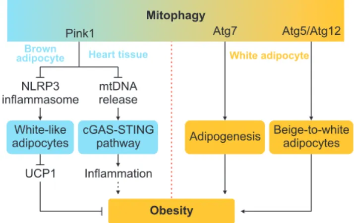
Knockout of Pink1 reverses BAT dysfunction found in the nlrp3-/- mice [50]. Autophagy-mediated mitochon- drial clearance, mediated by autophagy-related genes (atg), is also found to mediate beige-to-white adipocytes transition [51,52]. Adipocyte-specific depletion of atg5 or atg12 promotes beiging of white adipocytes, and pro- tects from DIO and IR [51]. Accordingly, adipocyte-spe- cific ablation of atg7 protects mice from HFD-induced obesity. Although atg7 deficiency induces defective au- tophagy, along with mitophagy, higher mitochondrial content and increased β-oxidation occur in adipose tis- sue. Inactivation of autophagy by atg7 ablation alters adipogenesis, leading to the production of adipose tis- sue with lots of anti-obesity and anti-diabetes features (Fig. 1) [53]. These contrasting results could be due to the distinct role of mitophagy in different adipocytes (brown versus white adipocytes). Distinguishing down- stream signaling pathways of those mitophagy compo- nent may also contribute to these inconsistent results.
Further investigations are needed to understand the role of mitophagy in obesity thoroughly.
It is well known that pattern recognition receptors (PRR) play an important role in mediating inflamma- tion and modulating obesity. PRR activation in brown adipocytes induces inflammation and significantly downregulates UCP1 expression and mitochondrial respiration [54]. Knockout of Toll-like receptor 4 (TLR4), a prominent family of PRR, increases body weight and decreases body temperature, while improves glucose metabolism in HFD feeding mice. TLR4 deficiency inhibits HFD-induced mitochondrial translocation of nuclear factor of activated T-cells 2 (NFATC2) in adipocytes, which reduces IL-1β expression and mito- chondrial oxidative stress leading to decreased adipose inflammation and ultimately ameliorated IR [55].
Taken together, HFD-induced obesity leads to mito- chondrial dysfunction in adipocytes, triggering innate immune response and regulating energy consumption, ultimately causing systematic inflammation and IR.
CROSS-REGULATION OF MITOCHONDRIAL FUNCTION BETWEEN MACROPHAGES AND ADIPOCYTES
As mitochondrial dysfunctions in both macrophages
Mitophagy
Pink1 Atg7 Atg5/Atg12
Adipogenesis Beige-to-white adipocytes Heart tissue
NLRP3 inflammasome
mtDNA release Brown
adipocyte
White-like adipocytes
cGAS-STING pathway UCP1 Inflammation
White adipocyte
Obesity
Fig. 1. The Yin-yang roles of mitophagy in obesity. PTEN-induced puta- tive kinase 1 (Pink1) deficiency activates nod-like receptor family pyrin domain containing 3 (NLRP3) inflammasome, leading to decreased uncoupling protein 1 (UCP1) expression and diminishes energy expen- diture. Pink1 deficiency also promotes mtDNA release and subsequent activation of the cyclic GMP-AMP synthase (cGAS)-STING pathway in heart tissue, leading to exacerbated systematic inflammation, which may ultimately contribute to obesity. Conversely, ablation of Atg5/
Atg12 inhibits mitophagy and restrains beige-to-white fat transition, revealing a negative role of mitophagy in energy consumption. Atg7 deficiency alleviates obesity by inhibiting adipogenesis.
and adipocytes are involved in obesity, their connection deserves some special attention. Adiponectin, which is well-known to activate AMPK, shifts macrophage po- larization from M1 to M2 and then suppresses adipose tissue inflammation [47]. Adipose-specific ablation of FUNDC1, a newly characterized mitophagy receptor in maintaining a healthy mitochondria pool, displays impaired mitophagy and decreases ATP levels accompa- nied by elevated total ROS and mtROS in white adipose tissue (WAT). FUNDC1 deficiency also facilitates M1 po- larization of ATMs via activating MAPK signaling and pro-inflammatory responses as obesity develops [56].
Activation of TLR4 and NLRP3 inflammasome induces IL-1β production in macrophages, which has been shown to attenuate thermogenesis by stimulating oxidative stress and decreasing mitochondrial mem- brane potential in adipocytes [57]. By activating p53 signaling, nitric oxide produced by M1 macrophages suppresses peroxisome proliferator-activated receptor γ (PPARγ) coactivator 1α (PGC-1α) to decrease mito- chondrial biogenesis in preadipocytes, which interrupts preadipocytes differentiate to mature adipocytes [58].
The expression of IL-25, also named IL-17E, is reduced in serum, liver and WAT in DIO animal model. The administration of exogenous IL-25 could stimulate M2 macrophages polarization and thereby improve the mitochondrial respiratory capacity in adipocytes as indicated by the increased expression of CPT1α, NAD+/ NADH ratio, ATP production and oxygen consumption rate. IL-25-educated macrophages also promotes lipoly- sis and inhibits lipogenesis in adipocytes to prevent from obesity [59].
Thus, obesity may induce mitochondrial dysfunction to decrease adiponectin production in adipocytes, caus- ing decreased M2 macrophages and increased adipose tissue inflammation. In turn, activated macrophages interrupt mitochondrial function in adipocytes to in- fluence obesity process.
DIABETES DRUGS TARGETING MITOCHONDRIAL FUNCTION
Metformin, a well-known diabetes drug, lowers gly- cemia level by reducing hepatic glucose production and improving glucose intake and utilization in the periph- eral tissues [60]. Emerging evidences support that met- formin exhibits immune-modulatory features, such as macrophage polarization [61]. For instance, metformin
could inhibit LPS-induced pro-IL-1β production in bone marrow-derived macrophage via suppressing mitochon- drial complex I and ROS production [62,63]. Rosigli- tazone, a PPARγ agonist to improve insulin sensitivity and glycemia [64], could promote mitochondrial biogen- esis, expression of mitochondrial protein in adipocytes to increase plasma adiponectin level [46]. Resveratrol (RSV), a polyphenolic phytoalexin, has many effects on metabolic diseases, such as obesity and diabetes. RSV could improve IR in HFD feeding rats, which may be linked with increased mitochondrial activity of brown adipocytes, but not white adipocytes [65]. In addition, RSV and metformin inhibits DRP1 activation via acti- vating AMPK, and then prevent mitochondrial fission in adipocytes [66]. TM5411, a novel orally active plas- minogen activator inhibitor-1 inhibitor, prevents HFD- induced body weight gain and IR. Of note, TM5411 re- stores HFD-induced downregulation of genes involved in mitochondrial biogenesis and function in adipocytes, indicating that it could maintain mitochondrial fit- ness to prevent obesity and obesity-related metabolic disorders [67]. These results show that improving mi- tochondrial functions is able to improve obesity and its related complications. Further investigations are needed to elucidate the detailed mechanisms by which mitochondrial dysfunction in innate immunity pro- motes obesity-related comorbidities, which may provide more efficient and precise treatment to choose.
CONCLUSIONS
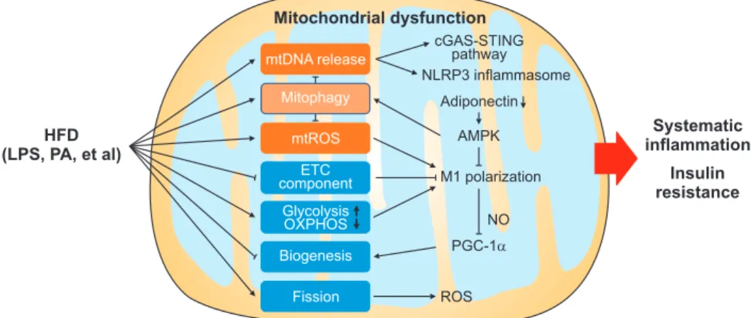
In this review, we have discussed how mitochondrial dysfunction contributes to obesity-related inflamma- tion and IR, focusing mainly on macrophages and adi- pocytes. HFD-induced DAMP and PAMP such as PA and LPS lead to excessive mtROS production in mac- rophages and subsequent pro-inflammatory immune responses and M1 macrophage polarization in adipose tissue. Mitochondrial fission, induced by HFD, also enhances ROS levels. Downregulated ETC components and altered energy support (upregulated glycolysis and downregulated OXPHOS) promote M1 polariza- tion, which disrupts mitochondrial biogenesis through inhibiting PGC-1α. Moreover, adiponectin and down- stream AMPK activation, which could suppress M1 po- larization but promote mitophagy, are both suppressed by obesity. Summarily, these HFD-induced macrophage and adipocyte mitochondrial alternations all contribute
to systematic inflammation and IR (Fig. 2). However, the effect of HFD on mitophagy remains to be fur- ther determined given that HFD feeding have been found to either promotes mitophagy via upregulating PINK1 expression [48] or suppressing mitophagy by downregulation of adiponectin expression [25,47] (Fig.
2). It is demonstrated that Parkin and PINK1 increase by about two-fold after 12 weeks of HFD [68]. By con- trast, adiponectin levels in mice are not significantly decreased until HFD feeding for 20 weeks [69]. Herein, it is possible that at the early stage of HFD feeding, increased mitophagy plays a major role in reducing mtDNA release and mtROS production to maintain ho- meostasis, while mitophagy is suppressed by decreased adiponectin at a late period of HFD feeding leading to metabolic imbalance.
Although much progress has been made on the un- derstanding of the regulation and roles of mitochondri- al function in macrophages and adipocytes in obesity, many questions remain to be explored. For example, it remains to be determined on whether the mitochon- drial-facilitated cross-talk between macrophages and adipocytes also exists in other metabolic tissues such as the intestine, liver, and heart. It is also unclear wheth- er and how altering mitochondrial function in either macrophages and adipocytes modulate their interac- tion with other tissue resident cells especially immune
cells. More studies would also be needed to verify the alternation of mitochondrial dysfunction in metabolic diseases in humans.
ACKNOWLEDGEMENTS
This work was supported by grants from the Na- tional Nature Science Foundation of China (Grant No. 81600671), and grants from the Nature Science Foundation of Hunan Province, China (Grant No.
2019JJ50867).
Conflict of Interest
The authors have nothing to disclose.
Author Contribution
Writing – original draft: LW. Writing – review & editing: all authors.
REFERENCES
1. World Health Organization. Obesity and overweight [Inter- net]. Geneva: World Health Organization; c2020 [cited 2020 Sep 15]. Available from: https://www.who.int/en/news-room/
fact-sheets/detail/obesity-and-overweight.
Mitochondrial dysfunction
HFD (LPS, PA, et al)
Systematic inflammation
Insulin resistance cGAS-STING
pathway NLRP3 inflammasome
Adiponectin
AMPK
M1 polarization
NO PGC-1 ROS mtDNA release
mtROS componentETC
Glycolysis OXPHOS Biogenesis
Fission Mitophagy
Fig. 2. Schematic diagram showing diet-induced mitochondrial dysfunction in obesity-related inflammation and insulin resistance. High-fat diet (HFD) could induce mitochondrial dysfunction via diet-derived lipopolysaccharides (LPS) and palmitic acid (PA). Subsequently, released mtDNA in the cytosol activates the cyclic GMP-AMP synthase (cGAS)-STING pathway and NLRP3 inflammasome, then triggering inflammatory response.
In addition, downregulated electron transport chain (ETC) component and the altered ratio of glycolysis to oxidative phosphorylation (OXPHOS) cause M1 polarization, then produce nitric oxide (NO) to prevent mitochondrial biogenesis by diminishing proliferator-activated receptor γ coact- ivator 1α (PGC-1α). Mitophagy is activated in obese individual. Of note, mitochondrial dysfunction inhibits adiponectin production, resulting in reduced AMP-activated protein kinase (AMPK) activation and consequently attenuated mitophagy and increased M1 polarization. HFD-induced fission enhances reactive oxygen species (ROS) level. All these mitochondrial dysfunctions contribute to obesity-related inflammation and insulin resistance.
2. Petrakis D, Margină D, Tsarouhas K, Tekos F, Stan M, Niki- tovic D, et al. Obesity - a risk factor for increased COVID-19 prevalence, severity and lethality (review). Mol Med Rep 2020;22:9-19.
3. Zhou Y, Chi J, Lv W, Wang Y. Obesity and diabetes as high- risk factors for severe coronavirus disease 2019 (Covid-19).
Diabetes Metab Res Rev 2020. doi: 10.1002/dmrr.3377 [Epub].
4. Prasad M, Chen EW, Toh SA, Gascoigne NRJ. Autoimmune responses and inflammation in type 2 diabetes. J Leukoc Biol 2020;107:739-48.
5. Lee YS, Wollam J, Olefsky JM. An integrated view of immu- nometabolism. Cell 2018;172:22-40.
6. Catrysse L, van Loo G. Adipose tissue macrophages and their polarization in health and obesity. Cell Immunol 2018;330:114-9.
7. Bournat JC, Brown CW. Mitochondrial dysfunction in obe- sity. Curr Opin Endocrinol Diabetes Obes 2010;17:446-52.
8. Khan MSH, Hegde V. Obesity and diabetes mediated chronic inflammation: a potential biomarker in Alzheimer's disease. J Pers Med 2020;10:42.
9. Liu R, Jin P, Yu L, Wang Y, Han L, Shi T, et al. Impaired mi- tochondrial dynamics and bioenergetics in diabetic skeletal muscle. PLoS One 2014;9:e92810.
10. Prasun P. Mitochondrial dysfunction in metabolic syndrome.
Biochim Biophys Acta Mol Basis Dis 2020;1866:165838.
11. Zhang P, Li T, Wu X, Nice EC, Huang C, Zhang Y. Oxidative stress and diabetes: antioxidative strategies. Front Med 2020.
doi: 10.1007/s11684-019-0729-1 [Epub].
12. Chen L, Na R, Gu M, Salmon AB, Liu Y, Liang H, et al. Re- duction of mitochondrial H2O2 by overexpressing perox- iredoxin 3 improves glucose tolerance in mice. Aging Cell 2008;7:866-78.
13. Netea MG, Balkwill F, Chonchol M, Cominelli F, Donath MY, Giamarellos-Bourboulis EJ, et al. A guiding map for inflam- mation. Nat Immunol 2017;18:826-31.
14. Kratz M, Coats BR, Hisert KB, Hagman D, Mutskov V, Peris E, et al. Metabolic dysfunction drives a mechanistically distinct proinflammatory phenotype in adipose tissue macrophages.
Cell Metab 2014;20:614-25.
15. Coats BR, Schoenfelt KQ, Barbosa-Lorenzi VC, Peris E, Cui C, Hoffman A, et al. Metabolically activated adipose tissue macrophages perform detrimental and beneficial functions during diet-induced obesity. Cell Rep 2017;20:3149-61.
16. Tiwari P, Blank A, Cui C, Schoenfelt KQ, Zhou G, Xu Y, et al.
Metabolically activated adipose tissue macrophages link obe- sity to triple-negative breast cancer. J Exp Med 2019;216:1345- 58.
17. Dalmas E, Clément K, Guerre-Millo M. Defining macrophage
phenotype and function in adipose tissue. Trends Immunol 2011;32:307-14.
18. Boscá L, González-Ramos S, Prieto P, Fernández-Velasco M, Mojena M, Martín-Sanz P, et al. Metabolic signatures linked to macrophage polarization: from glucose metabolism to oxi- dative phosphorylation. Biochem Soc Trans 2015;43:740-4.
19. O'Neill LA, Pearce EJ. Immunometabolism governs dendritic cell and macrophage function. J Exp Med 2016;213:15-23.
20. Mouton AJ, Li X, Hall ME, Hall JE. Obesity, hypertension, and cardiac dysfunction: novel roles of immunometabo- lism in macrophage activation and inflammation. Circ Res 2020;126:789-806.
21. Jung SB, Choi MJ, Ryu D, Yi HS, Lee SE, Chang JY, et al. Re- duced oxidative capacity in macrophages results in systemic insulin resistance. Nat Commun 2018;9:1551.
22. Kang YE, Kim HJ, Shong M. Regulation of systemic glucose homeostasis by T helper type 2 cytokines. Diabetes Metab J 2019;43:549-59.
23. Xu J, Chi F, Guo T, Punj V, Lee WN, French SW, et al.
NOTCH reprograms mitochondrial metabolism for proinflammatory macrophage activation. J Clin Invest 2015;125:1579-90.
24. Pan J, Ou Z, Cai C, Li P, Gong J, Ruan XZ, et al. Fatty acid activates NLRP3 inflammasomes in mouse Kupffer cells through mitochondrial DNA release. Cell Immunol 2018;332:111-20.
25. Wen H, Gris D, Lei Y, Jha S, Zhang L, Huang MT, et al. Fatty acid-induced NLRP3-ASC inflammasome activation inter- feres with insulin signaling. Nat Immunol 2011;12:408-15.
26. Zezina E, Snodgrass RG, Schreiber Y, Zukunft S, Schürmann C, Heringdorf DMZ, et al. Mitochondrial fragmentation in human macrophages attenuates palmitate-induced inflam- matory responses. Biochim Biophys Acta Mol Cell Biol Lipids 2018;1863:433-46.
27. Jheng HF, Tsai PJ, Guo SM, Kuo LH, Chang CS, Su IJ, et al.
Mitochondrial fission contributes to mitochondrial dysfunc- tion and insulin resistance in skeletal muscle. Mol Cell Biol 2012;32:309-19.
28. Lahera V, de Las Heras N, López-Farré A, Manucha W, Ferder L. Role of mitochondrial dysfunction in hypertension and obesity. Curr Hypertens Rep 2017;19:11.
29. Kabra UD, Pfuhlmann K, Migliorini A, Keipert S, Lamp D, Korsgren O, et al. Direct substrate delivery into mitochondri- al fission-deficient pancreatic islets rescues insulin secretion.
Diabetes 2017;66:1247-57.
30. Kulkarni SS, Joffraud M, Boutant M, Ratajczak J, Gao AW, Maclachlan C, et al. Mfn1 deficiency in the liver protects against diet-induced insulin resistance and enhances the hy-
poglycemic effect of metformin. Diabetes 2016;65:3552-60.
31. Chiurazzi M, Di Maro M, Cozzolino M, Colantuoni A. Mito- chondrial dynamics and microglia as new targets in metabo- lism regulation. Int J Mol Sci 2020;21:3450.
32. Kim JD, Yoon NA, Jin S, Diano S. Microglial UCP2 mediates inflammation and obesity induced by high-fat feeding. Cell Metab 2019;30:952-62.e5.
33. O'Connell J, Lynch L, Cawood TJ, Kwasnik A, Nolan N, Geoghegan J, et al. The relationship of omental and subcuta- neous adipocyte size to metabolic disease in severe obesity.
PLoS One 2010;5:e9997.
34. Guilherme A, Virbasius JV, Puri V, Czech MP. Adipocyte dysfunctions linking obesity to insulin resistance and type 2 diabetes. Nat Rev Mol Cell Biol 2008;9:367-77.
35. Demine S, Renard P, Arnould T. Mitochondrial uncoupling:
a key controller of biological processes in physiology and dis- eases. Cells 2019;8:795.
36. Heinonen S, Buzkova J, Muniandy M, Kaksonen R, Ollikain- en M, Ismail K, et al. Impaired mitochondrial biogenesis in adipose tissue in acquired obesity. Diabetes 2015;64:3135-45.
37. Zhou Z, Moore TM, Drew BG, Ribas V, Wanagat J, Civelek M, et al. Estrogen receptor α controls metabolism in white and brown adipocytes by regulating Polg1 and mitochondrial remodeling. Sci Transl Med 2020;12:eaax8096.
38. Ablasser A, Chen ZJ. cGAS in action: expanding roles in im- munity and inflammation. Science 2019;363:eaat8657.
39. Bai J, Cervantes C, Liu J, He S, Zhou H, Zhang B, et al.
DsbA-L prevents obesity-induced inflammation and insu- lin resistance by suppressing the mtDNA release-activated cGAS-cGAMP-STING pathway. Proc Natl Acad Sci U S A 2017;114:12196-201.
40. Bai J, Liu F. The cGAS-cGAMP-STING pathway: a mo- lecular link between immunity and metabolism. Diabetes 2019;68:1099-108.
41. Bai J, Cervantes C, He S, He J, Plasko GR, Wen J, et al. Mi- tochondrial stress-activated cGAS-STING pathway inhibits thermogenic program and contributes to overnutrition- induced obesity in mice. Commun Biol 2020;3:257.
42. Wang ZV, Scherer PE. Adiponectin, the past two decades. J Mol Cell Biol 2016;8:93-100.
43. Ruan H, Dong LQ. Adiponectin signaling and function in in- sulin target tissues. J Mol Cell Biol 2016;8:101-9.
44. Luo Y, Liu M. Adiponectin: a versatile player of innate immu- nity. J Mol Cell Biol 2016;8:120-8.
45. Hoffstedt J, Arvidsson E, Sjölin E, Wåhlén K, Arner P. Adi- pose tissue adiponectin production and adiponectin serum concentration in human obesity and insulin resistance. J Clin Endocrinol Metab 2004;89:1391-6.
46. Koh EH, Park JY, Park HS, Jeon MJ, Ryu JW, Kim M, et al. Es- sential role of mitochondrial function in adiponectin synthe- sis in adipocytes. Diabetes 2007;56:2973-81.
47. Woo CY, Jang JE, Lee SE, Koh EH, Lee KU. Mitochondrial dysfunction in adipocytes as a primary cause of adipose tissue inflammation. Diabetes Metab J 2019;43:247-56.
48. Cui C, Chen S, Qiao J, Qing L, Wang L, He T, et al. PINK1- Parkin alleviates metabolic stress induced by obesity in adi- pose tissue and in 3T3-L1 preadipocytes. Biochem Biophys Res Commun 2018;498:445-52.
49. Sliter DA, Martinez J, Hao L, Chen X, Sun N, Fischer TD, et al. Parkin and PINK1 mitigate STING-induced inflammation.
Nature 2018;561:258-62.
50. Ko MS, Yun JY, Baek IJ, Jang JE, Hwang JJ, Lee SE, et al. Mitophagy deficiency increases NLRP3 to induce brown fat dysfunction in mice. Autophagy 2020. doi:
10.1080/15548627.2020.1753002 [Epub].
51. Altshuler-Keylin S, Shinoda K, Hasegawa Y, Ikeda K, Hong H, Kang Q, et al. Beige adipocyte maintenance is regulated by autophagy-induced mitochondrial clearance. Cell Metab 2016;24:402-19.
52. Mizushima N. Autophagy: process and function. Genes Dev 2007;21:2861-73.
53. Zhang Y, Goldman S, Baerga R, Zhao Y, Komatsu M, Jin S.
Adipose-specific deletion of autophagy-related gene 7 (atg7) in mice reveals a role in adipogenesis. Proc Natl Acad Sci U S A 2009;106:19860-5.
54. Bae J, Ricciardi CJ, Esposito D, Komarnytsky S, Hu P, Curry BJ, et al. Activation of pattern recognition receptors in brown adipocytes induces inflammation and suppresses uncoupling protein 1 expression and mitochondrial respiration. Am J Physiol Cell Physiol 2014;306:C918-30.
55. Lin HY, Weng SW, Shen FC, Chang YH, Lian WS, Hsieh CH, et al. Abrogation of toll-like receptor 4 mitigates obesity- induced oxidative stress, proinflammation, and insulin resis- tance through metabolic reprogramming of mitochondria in adipose tissue. Antioxid Redox Signal 2020;33:66-86.
56. Wu H, Wang Y, Li W, Chen H, Du L, Liu D, et al. Deficiency of mitophagy receptor FUNDC1 impairs mitochondrial qual- ity and aggravates dietary-induced obesity and metabolic syndrome. Autophagy 2019;15:1882-98.
57. Okla M, Zaher W, Alfayez M, Chung S. Inhibitory effects of toll-like receptor 4, NLRP3 inflammasome, and interleukin- 1β on white adipocyte browning. Inflammation 2018;41:626- 42.
58. Jang JE, Ko MS, Yun JY, Kim MO, Kim JH, Park HS, et al. Ni- tric oxide produced by macrophages inhibits adipocyte differ- entiation and promotes profibrogenic responses in preadipo-
cytes to induce adipose tissue fibrosis. Diabetes 2016;65:2516- 28.
59. Feng J, Li L, Ou Z, Li Q, Gong B, Zhao Z, et al. IL-25 stimu- lates M2 macrophage polarization and thereby promotes mitochondrial respiratory capacity and lipolysis in adipose tissues against obesity. Cell Mol Immunol 2018;15:493-505.
60. Inzucchi SE, Maggs DG, Spollett GR, Page SL, Rife FS, Wal- ton V, et al. Efficacy and metabolic effects of metformin and troglitazone in type II diabetes mellitus. N Engl J Med 1998;338:867-72.
61. Ursini F, Russo E, Pellino G, D'Angelo S, Chiaravalloti A, De Sarro G, et al. Metformin and autoimmunity: a "new deal" of an old drug. Front Immunol 2018;9:1236.
62. Kelly B, Tannahill GM, Murphy MP, O'Neill LA. Metfor- min inhibits the production of reactive oxygen species from NADH:Ubiquinone oxidoreductase to limit induction of interleukin-1β (IL-1β) and boosts interleukin-10 (IL-10) in li- popolysaccharide (LPS)-activated macrophages. J Biol Chem 2015;290:20348-59.
63. Zatterale F, Longo M, Naderi J, Raciti GA, Desiderio A, Miele C, et al. Chronic adipose tissue inflammation linking obe- sity to insulin resistance and type 2 diabetes. Front Physiol 2020;10:1607.
64. Yki-Järvinen H. Thiazolidinediones. N Engl J Med 2004;351:1106-18.
65. Ku CR, Cho YH, Hong ZY, Lee H, Lee SJ, Hong SS, et al.
The effects of high fat diet and resveratrol on mitochondrial activity of brown adipocytes. Endocrinol Metab (Seoul) 2016;31:328-35.
66. Li A, Zhang S, Li J, Liu K, Huang F, Liu B. Metformin and resveratrol inhibit Drp1-mediated mitochondrial fission and prevent ER stress-associated NLRP3 inflammasome activa- tion in the adipose tissue of diabetic mice. Mol Cell Endocri- nol 2016;434:36-47.
67. Piao L, Jung I, Huh JY, Miyata T, Ha H. A novel plasminogen activator inhibitor-1 inhibitor, TM5441, protects against high- fat diet-induced obesity and adipocyte injury in mice. Br J Pharmacol 2016;173:2622-32.
68. Cummins TD, Holden CR, Sansbury BE, Gibb AA, Shah J, Zafar N, et al. Metabolic remodeling of white adipose tissue in obesity. Am J Physiol Endocrinol Metab 2014;307:E262-77.
69. Kwon EY, Shin SK, Cho YY, Jung UJ, Kim E, Park T, et al.
Time-course microarrays reveal early activation of the im- mune transcriptome and adipokine dysregulation leads to fi- brosis in visceral adipose depots during diet-induced obesity.
BMC Genomics 2012;13:450.