J O U R N A L O F
Veterinary Science
ABSTRACT
3)The ORF5 gene encodes a major envelope glycoprotein (GP5), which is one of the three major proteins of porcine reproductive and respiratory syndrome virus (PRRSV). The GP5 protein has been known to be a 24.5-26kDa N-glycosylated envelope protein. The GP5 is involved in inducing neutralizing antibodies. For this reason, the GP5 is primary can- didate for the PRRSV subunit vaccine. To produce the native form of GP5 in mammalian cells, we have cloned the ORF5 gene from PRRSV CNV-1 into the Semliki Forest virus (SFV)-based expression vector, resulting in recombinant pSFV-ORF5. By the infection with recombinant pSFV-ORF5 to BHK-21 cells, the GP5 expression was confirmed by immunocytochemistry and immunoblotting assay. The recombinant virus particle harboring ORF5 gene was infectious to BHK-21 and MARC-145. The RNA synthesis and expression of GP5 in the infected cell was also confirmed by RT-PCR.
Key words : PRRS virus, ORF5 gene, GP5, Semliki Forest virus-based expression vector, recombinant SFV particle.
Introduction
Porcine reproductive and respiratory syndrome (PRRS) is a disease found in swine farms worldwide and it is characterized by reproductive failures such as late-term abortions in sows and by respiratory illness and high mortality in young pig
1,2,5,6,7,8,10,11. PRRSV are related to those of a group of small, enveloped, positive-strand RNA viruses, including murine lactate dehydrogenase elevating virus (LDV), equine arteritis virus (EAV) and simian haemorrhagic fever virus, which are presently classified
*Corresponding author: Dr. Hyun-Soo Kim
Division of Preventive Medicine, College of Veterinary Medicine, Chungnam National University, Daejeon 305-764, Korea.
This work was supported by National Research Laboratory (NRL) Program Grant (2000-N-NL-01-C-171) from the Ministry of Science and Technology, Korea.
within the family Arteriviridae, order Nidovirales
12,13,14,15,16,17. The genome of PRRSV is about 15kb in length and contains eight open reading frames (ORFs) designed ORF1a, ORF1b, and ORFs 2 to 7. According to sequence data, ORFs 1a and 1b represent nearly 75% of the viral genome and code for functional proteins associated with virus replication
14,15,18,19,20. The virion contains three major structure proteins, 24.5- 26kDa enveloped glycoprotein (GP5), 18-19kDa unglycosylated membrane (M) protein and 15kDa nucleocapsid (N) protein, encoded by ORFs 5, 6 and 7, respectively
19,20,27. The GP5 is involved in inducing neutralizing antibodies, antigenic variability, apoptosis and possibly antibody-dependent enhancement phenomena
21,22,23,24,25,26. For this reason GP5 has been thought a primary candidate for the subunit vaccine.
The SFV is a small enveloped alphavirus that contains a 42S RNA genome replicating in the cytoplasm of the infected cells without the involvement of the nucleus.
Infection with SFV results in a suppression of host cell protein synthesis and enhances the production of viral proteins. This expression system is based on a genomic SFV cDNA inserted into an SP6 promoter plasmid and subsequently modified by deletion of the SFV structural genes to allow insertion of a heterologous gene as part of the SFV replicon. The helper RNA encoding the structural proteins of SFV is necessary for assembly and packaging of recombinant RNAs. The major advantages of this system, as comparing with other expression systems, are a broad range of susceptible host cells including those of insect, avian and mammalian origin and the high levels of RNA and protein produced in transfected cells by self- amplifying system
28,29,. In this study, Semliki Forest Virus (SFV)- based expression system to express GP5 in mammalian cells was adopted. We constructed pSFV-ORF5 plasmid vector by cloning ORF5 gene into the SFV replicon. Expression of GP5 and recombinat SFV particle harboring GP5 were demonstrated in the transfected BHK-21. The RNA synthesis and GP5 expression were also identified in the infected BHK-21 and MARC-145 cells with recombinant virus particles.
Expression of Open Reading Frame 5 Protein of Porcine Reproductive
and Respiratory Syndrome Virus Using Semliki Forest Virus Expression System
Hae-Sun Jung, In-Wook Hwang, Su-Mi Kim, Chul-Joong Kim, Kwang-Soon Shin and Hyun-Soo Kim
*College of Veterinary Medicine, Chungnam National University, Daejeon, 305-764, Korea
MATERIALS AND METHODS
Cell and virus culture
The BHK-21 cells and MARC -145 cells were cultured in minimum essential medium (α-MEM)(Sigma, U.S.A.) supplemented with 10% tryptose phosphate broth, 5% fetal bovine serum, 20 mM HEPES, 2 mM glutamine , penicillin (0.1 IU/ml) and streptomycin(0.1 ㎍/ml). The PRRSV CNV-1 was inoculated onto MARC-145 cell monolayers in cell culture plate
3,30. When cytopathic effect was observed on 70% of cell monolayer, the virus culture medium was harvested and stored at -70℃.
Reverse transcription-polymerase chain reaction (RT-PCR) of PRRSV RNA and cloning
The RNA of PRRSV was obtained from the PRRS virus CNV-1 culture supernatant by using viral RNA extraction kit (Qiagen, U.S.A.). The cDNA of the PRRSV ORF5 gene was synthesised by reverse transription (RT) and amplified by PCR. The reverse oligonucleotide primer for ORF5 gene(5'- TTCCCGGGCTAAGGACGACCCCATTG -3') was used for priming RT reaction. The Sma I restriction site was incorporated at 5' end of primer. 10 ㎕ of RT mixture consisting of 3㎕ of RNA template, 2㎕ of 5X buffer, 3㎕ of dNTP mixture(Roche, Germany), 1㎕ of reverse primer, 1㎕
of RNase inhibitor (Takara, Japan) and 1㎕ of Molony murine leukemia virus (M-MLV) reverse transcriptase (Promega, U.S.A.) was prepared in PCR tubes. After heat denaturation at 70℃ for 5min, RT was performed for 60min at 42℃ in a DNA thermal cycler (Perkin-Elmer, U.S.A.). For the PCR, the forward primer for ORF5 gene (5'- TTGGATTCCATGTTGGAGA AATGCTTG -3') and reverse primer were used. The forward primer specific for ORF5 gene contained BamH I restriction site at 5' end. After heat denaturation at 98℃ for 10 min, 88㎕ of PCR mixture consisting of 66㎕ of DEPC water, 10㎕ of 10X PCR buffer with MgCl
2, 4㎕ of forward primer, 4㎕ of reverse primer, 2
㎕ of dNTP mixture, and 2㎕ Taq polymerase (Roche, Germany) were added to PCR mixture. The PCR condition contained 40 cycles (composed of 1 min at 94℃ for denaturation, 1 min at 50℃ for annealing, and 1 min at 7 2℃ for extension) and l cycle (composed of 10 min ) at 72℃
for extension. The reaction was carried out in GeneAmp PCR system 2400 (Perkin Elmer, U.S.A.). The PCR product was cloned into the pSFV-1 plasmid which was digested with BamHⅠand Sma I and treated with calf intestine alkaline phosphatase (Promega, U.S.A) for 1hr at 37℃. The resulting recombinant pSFV plasmid was designated as pSFV-ORF5 and put into E. coli DH5α.
RNA synthesis and transfection of mammalian cells Recombinant pSFV-ORF5 plasmid and pSFV-helper were linearized with Spe I and capped RNA was synthesized in vitro at 37℃ for 1 hr in a total volume of 50㎕ containing 40 mM Tris-HCl(pH7.5), 6 mM MgCl
2, 2 mM spermidine, 5
mM DTT, 1 mM each of ATP, CTP and UTP, 0.5 mM GTP, 1 mM Cap analogue m7G (5')PPP(5')G, 40 units RNase inhibitor, and 50units SP6 RNA polymerase. Baby hamster kidney-21 (BHK-21) cells were trypsinized and washed twice with PBS and resuspended in PBS at concentration of 1×
10
7/ml. Cell suspension was mixed with 100㎕ of RNA transcripts (50㎕ of pSFV-helper RNA transcripts and 50㎕
of pSFV-ORF5 RNA transcripts). The mixture was used to electroporate 1×10
7cells by using Gene PulserⅡ(BioRad, U.S.A.). The cells were pulsed twice at 830v / 25μF at room temperature. The transfected cells were resuspended in 12 ml of culture medium(α-MEM) supplemented with 10%
tryptose phosphate broth, 5% fetal bovine serum, 20 mM HEPES, 2 mM glutamine, penicillin (0.1IU/ml) and streptomycin(0.1 ㎍/ml) and plated onto 100mm tissue culture dishes(Nunc, Denmark). The cells were incubated for 2 days in an atmosphere of 5% CO
2at 37℃. The culture fluid was harvested and clarified by centrifugation for 15 min at 3000 ×g. The supernatant was transferred to a fresh ultracentrifuge tube and centrifuged at 35,000 ×g (SW41 Ti rotor; Beckman, U.S.A.) for 3 hr to collect virus particles.
The virus pellet was resuspended in TNE buffer (50 mM Tris-HCl [pH7.4], 100 mM NaCl and 0.5 mM EDTA) and stored quickly in aliquots at -70℃.
Infection with recombinant virus particles
BHK-21 cells were seeded onto 24-well tissue culture plate and tissue culture dishes. Prior to infection, the cells were washed twice with PBS. The frozen recombinant virus particles were thawed quickly at room temperature. The virus particles were treated with 500 ㎍/ml chymotrypsin (Roche, Germany) and 0.5 mM CaCl
2,and then incubated on ice for 30 min. To inactivate chymotrypsin activity, 0.5 volume of aprotinin (2 ㎎/ml, Sigma, U.S.A.) was added. The activated vrus particles were diluted in MEM containing 1%
FBS and inoculated into BHK-21 cells. After 90 min incubation at 37℃ in an atmosphere of 5% CO
2, the inoculum was removed and fresh medium was added. At 24 hr postinfection, cells were washed three times with PBS and lysed with cell lysis buffer (1% NP-40, 50 mM Tris-HCl [pH7.6], 150 mM NaCl, 2 mM EDTA, 1 ㎍/ml PMSF) on ice for 1 hr. The cell lysates were centrifuged at 12,000×g at 4℃ for 5 min to remove insoluble materials and used as samples for sodium dodecyl sulfate-polyacrlyamide gel electrophoresis (SDS-PAGE) and RT-PCR.
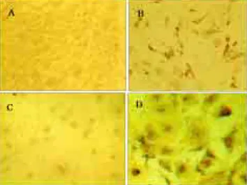
Immunocytochemistry
The cell monolayers were rinsed twice with PBS and fixed with ice-cold methanol at 4℃ for 15 min. The fixed cell monolayers were washed three times with PBS and incubated at room temperature for 1 hr with PBS containing 1%
gelatin to block nonspecific bindings. The PRRSV antibody
positive pig sera (1:500 dilution) were added and incubated at
room temperature for 3 hr. The cell monolayers were
washed with PBS three times and then treated with rabbit
anti-pig IgG conjugated with peroxidase (Sigma, U.S.A) at room temperature for 1 hr. The cells were finally washed with PBS and visualized by adding H
2O
2solution containing DAB (3,3'- diaminobenzidine), (Vector, U.S.A.).
Western immunoblotting
The SDS-PAGE was performed by the method of Laemmli
4,31. Cell lysates were mixed with SDS-PAGE sample buffer (62.5 mM Tris-Cl [pH6.8], 10% glycerol, 2% sodium dodecylsulfate [SDS], 5% 2-mercaptoethanol, 0.05% bromophenol blue) and then boiled at 100℃ for 5 min and chilled on ice. The denatured protein samples were electrophoretically separated on 12% polya- crylamide SDS- gels and transferred onto polyvinylidine difluoride (PVDF) membrane (Roche, Germany). The membrane was blocked for 1 hr with blocking buffer (50 mM Tris-Cl, 5%
skimmed milk, pH8.0). The blot was incubated for overnight at room temperature with PRRSV antibody positive pig sera(1:250 dilution) in shaking chamber. After washing three times in washing buffer (20 mM Tris-HCl, 150 mM NaCl, 0.05% Tween 20), the blot was treated with rabbit anti-pig IgG conjugated with peroxidase (Sigma, U.S.A.) at room temperature for 2 hr.
After rinsing blot in washing buffer, the specific protein band was visualized using DAB solution. The reaction was stopped by immersing the membrane in distilled water.
RT-PCR from infected cell with recombinant virus particles
The RNA from recombinant virus particles was obtained from cell culture media by using RNA extraction kit (Qiagen, U.S.A.). The cDNA synthesis of ORF5 gene and its amplification by RT-PCR were conducted by the method as described previously
30.
RESULTS
Construction of pSFV-ORF5
To express PRRSV GP5 in mammalian cells, we used the SFV-based expression system. Genomic RNAs of the PRRSV CNV-1 were extracted from virus culture media. The ORF5 gene was amplified by RT-PCR and cloned into the pSFV-1 vector. This cloning step resulted in pSFV-ORF5. The schematic diagram of pSFV-ORF5 and SFV- helper were illustrated in Fig.1.
Expression of the GP5 of PRRSV
The SFV expression system is provided with a packaging-deficient helper plasmid, pSFV-helper, which produces the structural proteins of SFV. When these proteins are produced in the same cells as RNA from pSFV 1-derived plasmids, the recombinant RNA is packaged into SFV particles that can be used for large scale infection of various mammalian cells.
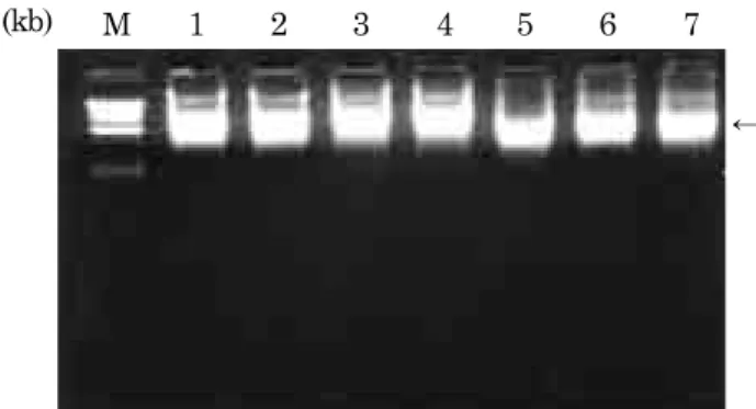
The capped RNAs (Fig.2) transcribed in vitro from the SP6 promoter on the linearized pSFV-ORF5 and pSFV- helper were cotransfeced into BHK-21 cells by electroporation.
Sacll 2327 EcoRV 278
pSp6 nsP1
nsP2
2Ss nsP4 nsP3 ORF5
pSFV-ORF5 Spel 8858
Smal 8003 Sacll 7423
BamHI 7400
Sacll 5055
Sphl 7760 Xbal 547
Sp6nsP1:nsP4
C
p62 6K
Spel 5425 E1
Ori Ap
pSFV-helper