28
Introduction
A coronary artery fistula (CAF) is an abnormal communica- tion between one of the coronary arteries and a cardiac cham- ber or vessels, including the coronary sinus, pulmonary artery, or superior vena cava.1-3) CAF is involved in 0.002% of general population and accounts for 0.4% of all cardiac malforma- tion.4) CAF mostly drains into the venous structures of circula- tion, such as the right-sided chambers, pulmonary artery, cor- onary sinus and superior vena cava, but drainage into the left- sided chambers is less frequent.5) We present a case of CAF which is originated from left coronary artery and drained into left ventricle.
Case
A 48-year-old male presented with abdominal pain that had lasted for 2 months. He had no cardiovascular risk factor, and no cardiovascular symptom such as chest pain or dyspnea.
He was diagnosed as gallbladder stone and admitted to our hospital for laparoscopic cholecystectomy. Preoperative elec- trocardiography was within normal limit, and chest X-ray showed no pathologic abnormality. He was consulted to car- diovascular department because surgeon heard a continuous cardiac murmur and ordered echocardiography for a preopera- tive evaluation. The echocardiography showed normal ejec- tion fraction (64%). The mitral inflow E/A ratio was 1.63,
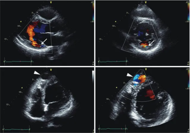
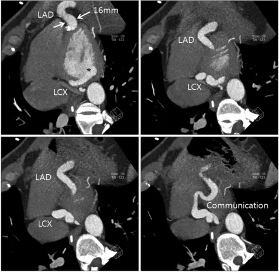
and E/E’ ratio was 12.0. The valvular morphology and func- tion was normal. However, there was abnormal color flow within left ventricle predominantly during diastole (Fig. 1, Supplementary movie 1-4). It was originated from basal pos- terior wall and drained into the left ventricular cavity. The maximal velocity of blood flow draining into left ventricle was approximately 3.0 m/s. In short-axis great arterial view, left coronary artery appeared dilated. Multiple tortuous dilated vascular structures with internal blood flows disclosed by color Doppler image were also found around left ventricular myo- cardium. Especially, dilated large echo-free vascular structure was detected in the apical area. The connection between those dilated vascular structures and left coronary artery was sus- pected, but clear visualization of the connection was limited in 2-dimensional echocardiography. Coronary artery comput- ed tomography (CT) scan was performed to confirm the pathologic anatomy of these abnormal findings. Coronary CT revealed markedly dilated (up to 16 mm) and serpentine whole left coronary arteries (Fig. 2). The left anterior descending ar- tery was communicating with left circumflex artery at the api- cal posterior epicardium, and was directly connected to the basal posterior side of left ventricular cavity (Fig. 3). He was finally diagnosed as having CAF.
Since the patient had no cardiovascular symptom, he under- went laparoscopic cholecystectomy without specific treatment
pISSN 1975-4612/ eISSN 2005-9655 Copyright © 2014 Korean Society of Echocardiography www.kse-jcu.org http://dx.doi.org/10.4250/jcu.2014.22.1.28
CASE REPORT J Cardiovasc Ultrasound 2014;22(1):28-31
Coronary Artery Fistula Draining into the Left Ventricle
Jihyun Sohn, MD, Jong-Min Song, MD, PhD, Jeong Yoon Jang, MD, Byung Joo Sun, MD, Dae-Hee Kim, MD, PhD, Duk-Hyun Kang, MD, PhD, and Jae-Kwan Song, MD, PhD
Division of Cardiology, Asan Medical Center, University of Ulsan College of Medicine, Seoul, Korea
We present a case of 48-year-old male who presented with coronary artery fistula draining into left ventricle. Transthoracic echocardiography showed abnormal blood flow draining into left ventricle, with enlarged coronary arteries and multiple vascular structures around ventricular myocardium. Coronary computed tomography revealed dilatation of entire left coronary artery which was wrapping around left ventricle, and draining into the posterior side of left ventricle. He did not undergo any invasive treatment, because he was not symptomatic.
KEY WORDS: Coronary artery fistula · Echocardiography · Computed tomography.
• Received: July 19, 2013 • Revised: January 10, 2014 • Accepted: February 18, 2014
• Address for Correspondence: Jong-Min Song, Division of Cardiology, Asan Medical Center, University of Ulsan College of Medicine, 88 Olympic-ro 43-gil, Songpa-gu, Seoul 138-736, Korea Tel: +82-2-3010-3168, Fax: +82-2-486-5918, E-mail: [email protected]
• This is an Open Access article distributed under the terms of the Creative Commons Attribution Non-Commercial License (http://creativecommons.org/licenses/by-nc/3.0) which permits unrestricted non-commercial use, distribution, and reproduction in any medium, provided the original work is properly cited.
Coronary Cameral Fistula | Jihyun Sohn, et al.
29 Fig. 1. Abnormal flow draining into the basal posterior portion of left ventricle (arrows) and dilated large echo-free vascular structure (arrowheads), revealed by transthoracic echocardiography and color Doppler images.
Fig. 2. Coronary computed tomography showed markedly dilated and serpentine whole left coronary arteries. LAD: left anterior descending artery, LCX: left circumflex artery.
Journal of Cardiovascular Ultrasound 22 | March 2014
30
of CAF. No clinical events occurred during his admission. The patient visited outpatient clinic a month after discharge with- out clinical events or symptoms.
Discussion
CAF communicating with the cardiac chambers are called coronary cameral fistula, as in our case. There have been many cases of coronary artery fistula. However, the incidence of coro- nary cameral fistula is very low and coronary cameral fistula in- volving left ventricle has been very rarely reported.4)5) This case is also unique because almost whole left coronary artery was di- lated and looked like cystic lesion in the apical portion, which could be clearly detected by transthoracic Doppler echocar- diography. The majority of coronary cameral fistula (up to 90%) drains into the right-sided chambers of the heart, while only 10% of coronary cameral fistula shows communications with the left-sided chambers or both left and right-sided chambers.6) Doppler echocardiography is a good screening im- aging tool for the diagnosis of CAF, but evaluation of whole anatomy of CAF can be best visualized by coronary CT image.
Clinical presentations generally depend on the hemody-
namic or anatomic significance of the lesion. The natural his- tory of CAF is variable. Patients can be asymptomatic during their entire life. Chest pain, exertional dyspnea and fatigue may develop, but many patients do not suffer from any symp- toms.7) The mechanism of symptoms appeared to be coronary steal phenomenon and diastolic overload.8) The most common complication is myocardial ischemia due to coronary steal phe- nomenon which occurs in 15% of patients with CAF.9) Conges- tive heart failure and arrhythmia could be caused by excessive loading of cardiac chambers. Intravascular thrombosis, infec- tive endocarditis, and rarely hemopericardium due to rupture of aneurysmal coronary artery might be complications.
The best therapy for CAF remains controversial. Surgical closure of CAF may have benefit for the patients with large, hemodynamically significant CAF.10) There were several rea- sons for not doing any surgical interventions for this patient.
First, this pathology was incidentally found without any car- diovascular symptom, and may carry only mild risk in under- going laparoscopic surgery. Therefore, the first thing we had to do for this patient was to let the surgeon not to postpone this necessary surgery because of the cardiac problem. Second,
Fig. 3. Coronary computed tomography revealed dilated left coronary arteries and the drainage site to the left ventricle (asterisk). LAD: left anterior descending artery, LCX: left circumflex artery.
Coronary Cameral Fistula | Jihyun Sohn, et al.
31 good functional status of this patient indicates low possibility
of coronary steal causing myocardial ischemia due to this pa- thology. Lastly, surgical treatment for this coronary cameral fistula was not technically easy, because almost entire portion of the coronary artery was dilated as noticed in coronary CT.
Supplementary movie legends
Movie 1. The parasternal long axis view revealed abnormal color flow within left ventricle predominantly during diastole.
Movie 2. The parasternal short axis view revealed diastole- dominant abnormal color flow draining from posterior left ventricular wall.
Movie 3. Dilated large vascular structure was detected in the apical area.
Movie 4. Abnormal vascular structure was originated from basal posterior wall and drained into the left ventricular cavity.
References
1. Ata Y, Turk T, Bicer M, Yalcin M, Ata F, Yavuz S. Coronary arterio- venous fistulas in the adults: natural history and management strategies. J Cardiothorac Surg 2009;4:62.
2. Fernandes ED, Kadivar H, Hallman GL, Reul GJ, Ott DA, Cooley DA. Congenital malformations of the coronary arteries: the Texas Heart In-
stitute experience. Ann Thorac Surg 1992;54:732-40.
3. Sommer RJ, Hijazi ZM, Rhodes JF Jr. Pathophysiology of congenital heart disease in the adult: part I: Shunt lesions. Circulation 2008;117:
1090-9.
4. Mangukia CV. Coronary artery fistula. Ann Thorac Surg 2012;93:
2084-92.
5. Mohanty SK, Ramanathan KR, Banakal S, Muralidhar K, Kumar P.
An interesting case of coronary cameral fistula. Ann Card Anaesth 2005;8:152-4.
6. Roberts WC. Major anomalies of coronary arterial origin seen in adult- hood. Am Heart J 1986;111:941-63.
7. Vavuranakis M, Bush CA, Boudoulas H. Coronary artery fistulas in adults: incidence, angiographic characteristics, natural history. Cathet Car- diovasc Diagn 1995;35:116-20.
8. Stierle U, Giannitsis E, Sheikhzadeh A, Potratz J. Myocardial isch- emia in generalized coronary artery-left ventricular microfistulae. Int J Car- diol 1998;63:47-52.
9. Valente AM, Lock JE, Gauvreau K, Rodriguez-Huertas E, Joyce C, Armsby L, Bacha EA, Landzberg MJ. Predictors of long-term adverse outcomes in patients with congenital coronary artery fistulae. Circ Cardio- vasc Interv 2010;3:134-9.
10. Schumacher G, Roithmaier A, Lorenz HP, Meisner H, Sauer U, Mül- ler KD, Sebening F, Bühlmeyer K. Congenital coronary artery fistula in infancy and childhood: diagnostic and therapeutic aspects. Thorac Cardio- vasc Surg 1997;45:287-94.