CASE REPORT pISSN 1225-7737/eISSN 2234-8042 http://dx.doi.org/10.12701/yujm.2014.31.1.13
Yeungnam Univ J Med 2014;31(1):13-16YUJM VOLUME 31, NUMBER 1, JUNE 2014 13
좌관상동맥 주간부와 우관상동에서 기원하는 이중 좌전하행동맥
김동휘1, 문건웅1, 김은희1, 우기현1, 신진경1, 장지연2, 하성은2, 이주영3
1가톨릭대학교 의과대학 수원 성빈센트병원 내과학교실; 2가톨릭대학교 의과대학 서울성모병원 내과학교실;
3서울 원자력병원 내과학교실
Dual left anterior descending coronary artery originating from left main stem and right coronary sinus
Dong-Hwi Kim
1, Keon-Woong Moon
1, Eun-Hee Kim
1, Gihyeon Woo
1, Jin-Kyeong Shin
1, Ji-Yeun Jang
2, Sungeun Ha
2, Joo-Young Lee
31
Department of Internal Medicine, St. Vincent’s Hospital, The Catholic University of Korea College of Medicine, Suwon;
2
Department of Internal Medicine, Seoul St. Mary’s Hospital, The Catholic University of Korea College of Medicine, Seoul;
3
Department of Internal Medicine, Korea Cancer Center Hospital, Korea Institute of Radiological & Medical Sciences, Seoul, Korea
Congenital abnormalities of the coronary arteries are found in 0.6% to 1.3% of patients in coronary angio- graphy. Dual left anterior descending coronary artery (LAD) is a rare coronary anomaly and is incidentally detected during coronary angiography. We report a case of a 65-year-old female with a rare coronary anom- aly who was diagnosed with dual LAD via coronary computed tomography and coronary angiography. The imaging studies revealed dual LAD originating from the left main stem and right coronary sinus. These angiographic findings were considered to be consistent with the type IV variety of dual LAD by Spindola- Franco classification. Recognition of dual LAD is important to prevent errors of interpretation of the coronary angiogram and for optimal surgery.
Keywords: Coronary vessel anomalies; Coronary angiography
Received: June 29, 2013; Revised: July 30, 2013;
Accepted: August 5, 2013
Corresponding Author: Keon-Woong Moon, Department of Internal Medicine, St. Vincent’s Hospital, The Catholic Uni- versity of Korea College of Medicine, 93 Jungbu-daero, Paldal-gu, Suwon 442-723, Korea
Tel: +82-31-249-7139, Fax: +82-31-247-7139 E-mail: [email protected]
서 론
좌전하행 관상동맥(left anterior descending artery)은 주행 이나 분포가 다른 혈관에 비해 일정하지만, 이중 좌전하행 관상동맥(dual left anterior descending artery)은 드문 선천 관상동맥 기형으로 알려져 있다. 이중 좌전하행 관상동맥 기형은 4가지 형태로 분류되어 왔으나, 최근 2가지 형태가 더 추가되어 6가지 형태로 분류할 수 있다. 6가지 중 type
IV의 경우 한 분지는 좌관상동맥(left coronary artery) 주간부 (main stem)에서 나머지 분지는 우대동맥굴(right aortic sinus) 또는 우관상동맥(right coronary artery)에서 이중으로 기원 하는 형태이다. Type IV는 매우 드문 형태로 알려져 있으며, 국내는 물론 국외에서도 거의 보고된 바가 없다. 이에 저자들 은 좌관상동맥 주간부와 우관상동(right coronary sinus)에서 기원하는 드문 형태의 이중 좌전하행 관상동맥 1예를 경험하 였기에 보고하는 바이다.
증 례
환 자: 여자, 65세
주 소: 아랫입술의 피부병변
현병력: 타 병원에서 아랫입술의 피부 종괴를 주소로 내원
Dong-Hwi Kim et al.
14 YUJM VOLUME 31, NUMBER 1, JUNE 2014
Fig. 1. Left anterior oblique cranial view of left coronary angio- gram shows a normal left circumflex artery and a short LAD, which terminated after first giving rise to septal and diagonal branches. LAD, left anterior descending artery; LCX, left circum- flex artery.
Fig. 2. Right coronary angiogram shows a normal right coronary artery and a long LAD originating from the right coronary sinus.
LAD, left anterior descending artery; RCA, right coronary artery.
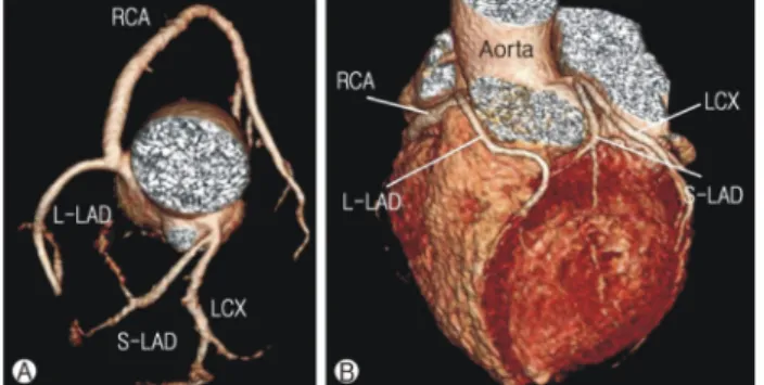
Fig. 3. Contrast enhanced multi-detector cardiac computed tomo- graphy. It showed that a L-LAD is originated from right coronary sinus and a S-LAD is originated from left main stem. (A) The LAD proper and S-LAD form a single very short vessel situated very high in the AIVS. The major septal perforator and diagonal branches are given off by this short vessel. The first portion of the L-LAD is formed by a transverse trunk which courses anteri- orly to the infundibulum of the right ventricle, making a sharp turn to descend on the AIVS (B). L-LAD, long-left anterior descen- ding artery; S-LAD, short-left anterior descending artery; AIVS, anterior interventricular sulcus; RCA, right coronary artery; LCX, left circumflex artery.
하였으며, 피부암이 의심되어 수술 전 심전도 검사에서 이상 소견으로 본과에 협진 의뢰되었다.
과거력: 5년 전 고혈압 진단 이외 특이사항 없음.
사회력: 특이사항 없음.
가족력: 특이사항 없음.
진찰 소견: 의식은 명료하였으며, 혈압 140/80 mmHg, 맥박 68회/분, 호흡 18회/분, 체온 36.5℃였다. 흉부 청진 상 심음 은 규칙적이고, 심잡음, 심낭마찰음 및 수포음은 청진되지 않았으며, 아랫입술의 종괴 이외에 다른 이학적 이상소견은 관찰되지 않았다.
심전도 소견: 심전도에서 정상 동리듬을 보였으며, V5-6에 서 ST 분절 하강이 관찰되었다.
심초음파 소견: 국소 심실벽운동 이상소견은 보이지 않았 고, 심박출량은 65%로 확인되었다.
검사실 소견: 말초혈액 검사 상 백혈구 7,400/mm3, 혈색소 13.1 g/dL, 혈소판 187,000/mm3였고, 혈청 생화학 검사 상 total protein 6.1 g/dL, aspartate transaminase/alanine trans- aminas 31/18 (IU/L), blood urea nitrogen/creatinine 12/0.8 (mg/dL), creatine phosphokinase/lactate dehydrogenase 27/280 (IU/L), creatine kinase MB/troponin-I 0.01/0.01 (ng/mL), sodi- um/potassium/chloride 141/3.7/95 (mmol/L)였다.
방사선 소견: 흉부단순촬영에서 심비대나 폐울혈은 관찰 되지 않았다.
경 과: 추가적인 검사를 위하여 관상동맥 전산화단층촬영 (computed tomography)과 관상동맥 조영술을 시행하였다.
관상동맥 조영술에서 유의한 관상동맥의 협착 소견은 없었 지만, 이중 좌전하행 관상동맥이 있는 것으로 확인되었다.
좌관상동맥 주간부에서 기원하는 하나의 좌전하행 관상동맥
은 짧은 주행을 가졌고, 중격분지(septal branch)를 내고 있었 으며(Fig. 1), 다른 하나는 우관상동맥 기시부(proximal)에서 기원하는 것이 확인되었다(Fig. 2). 우관상동맥이나 좌회선 동맥(left circumflex artery)의 기원과 주행에는 특이 이상소 견은 확인되지 않았다.
관상동맥 전산화단층촬영에서 주된 좌전하행 관상동맥 proper는 아주 짧은 형태를 보였으며, 앞심실사이고랑(ante- rior interventricular sulcus)에 높이 위치하고 있는 것이 확인 되었다. 주(major) 중격분지와 대각분지(diagonal branch)가 단좌전하행 관상동맥(short left anterior descending artery) 으로부터 분지되었다(Fig. 3). 환자는 관상동맥 기형 이외에
Coronary anomaly dual LAD type IV
YUJM VOLUME 31, NUMBER 1, JUNE 2014 15
Table 1. Classification of dual left anterior descending coronary artery
Type S-LAD L-LAD Origin of major
diagonal vessels
Origin Course Origin Course
I Proximal
LAD
Proximal AIVG
Proximal LAD
Epicardial course on the left ventricular side of the proximal AIVG, reentering the distal AIVG
Proximal LAD and/or L-LAD II Proximal
LAD
Proximal AIVG
Proximal LAD
Epicardial course on the right ventricular side of the proximal AIVG, reentering the distal AIVG
Proximal LAD III Proximal
LAD Proximal
AIVG Proximal
LAD Intramyocardial course in the proximal septum, then either emer- ging epicardially in the distal AIVG, or terminating intramyocar- dially as septal perforator arteries
Proximal LAD or S-LAD
IV LMCA Proximal
AIVG RCA Epicardial free wall course anterior to the infundibulum of the right ventricle traversing to the distal AIVG, or intramyocardial course within the septal crest emerging epicardially in the distal AIVG
S-LAD
V LCS Proximal
AIVG
RCS Intramyocardial course within the septal crest emerging epicardially in the distal AIVG
S-LAD
VI LMCA Proximal
AIVG RCA Underneath the RVOT in the area of the interventricular septum S-LAD Proximal LAD: the portion of the LAD just after bifurcation of the left main coronary artery into the LAD and left circumflex.
S-LAD, short-left anterior descending artery; L-LAD, long-left anterior descending artery; AIVG, anterior interventricular groove;
LMCA, left main coronary artery; RCA, right coronary artery; LCS, left coronary sinus; RCS, right coronary sinus; RVOT, right ventricular outflow tract.
는 특이 소견이 없었고, 전신마취 하에 피부암 수술을 시행하 였으며, 수술 후 합병증 없이 퇴원하였다.
고 찰
좌전하행 관상동맥은 정상적으로 좌관상동맥 주간부에서 기원하여 심첨부(apex)를 향하여 앞심실사이고랑을 따라 주 행하면서 좌우심실의 전측벽(anterolateral wall)과 심실 중격 에 분포하게 된다. 관상동맥 기형은 주로 관상동맥 조영술을 시행하며 우연히 발견되는 경우가 많으며, 이중 좌전하행 관상동맥은 드문 선천 관상동맥 기형으로 알려져 있다. 그 동안 발표된 문헌들에 따르면 관상동맥 기형의 발생률은 0.6-1.3%이며, 주로 남성에서 발생하는 것으로 보고되었다 [1,2]. 통상적인 관상동맥 조영술 시 이중 좌전하행 관상동맥 은 Morettin 등[3]과 Spindola-Franco 등[4]이 1% 정도에서 관찰되었다고 보고하였다.
그동안 이중 좌전하행 관상동맥 기형은 4가지 형태로 분류 하였으나[4], 최근 2가지 형태가 추가 보고됨에 따라 6가지의 형태로 나누고 있다(Table 1) [5]. Type I이 가장 흔하며, Spindola-Franco 등[4]은 23예 중 17예에서 관찰되었다고 보 고하였다. Type I 형태는 좌전하행 관상동맥 기시부에서 2개 의 좌전하행 관상동맥으로 분리되며, 단좌전하행 관상동맥 은 앞심실사이고랑 근위부를 주행하며, 장좌전하행 관상동맥 (long left anterior descending artery)은 좌심실의 심외막
(epicardium)을 통해 주행하다 원위부의 앞심실사이고랑으 로 합류하여 심첨부로 향하는 주행을 보인다.
Type II는 type I과 비슷한 형태를 보이지만 장좌전하행 관상동맥 근위부가 좌심실이 아닌 우심실의 전벽으로 주행 하는 것이 차이라고 할 수 있으며, 좌심실 전벽은 좌전하행 관상동맥 근위부에서 분지한 대각분지에 의해 혈류를 공급 받는다.
Type III는 앞에 기술한 type I, II와 유사한 형태이지만, 장좌전하행 관상동맥의 근위부가 심실 중격 속으로 주행하 며, 중간부위에서 앞심실고랑으로 나와 심첨부로 향한다. 장좌 전하행 관상동맥이 심실 중격 속으로 주행하기 때문에 관상 동맥 촬영 시 중간 부위에서 눌림현상(milking)을 보일 수 있다[4].
Type IV는 10% 이하에서 관찰되며, 발생률은 0.01-0.03%
정도로 아주 드문 것으로 알려져 있다. 앞에 기술한 type들과 는 달리 장좌전하행 관상동맥이 우관상동맥에서 기원하여 우심실 누두부(right ventricle infundibulum) 앞을 지나 앞심실 사이고랑으로 주행하며 중격분지와 대각분지를 낸다. 단좌 전하행 관상동맥은 매우 짧은 주행거리를 가지며 중격분지 와 대각분지를 낸다. 본 증례의 경우는 type IV와 동일한 형 태를 보였으며, 국내에서 이러한 type IV의 이중 좌전하행 관상동맥은 단 1예만이 문헌으로 보고되었다[6].
최근 Maroney 등[5]과 Manchanda 등[7]이 새로운 형태의
Dong-Hwi Kim et al.
16 YUJM VOLUME 31, NUMBER 1, JUNE 2014
이중 좌전하행 관상동맥을 보고하였으며, 이들은 type IV와 유사하며, type V는 두 좌전하행 관상동맥이 각각 좌관상굴 (left coronary sinus)과 우관상동굴(right coronary sinus)에서 직접 분지하는 형태이며, type VI는 장좌전하행 관상동맥의 주행이 심실사이 중격 부위의 우심실 유출로 바로 아래를 지나는 형태를 가지는 특징을 가진다.
임상적으로 이중 좌전하행 관상동맥은 관상동맥의 병변과 심초음파나 단일광전자단층촬영(single-photon emission com- puterized tomography)에서 보이는 국소 심실벽 운동장애 사이 의 불일치를 설명할 수 있다. Type I의 경우 단좌전하행 관상 동맥에 폐쇄를 보이고, 장좌전하행 관상동맥이 정상일 때 전심실 중격에만 무운동(akinesia)을 보이고, 좌심실 전벽과 심첨부의 운동은 정상으로 보일 수 있다. 또한 장좌전하행 관 상동맥이 완전폐쇄를 보이고 단좌전하행 관상동맥이 정상인 전벽심근경색 환자에서 전벽과 첨부에서는 무운동을 보이나 전심실 중격에서는 정상 운동을 보일 수 있다고 한다[1].
또한 이중 좌전하행 관상동맥 주행의 특징으로 인하여 관 상동맥 조영술 시 병변을 잘못 판단하거나 실제 병변을 놓칠 수 있다. Type III와 같은 경우는 근위부 장좌전하행 관상동 맥이 심실 중격속으로 주행하기 때문에 협착처럼 보일 수 있고, 수술적 치료 시 심근 절개 부위를 결정할 때 혈관주행 을 고려해야 한다. Type IV에서는 단좌전하행 관상동맥이 좌전하행 관상동맥의 완전 폐색으로 오진할 수 있기 때문에 앞심실사이고랑의 근위부에 짧거나 저형성된(hypoplastic) 좌전하행 관상동맥이 관찰될 경우, 반드시 type IV 이중 좌전
하행 관상동맥을 염두해 두어야 한다. 우관상동맥에서 분지 하는 장좌전하행 관상동맥은 중격분지나 대각분지를 가진 동맥원뿔가지로 오인될 수 있다. 이러한 이중 좌전하행 관상 동맥이 확인될 경우, 정확한 관상동맥질환의 진단과 관상동 맥 우회술과 같은 수술적 치료를 계획하기 위하여 세심하게 혈관 분포를 파악하고 인지하는 것이 중요할 것으로 보인다.
REFERENCES