Case Reports Korean Circulation J 1998;;;;28((((12))))::::2056-2060
좌Valsalva동에서 비정상 기시한 우관상동맥에 대한 요골접근법에 의한 스텐트 시술
동아대학교병원 순환기내과
차광수·금동주·박형렬·김봉근·김무현·김영대·김종성
Transradial Stenting of an Anomalous Right Coronary Artery Originating from the Left Sinus of Valsalva
Kwang Soo Cha, MD, Dong Joo Keum, MD, Hyung Ryul Park, MD, Bong Keun Kim, MD, Moo Hyun Kim, MD, Young Dae Kim, MD and Jong Seong Kim, MD
Cardiology Division, Department of Internal Medicine, Dong-A University Hospital, Pusan, Korea
ABSTRACT
Anomalous right coronary artery arising from the left sinus of Valsalva is rare, but not protected from ather- osclerotic disease. Major factor determining successful angioplasty is the selection of the appropriate guiding catheter to provide optimal coaxial backup support. We report the first case of successful transradial stenting of an anomalous right coronary artery originating from the left sinus of Valsalva. ((((Korean Circulation J 1998;28 (
((
(12)))):2056-2060))))
KEY WORDS:Anomalous right coronary artery·Stents·Transradial approach.
서 론
관상동맥 기형은 성인에서 약 1%1)로 드물지만 이들 에 대한 관상동맥 풍선확장술의 보고2-4)가 증가되고 있 다. 좌Valsalva동에서 비정상으로 기시하는 우관상동맥 은 그 빈도가 0.17%로 보고되며5) 서구에서는 좌회선 동맥의 비정상 기시 다음으로 흔한 형태로서 관상동맥 기 형중 26%,6) 22%1)의 빈도로 알려지고 있으나 일본의 연구7)에서는 79%로 가장 흔한 기형 형태이다.
최근 비정상으로 기시한 우관상동맥에 대하여 풍선확
장술8-14)과 스텐트 시술15)16)이 적지 않게 행하여지고 있
다. 저자들은 급성 하벽 심근경색증 환자에서 요골접근법 에 의한 관상동맥 조영술로 발견된 좌Valsalva동에서 비 정상 기시한 우관상동맥의 병변에 대하여 요골접근법에 의하여 스텐트 시술을 성공적으로 시행하였던 1예를 보 고하는 바이다.
증 례
62세 여자 환자가 내원 일주일전에 격심한 흉통이 발 생하였으나 그냥 지내다가 내원 당일 흉통이 재발한 후 지속되어 내원하였다. 내원 2년전 당뇨병을 진단받고 식 이 및 운동 치료를 하였으며, 평소에 흉통이 있을 때 nif- edipine 설하정으로 조절하였다고 하였다. 심전도의 Ⅲ, 논문접수일:1998년 11월 20일
심사완료일:1999년 2월 5일
교신저자:차광수, 602-715 부산광역시 서구 동대신동 3가 1 동아대학교병원 순환기내과
전화:(051) 240-5620,21・전송:(051) 242-1449 E-mail:[email protected]
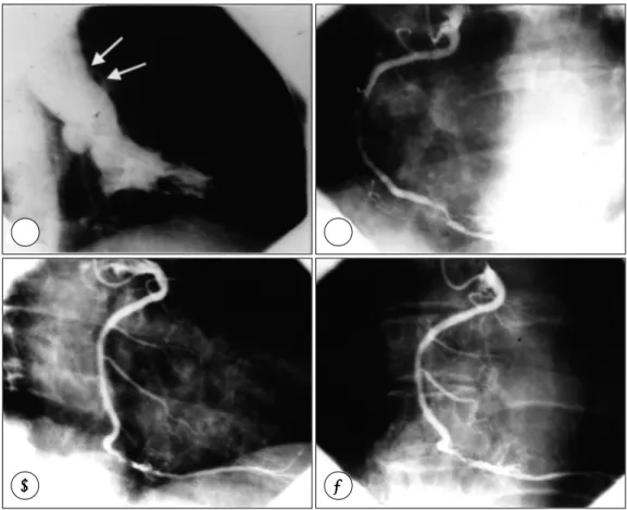
Fig. 1. A (Right anterior oblique view of left ventriculogram):Origin of aberrant vessel (arrows) is visualized just above the left sinus of valsalva. B (Left anterior oblique view), C (Right anterior oblique view), D (Anteroposterior view with cranial angulation):The contour of the left sinus of valsalva is opacified by the dye reflux. Ectopic vessel is opacified through 6 French Amplatz right 1 diagnostic catheter. Note the direction of the catheter to the left sinus and the pres- ence of critical stenosis in the distal segment.
aVF 유도에서 ST절의 상승과 Q파가 있었고, Ⅰ, aVL 유 도에서 ST절의 하강이 있었다. CK치와 CK-MB치는 각 각 238 U/L, 16 U/L이었다.
입원 2일째 증상이 완화된 후 요골접근법에 의하여 관 상동맥 및 좌심실 조영술을 시행하였다. Multipurpose 도자(5 French MPA-1, USCI, USA)를 이용하여 먼 저 우관상동맥의 삽관(cannulation)을 시도하였으나 우 Valsalva동에서 보이지 않았고 먼저 시행한 좌심실 조영 술에서 정상 벽운동, 60%의 구혈율을 보였고 우관상동 맥이 좌Valsalva동의 상방에서 비정상으로 기시하는 형태 임을 알 수 있었다(Fig. 1A). 좌관상동맥 조영술은 Amp- latz Left 1 도자(5 French, USCI, USA)를 사용하여 쉽게 삽관되었고 원위부 좌회선동맥의 분지부위에 90%
협착이 관찰되었다. 비정상 기시의 우관상동맥은 Am- platz Right 2 도자(5 French, USCI, USA)로 쉽게 삽
관되었고 여러면에서 확인한 후 좌Valsalva동에서 기시 함을 알 수 있었다(Fig. 1B, C and D). 후하행동맥이 기 시하는 원위부 우관상동맥에 90%의 협착이 확인되었다.
Amplatz Left 1 유도도관(guiding catheter;6 Fre- nch, Cordis, USA)을 선택하여 삽관한 후 지지가 좋도 록 비정상으로 기시한 우관상동맥의 각이 진 개구부와 연 결이 좋도록 조절하였다(Fig. 2A). 병변은 Over-The- Wire용 Bandit balloon(1.5×20 mm, Scimed, USA) 을 이용하여 0.014″ Supersoft Wisdom wire(Cordis, USA)가 쉽게 통과되었고, 3회의 사전확장(6, 8, 8기압, 총 110초)후 NIR Primo stent(3.0×16 mm, Scimed, USA)를 위치시킨 후 12기압으로 전개하였다. 스텐트 원 위부의 연결부에 흐린 음영(hazness)이 보여서(Fig. 2B) 추가적인 확장(5기압, 20초)후 비폐쇄성 박리가 생겼다 (Fig. 2C). Jo stent(16 mm, JoMED, Germany)를 Wo-
A A
AA BBBB
CC
CC DDDD
Fig. 2. A (Anteroposterior view with cranial angulation):Stable guiding catheter position and good coaxial alignment (arrow) was achieved with 6 French AL1, B (Anteroposterior view with cranial angulation):After initial stent deploym- ent, residual haziness (arrow) was seen near distal margin of stent, C (Anteroposterior view with caudal angulation):
After additional balloon inflation, nonocclusive dissection was developed (arrows), D (Anteroposterior view with cran- ial angulation):After second stent deployment, good final result was achieved.
rldpass balloon(2.5×20 mm, Cordis, USA)에 손으로 말아서 박리 부위에 위치시켜 전개한 후(13기압, 25초) 3.0 mm NIR stent balloon을 이용하여 추가적인 팽창 후(12기압, 10초) 좋은 결과(잔여 협착 -5%)를 얻었다 (Fig. 2D). 이후 좌회선동맥의 병변에 풍선확장술을 시 행하여 양호한 결과(잔여 협착 10%)를 얻었다. 시술후 더 이상의 흉통은 없었고 임상 경과는 양호하였으며 as- pirin(100 mg/day)과 ticlopidine(500 mg/day), capt- opril(25 mg/day), dinitrate isosorbide(80 mg/day), tenormin(50 mg/day)를 투약하여 다음날 퇴원시켰고 외래에서 경과 관찰중이다.
고 찰
좌Valsalva동에서 비정상 기시한 우관상동맥의 병변
에 대한 관상동맥 중재술은 협심증 또는 허혈이 시술하 고자 병변에 의하여 초래되는지에 대한 정확한 평가가 선행되어야 한다. 왜냐하면 우관상동맥의 비정상적 주 행(즉, 좌Valsalva동에서 기시하여 대동맥과 우심실유 출로 사이에 끼이는 주행)에 의한 압박과 slit처럼 좁아 져 있는 개구부에 의하여17) 동맥경화가 없더라도 심각 한 허혈성 합병증(협심증,18) 심근경색증,19) 급사20))이 초래될 수 있기 때문이다. 본 증례는 후하행동맥이 기 시하는 원위부 우관상동맥이 완전 폐쇄된 급성 하벽 심 근경색증으로서 상기의 두 가지 기전 즉, 개구부의 꼬임 (kinking)과 대혈관사이의 압박에 기인하였을 가능성은 적었다.
비정상 관상동맥에서 풍선확장술의 기술적인(특히, 장 비 및 기구의 선택) 문제점은 Topaz 등2)과 Ilia3)에 의 하여 지적되었다. 가장 중요한 인자는 적절한 유도도관 A
AA
A BBBB
C C
CC DDDD
을 선택하고 관상동맥과의 연결이 좋게 하여 최적의 지 지(optimal coaxial backup support)를 얻게 하는 것이 라고 지적하였다. 이를 위하여 진단적 관상동맥 조영술에 서 개구부의 모양, 비정상 관상동맥의 굴곡 정도(ang- ulation)와 주행, 협착 병변의 위치와 심한 정도를 잘 살 필 것을 권고하였다. 이전에 보고되었던 비정상 기시의 우관상동맥에서 9예의 풍선확장술과 1예의 스텐트 시술 에서 사용된 유도도관은 AL1(5예),2)9)12)13)15)
FLG4(3 예),8)11) JL(1예),14) Williams LR(1예)10)의 순서로서 AL1형이 가장 흔히 선택되었다.
비정상 기시의 우관상동맥에서 새로운 중재 기기의 적 용에 대하여는 Bass 등21)이 풍선확장술후 재협착 병변 에서 1.75~2.15 mm의 두 개의 bur를 이용하여 성공 적인 죽상반 제거술(rotational atherectomy)을 보고 하였고, Olympios 등15)은 대퇴접근법에 의한 풍선확장 술중 발생한 비폐쇄성 박리(nonocclusive dissection) 에 대하여 시행한 3.5 mm MICRO Ⅱ 스텐트 시술을 보고하였다. Wong 등16)은 Multi-Link stent의 경험을 보고하면서 비정상 기시의 우관상동맥에서 세 개의 연속 적인 스텐트를 사용하였다고 하였다.
본 증례는 대퇴접근법으로써 8 또는 7 French의 유 도도관을 사용하여 시술하였던 이전의 보고8-14)16)21)
된 경험들이 요골접근법에서도 동일하게 가능하였음을 보 여주었다. 특히 개구부가 slit같이 좁고 굴곡이 심한 비정 상 기시의 우관상동맥 원위부 병변에서는 최적의 지지를 얻기 위하여 Amplatz형 유도도관의 선택이 중요하며 개 구부의 손상 위험을 줄이고 필요에 따라 안정된 유도도 관의 위치 확보를 위해 유도도관을 깊게 삽관할 수 있는 6 French 유도도관의 사용이 유리할 것이다.
요 약
좌Valsalva동에서 비정상으로 기시한 우관상동맥은 드 물지만 동맥경화성 질환으로부터 보호되지는 않는다. 성 공적인 혈관성형술을 결정하는 중요 인자는 적절한 유도 도관을 선택하여 최적의 지지를 확보하는 것이다.
저자들은 좌Valsalva동에서 비정상 기시한 우관상동맥 에 대하여 요골접근법에 의하여 성공적으로 스텐트 시술 을 하였던 첫 보고를 하는 바이다.
중심 단어:비정상 우관상동맥・스텐트・요골접근법.
REFERENCES
1) Yamanaka O, Hobbs RE. Coronary artery anomalies in 126, 595 patients undergoing coronary arteriography. Cathet Cardiovasc Diagn 1990;21:28-40.
2) Topaz O, DiSciascio G, Goudreau E, Cowley MJ, Nath A, Kohli RS, et al. Coronary angioplasty of anomalous cor- onary arteries: Notes on technical aspects. Cathet Cardi- ovasc Diagn 1990;21:106-11.
3) Ilia R. Percutaneous transluminal angioplasty of coronary arteries with anomalous origin. Cathet Cardiovasc Diagn 1995;35:36-41.
4) Olympios CD, Fakiolas CN, Sifaki MD, Foussas SG.
Percutaneous transluminal coronary angioplasty of single coronary artery. J Intervent Cardiol 1996;9:297-9.
5) Chaitman BR, Lesperance J, Saltiel J, Bourassa MG. Clini- cal, angiographic, and hemodynamic findings in patients with anomalous origin of the coronary arteries. Circulat- ion 1976;53:122-31.
6) Kimbiris D, Iskandrian AS, Segal BL, Bemis CE. Anomal- ous aortic origin of coronary arteries. Circulation 1978;
58:606-15.
7) Kafka B, Shimmied M, Worship H, Rno H, Micron S, Kansan H, et al. Clinical features and prognosis of Japa- nese patients with anomalous origin of the coronary artery.
Jpn Circ J 1996;60:731-41.
8) Mooss AN, Heintz MH. Percutaneous transluminal ang- ioplasty of anomalous right coronary artery. Cathet Car- diovasc Diagn 1989;16:16-8.
9) Musial B, Schob A, Marchena E, Kessler KM. Percuta- neous transluminal coronary angioplasty of anomalous right coronary artery. Cathet Cardiovasc Diagn 1991;22:
39-41.
10) Fournier JA, Gonzalez-Barrero A, Fernandez-Cortacero JAP, Sanchez A. Coronary angioplasty of anomalous ri- ght coronary artery originating from the left sinus of Val- salva. Int J Cardiol 1995;49:284-6.
11) Yabe Y, Tsukahara R. Percutaneous transluminal coronary angioplasty for culprit lesions in patients postmyocardial infarction angina based on dextrocardia and anomalous coronary arteries. Case reports and methods. Angiology 1995;46:431-40.
12) Charney R, Spindola-Franco H, Gross R. Coronary ang- ioplasty of anomalous right coronary arteries. Cathet Car- diovasc Diagn 1993;29:233-5.
13) Jeoh JK, Ling LH, Maurice C. Percutaneous translumi- nal angioplasty of anomalous right coronary artery arising from the ascending thoracic aorta. Cathet Cardiovasc Dia- gn 1994;32:254-6.
14) Chakraborty B, Chan CN, Tan A. Percutaneous translu- minal coronary angioplasty of an anomalous right coron- ary artery arising from a separate ostium in the left sinus of Valsalva. A case report. Angiology 1995;46:629-32.
15) Olympios CD, Sifaki MD, Lembidakis EG, Pissimissis EG, Fakiolas CN. Coronary stenting of an anomalous ri- ght coronary artery. J Invas Cardiol 1998;10:342-5.
16) Wong P, Wong CM, Cheng CH, et al. Early clinical exp- erience with the Multi-Link coronary stent. Cathet Cardi- ovasc Diagn 1996;39:413-419.
17) Roberts WC, Siegel RJ, Zipes DP. Origin of the right
coronary artery from the left sinus of Valsalva and its fu- nctional consequences: Analysis of 10 necropsy patients.
Am J Cardiol 1982;49:863-70.
18) Bloomfield P, Erhlich C, Folland E, et al. Anomalous ri- ght coronary artery: A surgically correctable cause of angina pectoris. Am J Cardiol 1983;51:1235-7.
19) Benge W, Martins J, Funk D. Morbidity associated with anomalous origin of the right coronary artery from the
left sinus of Valsalva. Am Heart J 1980;99:96-100.
20) Isner J, Shen E, Martin E, Fortin R. Sudden unexpected death as a result of anomalous origin of the right coronary artery from the left sinus of Valsalva. Am J Med 1984;76:
155-8.
21) Bass TA, Gilmore PS, Ceitami EL. Rotational atherectomy in anomalous coronary arteries. Cathet Cardiovasc Diagn 1992;17:322-4.