Kainic Acid-induced Neuronal Death is Attenuated by Aminoguanidine but Aggravated by L-NAME in Mouse Hippocampus
7
0
0
전체 글
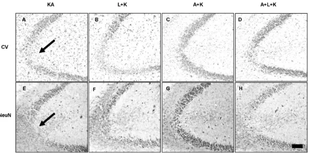
(2) 266. JS Byun, et al. METHODS Animals and reagents Male ICR mice weighing 23∼25 g were obtained from Folas-International, Ltd. (Seoul, Korea). Inducible NOS −/− knockout (iNOS ) and littermate control mice were purchased from Jackson Laboratory (Bar Harbor, ME, USA). All of the animal experiments were conducted in accordance with the animal care guidelines of the National Institutes of Health (NIH) and Korean Academy of Medical Sciences (KAMS). Mice were housed five per cage in a room maino tained at 22±2 C with an alternating 12/12 h light/dark cycle. Food and water were available ad libitum. KA, L-NAME, and aminoguanidine were obtained from Sigma Chemical Co. (St. Louis, MO, USA). KA was prepared as a stock solution at 1 mg/ml in sterile 0.1 M phosphate-buffered saline (PBS, pH 7.4), and aliquots were stored at −20oC until use. L-NAME (50 mg/kg) or aminoguanidine (200 mg/kg) was administered intraperitoneally 1 h prior to KA injection. Intracerebroventricular (i.c.v.) injection of KA The administration of KA (0.1 μg/5 μl) was performed. according to the procedure established by Laursen and Belknap (Laursen and Belknap, 1986). Briefly, KA was injected at the bregma with a 50-μl Hamilton microsyringe fitted with a 26-gauge needle that was inserted to a depth of 2.4 mm. In situ labeling of DNA fragmentation DNA fragmentation analysis, which indicates apoptosis as previously described (Henshall et al., 2001), was performed according to the manufacturer’s instructions using terminal deoxynucleotidyl transferase with peroxide-12-UTP nick-end labeling (TUNEL) (Roche Molecular Biochemicals, Indianapolis, IN, USA). The percentage of TUNEL-positive cells (bright fluorescent green) was assessed by analysis of digitized images from 5 or more microscopic fields of TUNEL-stained cells from TIFF files (Adobe Photoshop). Immunohistochemistry All mice were sacrificed at 6 h or 24 h after KA injection. The mice were transcardially perfused and post-fixed for 4 h in 4% paraformaldehyde. The brains were cryoprotected in 30% sucrose, sectioned coronally (40 μm) on a freezing microtome, and collected in cryoprotectant for storage at. Fig. 1. Effects of NOS inhibitors on KA-induced neuronal cell death in the CA3 region of mouse hippocampus. Neuronal cell death was determined with cresyl violet staining. KA injection produced a marked loss of pyramidal neurons (IA, E) and L-NAME treatment prior to KA (IB, F) potentiated this KA-induced neuronal loss. Aminoguanidine pretreatment (IC, G) markedly attenuated KA-induced and L-NAME-aggravated (ID, H) neuronal death. Neuronal cell death was not observed in vehicle-, aminoguanidine-only-, or L-NAMEonly-treated mice (II). Images in the upper right corner show the entire hippocampus and boxes in these images indicate the enlarged views of the CA3 region. Arrows indicate areas where most neuronal death appears. KA & K stand for kainic acid, L for L-NAME, and A for aminoguanidine. Scale bar: 100 μm..
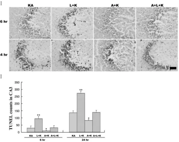
(3) Aminoguanidine Attenuates KA-induced Neuronal Death but L-NAME Aggravates. −20oC. For cresyl violet staining, the sections were mounted in gelatin-coated slides and allowed to air-dry overnight. The mounted sections were submerged in 0.1% cresyl violet solution for 5 min. The sections were rinsed in 70% ethanol and dehydrated in graded series of ethanol, immersed in xylene, mounted in Permount (Fisher Scientific, NJ, USA), and cover slipped. The free-floating immunohistochemistry of the brain sections was processed as previously described (Baker and Farbman, 1993). Sections collected from cryoprotectant were washed with PBS, pre-incubated for 30 min in 0.1 M PBS with 1% bovine serum albumin and 0.2% o Triton X-100, and incubated for 2 days at 4 C with the following primary antibodies: iNOS (1:1,000; BD Pharmingen, San Jose, CA, USA), OX-6 (1:1,000; BD Pharmingen), or NeuN (1:1,000; Santa Cruz, Santa Cruz, CA, USA). The antigens were then detected with 3,3-diaminobenzidine tetrahydrochloride using an Elite ABC kit (Vector, Burlingame, CA, USA). The sections were mounted, air-dried, dehydrated through graded ethanol, cleared in histoclear, and cover slipped using Permount. Statistical analysis The data were analyzed with the Mann-Whitney U test. 267. using SPSS software 12K (SPSS, Chicago, IL, USA) for independent samples to compare KA-only and treatment groups. A value of p<0.05 was accepted as statistically significant. The results are expressed as mean±SEM values.. RESULTS L-NAME aggravates KA-induced neuronal death and aminoguanidine attenuates both KA-induced and L-NAME-aggravated neuronal death The i.c.v. injection of KA time-dependently killed hippocampal neurons in the CA3 region (Fig. 1IA, E); neuronal death was negligible in vehicle-injected mice (Fig. 1II). L-NAME treatment prior to the KA injection further increased the KA-induced neuronal death (Fig. 1IB, F), whereas pretreatment with aminoguanidine significantly attenuated the KA-induced neuronal loss (Fig. 1IC, G). Co-administration of aminoguanidine with L-NAME showed reduced neuronal death compared with L-NAME alone (Fig. 1ID, H). Similarly, L-NAME pretreatment increased the number of early TUNEL-positive neurons in the CA3 region compared with KA alone (Fig. 2), and aminoguanidine re-. Fig. 2. Representative (I) and quantitative (II) neuronal death measured with Terminal deoxytransferase-mediated dUTP-nick end labeling (TUNEL) assay. KA produces TUNEL-positive neurons within 24 h, and L-NAME (L+KA) treatment potentiates this induction. Aminoguanidine (A+KA) reduces the numbers of TUNEL-positive cells compared with KA-only (KA) and L-NAME-aggravated (A+L+KA) groups. L-NAME and aminoguanidine alone did not affect cell survival (data not shown). Quantitative data represent three independent experiments and are expressed as mean±SEM. *p<0.05, **p<0.01 versus the KA-only treated group. KA & K stand for kainic acid, L for L-NAME, and A for aminoguanidine..
(4) 268. JS Byun, et al. duced both KA-induced and L-NAME-aggravated TUNEL staining. Vehicle-treated mice showed no TUNEL staining (i.p. administration of saline as pretreatment and i.c.v. injection of PBS instead of KA) In addition, no TUNEL staining was observed with L-NAME or aminoguanidine alone, respectively (data not shown).. munoreactivity for up to 2 weeks after the KA injection. Neuron number did not change dramatically between 24 h and 2 weeks after KA and aminoguanidine treatment, suggesting that the neuroprotective effects of aminoguanidine were permanent (Fig. 3).. of. L-NAME potentiates KA-induced iNOS expression in activated microglia. To test the time-dependency of the neuroprotective effects of aminoguanidine, we measured levels of viable neurons in the CA3 region with cresyl violet staining and NeuN im-. KA induced microglial activation (Fig. 4E) as determined by immunostaining of the microglial marker, OX-6, and the subsequent induction of inducible iNOS (Fig. 4A) in the region of neuronal death. iNOS expression was localized to. Aminoguanidine produces hippocampal CA3 neurons. prolonged. survival. Fig. 3. Aminoguanidine produces persistent neuronal survival against KA and L-NAME injury. Pyramidal neurons were measured with cresyl violet staining (Top panels) and NeuN immunoreactivity (Bottom panels) at 2 weeks after KA injection. Arrows indicate areas with highest neuronal death. KA & K stand for kainic acid, L for L-NAME, A for aminoguanidine.. Fig. 4. Effects of NOS inhibitors on KA-induced microglial activation and subsequent iNOS expression. Expression of iNOS (top panels) and OX-6, a microglial marker, (bottom panels) within the CA3 was determined with immunohistochemical analysis at 24 h after treatment. KA increased iNOS expression (A) and microglial activation (E), and these phenomena were attenuated with aminoguanidine pretreatment (C, G). L-NAME pretreatment (B, F) potentiated the KA effect, but aminoguanidine reduced this potentiation (D, H). L-NAME and aminoguanidine alone did not affect microglial activation and iNOS expression (data not shown). KA & K stand for kainic acid, L for L-NAME, and A for aminoguanidine..
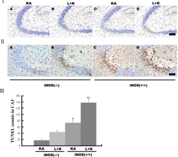
(5) Aminoguanidine Attenuates KA-induced Neuronal Death but L-NAME Aggravates. activated microglia. L-NAME treatment prior to KA produced earlier (data not shown) and increased (Fig. 4B) expression of iNOS in activated microglia. However, pre-treatment with aminoguanidine markedly attenuated the KA-induced microglial activation and iNOS expression (Fig. 4C, G). Aminoguanidine also attenuated the L-NAME-potentiated increases in microglial activation and iNOS expression to the level of the KA-only group (Fig. 4D, H). iNOS knockout mice (iNOS − /− ) are resistant to KA-induced neuronal death To further delineate the role of iNOS in KA-induced neuronal death, we next tested KA-induced neuronal death in −/− iNOS knockout mice (iNOS ). KA injection was less toxic −/− in iNOS mice than in littermates with the iNOS gene (Fig. 5I), but L-NAME still potentiated this toxicity (Fig. 5I, II). Similarly, KA treatment produced fewer TUNEL−/− mice than wild-type litterpositive neurons in iNOS mates (Fig. 5II, III). 269. DISCUSSION In the present study, we demonstrated that inhibiting NOS activity with L-NAME aggravated KA-induced hippocampal neuronal loss in the CA3 region, but that the selective inhibition of iNOS with aminoguanidine significantly attenuated this KA-induced death. Glutamate and other excitatory amino acids induce neuronal cell death by excitotoxicity, which may contribute to the neuronal cell loss caused by acute insults and chronic degeneration in the central nervous system (Choi, 1988; Coyle and Puttfarcken, 1993; Lee et al., 1999; McNamara, 1999). However, the role of NO in brain excitotoxicity is controversial. L-NAME has a 20-fold selectivity for the constitutive isoforms of NOS (n- and e-NOS) (Boer et al., 2000) and aminoguanidine has a 5.5 (iNOS/nNOS)- or 11 (iNOS/eNOS)-fold selectivity (Alderton et al., 2001). Therefore, NO originating from iNOS could cause neuronal death, whereas NO from eNOS and nNOS are protective. NO produced from iNOS can cause neuronal damage,. Fig. 5. Decreased neuronal death after KA in iNOS knockout (iNOS−/−) mice. Neuronal death was measured with cresy violet (I) and −/− TUNEL staining (II&III). KA produced less neuronal death in iNOS mice (I&II-As) than littermates (I&II-Cs). L-NAME pretreatment potentiated neuronal death in both littermates (I&II-Ds) and iNOS−/− mice (I&II-Bs) compared to KA alone, but iNOS−/− mice showed less potentiation (III). Quantitative data represent three independent experiments and are expressed as mean±SEM. **p<0.01 versus −/− KA-only treated iNOS mice. Images in the upper right corner show the entire hippocampus and boxes in these images indicate the enlarged views of the CA3 region. KA & K stand for kainic acid, L for L-NAME, and A for aminoguanidine. Scale bars: I, 100 μm; II, 50 μm..
(6) 270. JS Byun, et al. such as neurological disorders (Koprowski et al., 1993; Sugimoto and Iadecola, 2002) and apoptotic cell death (Kim et al., 1999). Inhibiting iNOS activity decreases glutamate release and improves stroke outcomes after experimental ischemia (Perez-Asensio et al., 2005). Similarly, we found that blockade of NO production from iNOS either with a specific inhibitor, aminoguanidine, or in an iNOS gene knockout significantly attenuated KA-induced neuronal death. However, the role of eNOS inhibition is still controversial. L-NAME protects hippocampal pyramidal neurons against kainite-induced excitotoxicity in rats (Jones et al., 1998) and in newborn rabbits (Takei et al., 2001), but eNOS inhibition can also aggravate seizures in animal models (Haberny et al., 1992; Starr and Starr, 1993; Penix et al., 1994; Tsuda et al., 1997). For example, L-NAME treatment facilitated pilocarpine-induced seizures (Starr and Starr, 1993) and seizures induced by DMCM (methyl6,7-dimethoxy-4-ethyl-β-carboline-3-carboxylate), an inverse agonist for the GABAA receptor benzodiazepine binding site, in mice (Tsuda et al., 1997). Thus, endogenous NO originating from eNOS may protect against epileptogenesis by excitotoxins. Chronic L-NAME treatment aggravates animal mortality and neuronal damage in KA-induced excitotoxic brain injury (Ciani et al., 2001). Relatively low doses of L-NAME, up to 20 mg/kg, were beneficial against excitotoxic neuronal damage (Jones et al., 1998; Takei et al., 2001), whereas higher doses, ranging from 50 mg/kg to 125 mg/kg, were detrimental (Starr and Starr, 1993; Tsuda et al., 1997). We therefore used a relatively high dose of L-NAME, 50 mg/kg, which aggravated KA-induced death of pyramidal neurons in the CA3 region. NARG (N-nitro-L-arginine), an irreversible NOS inhibitor, exhibits biphasic dosing effects during transient forebrain ischemia in gerbils (Shapira et al., 1994). Low levels of NO from eNOS and/or nNOS, which may not be completely blocked by low doses of L-NAME, could protect brain tissue. However, higher doses of L-NAME, which may completely block these enzymes, produces neuronal death. In conclusion, the present data demonstrate that NOS isoforms play a different role in KA-induced excitotoxicity. Pathological levels of NO originating from iNOS play a detrimental role, whereas NO from eNOS and/or nNOS can be beneficial depending on the magnitude of inhibition. Careful consideration should be applied in selecting NOS inhibitors for pharmacological approaches to treating and preventing diseases associated with excitotoxic insults such as epilepsy, ischemia, and traumatic brain injury.. ACKNOWLEDGEMENTS This study was supported by the Rural Development Administration of the Bio-Green 21 project (20080701-034003-009-02-00).. REFERENCES Alderton WK, Cooper CE, Knowles RG. Nitric oxide synthases: structure, function and inhibition. Biochem J 357: 593−615, 2001. Baker H, Farbman AI. Olfactory afferent regulation of the dopamine phenotype in the fetal rat olfactory system. Neuroscience 52: 115−134, 1993. Boer R UMPW, Klein T, Mirau B, Haas S, Baur I. The inhibitory. potency and selectivity of arginine substrate site nitric-oxide synthase inhibitors is solely determined by their affinity toward the different isoenzymes. Mol Pharmacol 58: 1026−1034, 2000. Bredt DS, Snyder SH. Nitric oxide, a novel neuronal messenger. Neuron 8: 3−11, 1992. Buisson A, Lakhmeche N, Verrecchia C, Plotkine M, Boulu RG. Nitric oxide: an endogenous anticonvulsant substance. Neuroreport 4: 444−446, 1993a. Buisson A, Margaill I, Callebert J, Plotkine M, Boulu RG. Mechanisms involved in the neuroprotective activity of a nitric oxide synthase inhibitor during focal cerebral ischemia. J Neurochem 61: 690−696, 1993b. Choi DW. Glutamate neurotoxicity and diseases of the nervous system. Neuron 1: 623−634, 1988. Ciani E, Baldinotti I, Contestabile A. Sustained, long-lasting inhibition of nitric oxide synthase aggravates the neural damage in some models of excitotoxic brain injury. Brain Res Bull 56: 29−35, 2001. Coyle JT, Puttfarcken P. Oxidative stress, glutamate, and neurodegenerative disorders. Science 262: 689−695, 1993. Danielisova V, Nemethova M, Burda J. The protective effect of aminoguanidine on cerebral ischemic damage in the rat brain. Physiol Res 53: 533−540, 2004. Du W, Harvey JA. The nitric oxide synthesis inhibitor L-NAME facilitates associative learning. Prog Neuropsychopharmacol Biol Psychiatry 20: 1183−1195, 1996. Du W, Weiss H, Harvey JA. Associative learning is enhanced by selective neuronal nitric oxide synthase inhibitors and retarded by a nitric oxide donor in the rabbit. Psychopharmacology (Berl) 150: 264−271, 2000. Garthwaite J, Boulton CL. Nitric oxide signaling in the central nervous system. Annu Rev Physiol 57: 683−706, 1995. Gross SS, Wolin MS. Nitric oxide: pathophysiological mechanisms. Annu Rev Physiol 57: 737−769, 1995. Haberny KA, Pou S, Eccles CU. Potentiation of quinolinate-induced hippocampal lesions by inhibition of NO synthesis. Neurosci Lett 146: 187−190, 1992. Henshall DC, Bonislawski DP, Skradski SL, Araki T, Lan JQ, Schindler CK, Meller R, Simon RP. Formation of the Apaf-1/cytochrome c complex precedes activation of caspase-9 during seizure-induced neuronal death. Cell Death Differ 8: 1169− 1181, 2001. Jones PA, Smith RA, Stone TW. Nitric oxide synthase inhibitors L-NAME and 7-nitroindazole protect rat hippocampus against kainate-induced excitotoxicity. Neurosci Lett 249: 75−78, 1998. Kiedrowski L, Costa E, Wroblewski JT. Glutamate receptor agonists stimulate nitric oxide synthase in primary cultures of cerebellar granule cells. J Neurochem 58: 335−341, 1992. Kim YM, Bombeck CA, Billiar TR. Nitric oxide as a bifunctional regulator of apoptosis. Circ Res 84: 253−256, 1999. Koprowski H, Zheng YM, Heber-Katz E, Fraser N, Rorke L, Fu ZF, Hanlon C, Dietzschold B. In vivo expression of inducible nitric oxide synthase in experimentally induced neurologic diseases. Proc Natl Acad Sci USA 90: 3024−3027, 1993. Laursen SE, Belknap JK. Intracerebroventricular injections in mice. Some methodological refinements. J Pharmacol Methods 16: 355−357, 1986. Lee JM, Zipfel GJ, Choi DW. The changing landscape of ischaemic brain injury mechanisms. Nature 399: A7−14, 1999. McNamara JO. Emerging insights into the genesis of epilepsy. Nature 399: A15−22, 1999. Moncada S, Higgs A, Furchgott R. International Union of Pharmacology Nomenclature in Nitric Oxide Research. Pharmacol Rev 49: 137−142, 1997. Mulsch A, Busse R, Mordvintcev PI, Vanin AF, Nielsen EO, Scheel-Kruger J, Olesen SP. Nitric oxide promotes seizure activity in kainate-treated rats. Neuroreport 5: 2325−2328, 1994. Penix LP, Davis W, Subramaniam S. Inhibition of NO synthase increases the severity of kainic acid-induced seizures in rodents. Epilepsy Res 18: 177−184, 1994. Perez-Asensio FJ, Hurtado O, Burguete MC, Moro MA, Salom JB,.
(7) Aminoguanidine Attenuates KA-induced Neuronal Death but L-NAME Aggravates. Lizasoain I, Torregrosa G, Leza JC, Alborch E, Castillo J, Knowles RG, Lorenzo P. Inhibition of iNOS activity by 1400W decreases glutamate release and ameliorates stroke outcome after experimental ischemia. Neurobiol Dis 18: 375−384, 2005. Rondouin G, Bockaert J, Lerner-Natoli M. L-nitroarginine, an inhibitor of NO synthase, dramatically worsens limbic epilepsy in rats. Neuroreport 4: 1187−1190, 1993. Schulz JB, Matthews RT, Jenkins BG, Ferrante RJ, Siwek D, Henshaw DR, Cipolloni PB, Mecocci P, Kowall NW, Rosen BR. Blockade of neuronal nitric oxide synthase protects against excitotoxicity in vivo. J Neurosci 15: 8419−8429, 1995. Shapira S, Kadar T, Weissman BA. Dose-dependent effect of nitric oxide synthase inhibition following transient forebrain ischemia in gerbils. Brain Res 668: 80−84, 1994. Southam E, Garthwaite J. The nitric oxide-cyclic GMP signalling pathway in rat brain. Neuropharmacology 32: 1267−1277, 1993. Starr MS, Starr BS. Paradoxical facilitation of pilocarpine-induced seizures in the mouse by MK-801 and the nitric oxide synthesis inhibitor L-NAME. Pharmacol Biochem Behav 45: 321−325, 1993.. 271. Sugimoto K, Iadecola C. Effects of aminoguanidine on cerebral ischemia in mice: comparison between mice with and without inducible nitric oxide synthase gene. Neurosci Lett 331: 25−28, 2002. Takei Y, Takashima S, Ohyu J, Matsuura K, Katoh N, Takami T, Miyajima T, Hoshika A. Different effects between 7-nitroindazole and L-NAME on cerebral hemodynamics and hippocampal lesions during kainic acid-induced seizures in newborn rabbits. Brain Dev 23: 406−413, 2001. Takei Y, Takashima S, Ohyu J, Takami T, Miyajima T, Hoshika A. Effects of nitric oxide synthase inhibition on the cerebral circulation and brain damage during kainic acid-induced seizures in newborn rabbits. Brain Dev 21: 253−259, 1999. Tsuda M, Suzuki T, Misawa M. Aggravation of DMCM-induced seizure by nitric oxide synthase inhibitors in mice. Life Sci 60: 339−343, 1997. Yamamoto S, Golanov EV, Berger SB, Reis DJ. Inhibition of nitric oxide synthesis increases focal ischemic infarction in rat. J Cereb Blood Flow Metab 12: 717−726, 1992..
(8)
수치

관련 문서
Compounds 2 and 3 showed marked inhibitory activities against inducible nitric oxide synthase (iNOS) on the lipopolysaccharide and interferon- γ - stimulated mouse
The levels of nuclear factor-κB (NF-κB), sterol regulatory element-binding transcription protein 1 (SREBP-1) activity and cyclooxygenase-2 (COX-2), inducible nitric oxide
The effect of pretreatment with nonspecific NOS inhibitor Nw-nitro-Larginine methyl ester L-NAME 10–4 M, NO scavanger OHB12 10–3 M, non-specific K+ channel blocker
Control group (CON) was injected with i.c.v. The mice number of each group was 3. Experiments were conducted inde- pendently three times, giving a total of nine per group in
To achieve this purpose, the present study investigated effects of L-NAME, a nitric oxide synthase (NOS) inhibitor, on IL-1 β-induced mechanical allodynia and examined the
LPS-stimulated BV-2 microglia were used to study the expression and production of inflammatory mediators such as nitric oxide (NO), inducible NO synthase
Anti-inflammatory Properties of Meso-dihydroguaiaretic Acid in Lipopolysaccharide-induced Macrophage
The inhibition of NO generation by MDGA was consistent with the inhibitory effect on the expression of inducible nitric oxide synthase (iNOS).. In addition, MDGA
After pretreatment of L-NAME (N-Nitro-L- arginine methyl ester), which is an NO inhibitor, adenosine-induced relaxation was not affected, but ATP-induced relax- ation
