INTRODUCTION
Kidney is the main site of endogenous L-arginine synthe- sis (1, 2). L-arginine used in the protein synthesis is a mate- rial for transport, storage and excretion of nitrous products.
Especially, L-arginine is main substance of guanidine prod- ucts such as creatinine and methylguanidine which are con- sidered as uremic toxins. All of these L-arginine metabolites are involved in renal pathophysiology.
Nitric oxide (NO), as a secondary messenger, is formed in the process of oxidation of L-arginine guanidine nitrogen to L- citrulline in cytoplasm. The role of NO in kidney is of impor- tance not only in the modulation of renal hemodynamics but also in the regulation of renal tubular and glomerular functions (3-5). However, inducible nitric oxide synthase (iNOS) path- ways which exist in the glomerular mesangial cells, the mono- cyte, the macrophage and vascular smooth muscle cells are activated by various cytokines in pathologic conditions. The iNOS, induced by many cytokines such as interleukin-1, tumor necrosis factor and lipopolysaccharide, is a mediator to outside injury or inflammation in immune defense mechanism (6).
Excess NO is locally formed and damages the kidney. The NO produced by excess arginine as well as the creatine and
methylguanidin is the major factor to kidney injury (7, 8).
More and more attentions have been paid to the protective effects of natural antioxidants against drug-induced toxicities especially when free radical generations are involved. Tea has many advantages over chemicals since it contains several an- tioxidants including polyphenols of catechin and theaflavin (9). Green tea has received a lot of attention owing to its role as an antioxidant and free radicals scavenger (10, 11). It is well known that green tea has anti-cancer, anti-diabetics and anti- hypertensive actions in the clinical studies (12).
In this study, we investigated the antioxidative effects of green tea extract on human mesangial cells with L-arginine induced toxicity.
MATERIALS AND METHODS Green tea extract (GTE)
GTE was prepared from a hot-water extract of green tea (Boseong, Gwangju, Korea) as described by Maity et al. (13).
Ten g of green tea leaves were soaked in 100 mL of boiling distilled water for 5 min and filtered.
S204
Byung Chul Shin, Hyun Ho Ryu*, Jong Hoon Chung�, Byoung Rai Lee�, and Hyun Lee Kim�
Department of Internal Medicine, Seonam University College of Medicine, Gwangju; Department of Emergency Medicine*, Chonnam National University Hospital, Gwangju; Departments of Internal Medicine�, Biochemistry�, Chosun University College of Medicine, Gwangju, Korea.
Address for correspondence Hyun Lee Kim, M.D.
Department of Internal Medicine, Chosun University, College of Medicine, 375 Seoseok-dong, Dong-gu, Gwangju 501-759, Korea
Tel : +82.62-220-3178, Fax : +82.62-234-9653 E-mail : [email protected]
*This study was supported by grants from the Clinical Medicine Research Institute at Chosun University Hospital, 2007.
DOI: 10.3346/jkms.2009.24.S1.S204
The Protective Effects of Green Tea Extract against L-arginine Toxicity to Cultured Human Mesangial Cells
The aim of this study was to investigate whether green tea extract (GTE) has the protective effects on excess L-arginine induced toxicity in human mesangial cell.
Human mesangial cells treated with L-arginine were cultured on Dulbecco’s modi- fied eagle medium in the presence and absence of inducible nitric oxide synthase (iNOS) inhibitor and GTE. The cell proliferation was determined by 3 (4,5-dimethylth- iazol-2-yl)-2, 5-diphengltetrqzolium bromide, a tetrazole assay. The iNOS mRNA and its protein expression were detected by reverse transcription polymerase chain reaction and Western blot, respectively. The concentration of nitric oxide (NO) was measured by NO enzyme-linced immuno sorbent assay kit. L-arginine significantly inhibited the proliferation of human mesangial cells, and induced the secretion of NO to the media. NO production by L-arginine was significantly suppressed by GTE and iNOS inhibitor (p<0.01). The expression level of iNOS mRNA and its protein that was significantly increased by L-arginine was decreased by iNOS inhibitor but not by GTE. GTE protected the mesangial cells from the NO-mediated cytotoxicity by scavenging the NO rather than by iNOS gene expression. Therefore, we con- clude that GTE has some protective effect for renal cells against oxidative injury possibly by polyphenols contained in GTE.
Key Words : Green Tea Extract; Arginine; Nitric Oxide; Polyphenols
Received : 12 June 2008 Accepted : 14 January 2009
Cell line and culture
Human mesangial cell line SV 40 MES 13 (ATCC catalog number CRL-1927) were cultured in Dulbecco’s modified eagle medium (DMEM) containing 10% fetal bovine serum (Gibco, Carlsbad, CA, U.S.A.), guanidine 2 mM/L, benzylpeni- cillin 100,000 U/L, streptomycin 0.1 g/L, human transfer- rin 2.5 kg/L, sodium selenita 5 μg/L in 5% CO2incubator at 37℃.
Mesangial cell preperation
Mesangial cells were cultured with 1×10⁴density in 96- well microplates. The mesangial cells were divided into four groups. In group A (control group), mesangial cells were cul- tured without L-arginine, and group B (L-arginine group), L-arginine was given 100 μM/L on 200 μL media with dilu- tion ratio of 4:1. In group C (iNOS inhibitor group), mesan- gial cells were cultured with L-arginine 100 μM/L with seri- al dilution plus iNOS inhibitor (L-NAME, 3, 4, and 5 mM/L).
In group D (GTE group), mesangial cells were cultured with L-arginine 100 μM/L with serial dilution plus GTE 0.1g (10, 20, and 30 μL) with 10 mL PBS (NaCl 8.0, KCl 0.2, Na2HPO4 12H2O 3.58, and KH2PO40.24 g/L). The mesangial cells were cultured for 48 hr.
Cell viability assay
Cell viability of mesangial cells was measured by 3-(4,5- dimethylthiazol-2-yl)-2, 5-diphenyltetrazolium bromide (MTT) assay. The incubation medium was completely aspirat- ed upon treatment with L-arginine 0, 26, 33, 41, 51, 64, 80, and 100μM/L for 2 days and medium was completely removed. The MTT-formazon was added to the cells, and further incubated for 4hr at 37℃. The cells were washed out, and then lysed with 100 μL of fresh dimethyl sulfoxide (DMSO). The absorbance was calculated at 540 nm (A540) using a microplate autoreader (Bio-TEK Instruments, Winoos- ki, VT, U.S.A.).
NO measurement
Tissue nitrite (NO-2) and nitrate (NO-3) were assessed as an index for NO production. Total NO (nitrite plus nitrate) was measured after conversion of nitrate to nitrite by nitrate reduc- tase. The method for measuring nitrite and nitrate levels was based on the Griess reaction (14) and determined by Hsp60 ELISA kit (StressXpressTM, Stressgen Biotechnologies Corp., Victoria, BC, Canada).
iNOS mRNA expression by reverse transcription polymerase chain reaction (RT-PCR)
Total RNA was isolated from mesangial cells using the
TRIzol one step method (Gibco, Bethesda, MD, U.S.A.). One μg of total RNA was heated at 70℃for 10 min and reversely transcribed using reverse transcriptase 200 U (Promega, Madi- son, WI, USA). The mixture was then incubated at 37℃ for 60 min, heated at 95℃for 10 min and stored at -20℃ until use. Selected sequences in 5 μL aliquots of cDNA were amplified by PCR 32 cycles using primers for iNOS and glyc- eraldehyde-3-phosphate dehydrogenase (GAPDH) as an inter- nal standard. Cycles consisted of 45 sec of denaturation at 95℃, 45 sec of annealing at 63℃, 1 min of extension at 72℃, fol- lowed by 7 min of elongation at 72℃. Primers were designed with computer assistance according to the gene bank. The sequence of the primers are as follows; iNOS sense primer 5′-AGC ATC ACC CCT GTG TTC CAC CA-3′, and iNOS anti-sense 5′-TGG GGC AGT AGT CTC CAT TGC CA-3′, (Genemed Synthesis Inc., South San Francisco, CA, U.S.A.);
GAPDH sense 5′-CCA TCA CCA TCT TCC AGG AGC GAG-3′, and GAPDH antisense 5′-TGC CAG TGA GCT TCC CGT TCA GCT C-3′. The iNOS cDNA was normal- ized by comparison with GAPDH cDNA.
Western blot analysis
iNOS protein expression was determined by western blot analysis. For the detection of iNOS protein, cells were harvest- ed and incubated for 20 min on ice in lysis buffer and were centrifuged at 14,000 rpm for 20 min.
The samples were subjected to the 10% polyacrylamide gels and transferred to a nitrocellulose membrane at 20 V overnight.
Membranes were incubated with anti-iNOS antibodies (1:
1,000, Sigma, St. Louis, MO, U.S.A.) for two hours at room temperature. The membranes were then incubated with a conjugated horseradish peroxidase anti-mouse antibody (R&D system, Minneapolis, MN, U.S.A.) for two hours. They were exposed to a radiography film by LumiGLO chemilumines- cent substrate and band density were detected by densitom- etry (LabWorks 4.0, UVP Inc., Upland, CA, U.S.A.).
Statistical analysis
Results are expressed as the means value±SD. Differences between the control and the experimental groups were ana- lyzed using the Mann-Whitney test and the Kruskal-Wallis test (SPSS, Statistical Package for Social Science 10.0). p val- ues of <0.05 were considered significant.
RESULTS
Toxicity of L-arginine on mesangial cell proliferation
L-arginine (26, 33, 41, 51, 64, 80, and 100 μM/L) signif- icantly inhibited human mesangial cells proliferation in a con- centration-dependent manner compared with that of control
group (Fig. 1).
Effect of iNOS inhibitor on L-arginine induced mesangial cell toxicity
Treatment with L-NAME (iNOS inhibitor, 3, 4, and 5 mM) significantly inhibited the anti-proliferative effect of L-argi- nine (51 μM/L) in a concentration-dependent manner (Fig. 2).
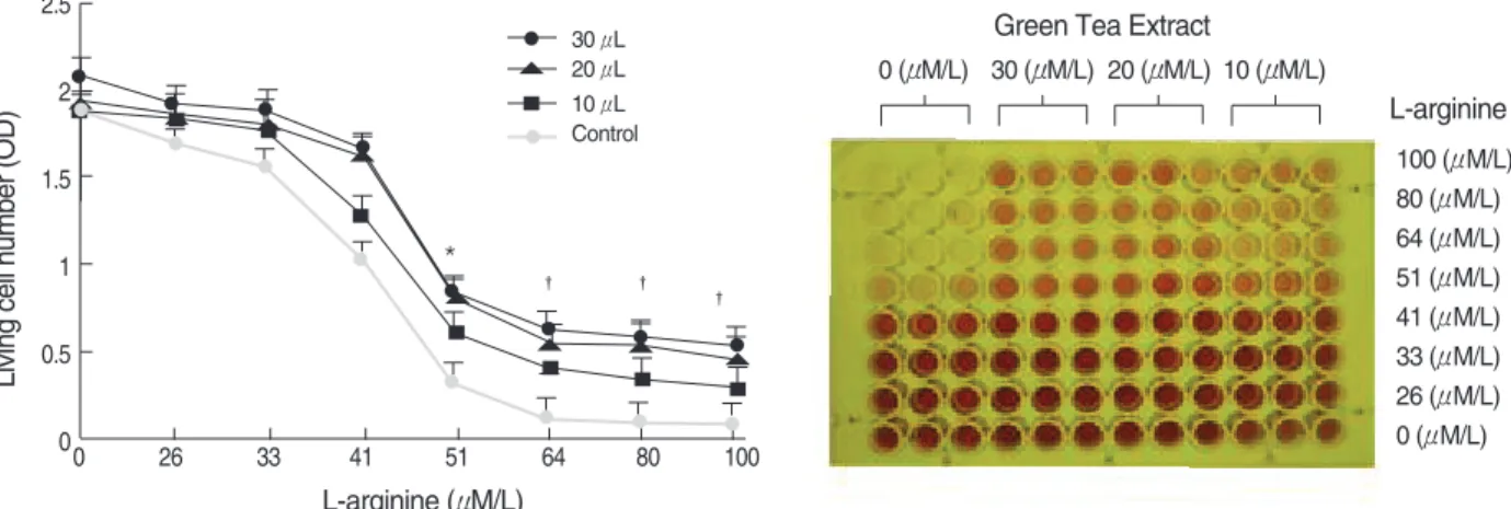
Effect of GTE on L-arginine induced mesangial cell toxicity
To determine whether TGE blocks L-arginine-induced tox- icity in mesangial cell, cells were cultured with L-arginine treated with GTE. Treatment with GTE (10, 20, and 30 μL) significantly inhibited the anti-proliferative effect L-arginine
in a concentration-dependant manner (Fig. 3).
Expression of iNOS mRNA by RT-PCR in mesangial cell
The expression of iNOS mRNA was significantly higher in the L-arginine group than in the control group and lower in the L-NAME group than in the L-arginine group (p<0.05).
GTE had no effect on the expression of iNOS mRNA (Fig. 4).
Expression of iNOS protein by Western blot analysis in mesangial cell
Total amount of iNOS protein expression was significantly higher in the L-arginine group than in the control group and was lower in the L-NAME group than in the L-arginine group (p<0.05), but was not reduced in the GTE group (Fig. 4).
Concentration of NO
Treatment with L-arginine caused a significant increase in the concentration of NO versus the control group (0.139± 0.047 μM/g vs. control 0.047±0.026 μM/g, p<0.01). Increas- es in NO concentration reduced by L-arginine were signifi- cantly suppressed by L-NAME (0.049±0.022 μM/g, p<0.01) and by GTE (0.052±0.032 μM/g, p<0.01, Fig. 5).
DISCUSSION
Our study revealed that excess L-arginine inhibited the pro- liferation of mesangial cells by increasing the productions of NO and iNOS. We investigated the effects of GTE on L-argi- nine-induced cytotoxicity in the mesangial cell, and found that GTE protected the human mesangial cells from L-argi-
Fig. 1. Toxicity of L-arginine on the proliferation of human mesan- gial cells. Cell viabilities were measured by MTT assay. Indicated amount of L-arginine were added to mesangial cell for 48 hr (n=10 wells. Values expressed as mean±SD. *p<0.05 and �p<0.01 as compared to control group. OD, Optical density.
Living cell number (OD)
3.5 3 2.5 2 1.5 1 0.5 0
0 26 33 41 51 64 80 100
L-arginine (μM/L)
*
� � �
Fig. 2. Effect of iNOS inhibitor (L-NAME) on L-arginine induced human mesangial cells toxicity (96-well microplates). Cell viabilities were measured by MTT assay. Mesangial cells were treated with indicated amounts of L-arginine in the presence and absence of iNOS inhibitor (L-NAME, 3 mM/L, 4 mM/L, and 5 mM/L) for 48 hr (n=9 wells). Values expressed as mean±SD. *p<0.05 as compared to L-arginine group.
OD, Optical density.
Living cell number (OD)
3
2.5
2
1.5
1
0.5
00 26 33 41 51 64 80 100
L-arginine (μM/L)
iNOS inhibitor (L-NAME)
3 mM
100 (μM/L) 80 (μM/L) 64 (μM/L) 51 (μM/L) 41 (μM/L) 33 (μM/L) 26 (μM/L) 0 (μM/L) 0 (μM/L) 3 (μM/L) 4 (μM/L) 5 (μM/L)
L-arginine
4 mM 5 mM Control
*
nine-induced cytotoxicity by scavenging the NO.
Green tea is produced from the dried leaves of the plant Camellia sinensis and contains several polyphenolic compo- nents, such as, (-)-epigallocatechin 3-O-gallate (EGCG), (-)- gallocatechin 3-O-gallate, (-)-epicatechin 3-O-gallate, (-)- epigallocatechin, (+)-gallocatechin, (-)-epicatechin, and (+)- catechin (15). In this study, the components of GTE were EGCG (28% by weight), epigallocatechin (15%), gallocate- chin (15%), gallocatechin gallate (10%), epicatechin (7%), epicatechin-3-gallate (5%), catechin (4%). EGCG, the main polyphenol in green tea, has been suggested to participate in the elimination of the uremic toxins, and thus, ameliorate renal disorders (10). The mechanisms of green tea polyphenol were suggested as follows. First, green tea polyphenols increase the bioactivities of superoxide dismutase (SOD) and glutathione peroxidase, which inactivate free oxygen radicals like O2-. Fur- thermore, because O2-can react with NO by forming ONOO2-, green tea polyphenols may reduce NO loss and maintain NO physical function (16, 17). Second, green tea polyphenol may
Fig. 4. The expression of iNOS mRNA and its protein in human mesangial cells by RT-PCR and Western blot. (A) Mesangial cell pretreated with L-arginine were cultured with iNOS inhibitor (L-NAME) or GTE and were analyzed by RT-PCR for iNOS mRNA. M, molecular marker;
C, control; A1, L-arginine 25 μM/L; A2, L-arginine 50 μM/L; A3, L-arginine 100 μM/L; I, iNOS inhibitor 5 mM; G1, GTE 10 μL; G2, GTE 20 μL;
G3, GTE 30 μL. (B) Mesangial cell pretreated with L-arginine (50 μM/L) were cultured with iNOS inhibitor (L-NAME, 5 mM) or GTE (30 μL) and were analyzed by Western blot. C, control; A, L-arginine 50 μM/L; A+I, L-arginine 50 μM/L+iNOS inhibitor 5 mM; A+G, L-arginine 50 μM/L+GTE 30 μL.
A B
iNOS iNOS
β-actin GAPDH
M C A1 A2 A3 I G1 G2 G3
C A A+I A+G
Fig. 5. Concentration of NO (nitric oxide). Mesangial cell pretreat- ed with L-arginine (50 μM/L) were cultured with iNOS inhibitor (L- NAME, 5 mM) or GTE (30 μL) and were analyzed by NO ELISA kit. *p<0.01 as compared to control group; �p<0.01 as compared to L-arginine group.
NO level (μM/g) 0.16 0.14 0.12 0.1 0.08 0.06 0.04 0.02
0 Control L-arg L-NAME GTE
*
� �
Fig. 3. Effect of green tea extract on proliferation of human mesangial cells (96-well microplates). Cell viabilities were measured by MTT assay.
Mesangial cells were treated with indicated amounts of L-arginine in the presence and absence of GTE (10 μL, 20 μL, and 30 μL) for 48 hr (n=9 wells). Values expressed as mean±SD. *p<0.05 and �p<0.01 as compared to L-arginine group. OD, Optical density.
Living cell number (OD)
2.5
2
1.5
1
0.5
00 26 33 41 51 64 80 100
L-arginine (μM/L)
Green Tea Extract
30 μL
100 (μM/L) 80 (μM/L) 64 (μM/L) 51 (μM/L) 41 (μM/L) 33 (μM/L) 26 (μM/L) 0 (μM/L) 0 (μM/L) 30 (μM/L) 20 (μM/L) 10 (μM/L)
L-arginine
20 μL 10 μL Control
*
� �
�
inhibit the synthesis of thromboxane A2(TXA2) and leuko- trienes (18). The potent vasoconstrictive effects of TXA2and leukotrienes contribute to the activation of the renin-angio- tensin system. There are intrarenal angiotensin II deposits and excessive expression of angiotensin II type I receptor expres- sion has been observed in the renal medulla (19). Future stud- ies are needed to demonstrate the other mechanisms, such as effects of renin-angiotensin systems, of GTE in vitro and vivo.
Excessive dietary arginine evokes renal failure by increas- ing the production of NO in the kidney. Lui et al. (20) showed that L-arginine could exert in inhibitory effect on the prolif- eration of human mesangial cells and the production of extra- cellular components. In the present study, the administration of excess L-arginine inhibited the proliferation of mesangial cells and increased the concentration of NO. Noris et al. (21) reported that arginine levels and NO synthesis were higher in uremic patients than in healthy volunteers, suggesting an explanation for the increased NO synthesis in uremia. Also, in the present study, combined NO2-and NO3-levels was high- er in arginine-given mesangial cells than in arginine-free mesan- gial cells. It raises the possibility that the increase in NO pro- duction may be attributable to dietary arginine and that it may cause renal injury.
Therefore, green tea polyphenol suppresses the production of NO and it would be expected to ameliorate the renal injury induced by excessive arginine. Furthermore, green tea polyphe- nol has a potent scavenging effect through the inhibition of oxidative stress-induced apoptosis in cell culture (22). Yokoza- wa et al. (23) suggested that excessive arginine affected the activity of antioxidative enzymes in the renal peroxisomes and the reductions in the SOD and catalase activities induced by arginine imply that oxygen-derived free radicals were generat- ed and the biological defense system was weakened but the administration of green tea polyphenol increased the activi- ties of SOD and catalase. In the present study, GTE increased the inhibited proliferation of mesangial cells by L-arginine and suppressed the increase in NO concentration after L-argi- nine treatment.
In summary, GTE might play a crucial role of NO inhibition as free radical scavenging effect rather than iNOS inhibition.
In addition, it was found to ameliorate anti-proliferative effect of L-arginine in mesangial cells, which suggests that GTE, and probably its polyphenols, can protect renal cells against oxidative injury. Future studies are needed to demonstrate the antioxidant effects and other effects of GTE on renal diseases.
REFERENCES
1. Visek WJ. Arginine needs physiological state and usual diets. A reeval- uation. J Nutr 1986; 116: 34-46.
2. Natelson S, Sherwin JE. Proposed mechanism for urea nitrogen re- utilization: relationship between urea and proposed guanidine cycles.
Clin Chem 1979; 25: 1343-4.
3. Ito S, Ren Y. Evidence for the role of nitric oxide in the macula densa control of glomerular hemodynamics. J Clin Invest 1993; 92: 1093-8.
4. Radermacher J, Klanke B, Schurek HJ, Stolte HF, Frolich JC. Impor- tance of NO/EDRF for glomerular and tubular function: studies in the isolated perfused rat kidney. Kidney Int 1992; 41: 1549-59.
5. Shultz PJ, Schorer AE, Raij L. Effects of endothelium-derived relax- ing factor on rat mesangial cells. Am J physiol 1990; 258: F162-7.
6. Raij L, Shultz PJ. Endothelium-derived relaxing factor, nitric oxide:
Effects on and production of mesangial cell and glomerulus. J Am Soc Nephrol 1993; 3: 1435-41.
7. Orita Y, Tsubakihara Y, Ando A, Nakata K, Takamitsu Y, Fukuhara Y, Abe H. Effect of arginine or creatinine administration on urinary excretion of methylguanidine. Nephron 1978; 22: 328-36.
8. Paller MS, Hoidal JR, Ferris TF. Oxygen free radicals in ischemic acute renal failure in the rat. J Clin Invest 1984; 74: 1156-64.
9. Mohamadin AM, El-Beshbishy HA, El-Mahdy MA. Green tea extract attenuates cyclosporine A-induced oxidative stress in rats. Pharma- col Res 2005; 51: 51-7.
10. Jung YD, Kim MS, Shin BA, Chay KO, Ahn BW, Liu W, Bucana CD, Gallick GE, Ellis LM. EGCG, a major component of green tea, inhibits tumor growth by inhibiting VEGF induction in human colon carcinoma cells. Br J Cancer 2001; 84: 844-50.
11. Tsubono Y, Nishino Y, Komatsu S, Hsieh CC, Kanemura S, Tsuji I, Nakatsuka H, Fukao A, Satoh H, Hisamichi S. Green tea and the risk of gastric cancer in Japan. N Engl J Med 2001; 344: 632-6.
12. Ji BT, Chow WH, Hsing AW, McLaughlin JK, Dai Q, Gao YT, Blot WJ, Fraumeni JF Jr. Green tea consumption and the risk of pancreas and colorectal cancer. Int J Cancer 1997; 70: 225-58.
13. Maity S, Vedasiromoni JR, Ganguly DK. Role of glutathione in the antiulcer effect of hot water extract of black tea (Camellia sinensis).
Jpn J Pharmacol 1998; 78: 285-92.
14. Cortas NK, Wakid NW. Determination of inorganic nitrate in serum and urine by a kinetic cadmium reduction method. Clin Chem 1990;
36: 1440-3.
15. Zhang Q, Kelly AP, Wang L, French SW, Tang X, Duong HS, Mes- sadi DV, Le AD. Green tea extract and (-)-epigallocatechin-3-gal- late inhibit mast cell-stimulated type I collagen expression in keloid fibroblasts via blocking PI-3K/AkT signaling pathways. J Invest Der- matol 2006; 126: 2607-13.
16. Morrisey JJ, Ishidoya S, McCracken R, Klahr S. Nitric oxide generation ameliorates the tubulointerstitial fibrosis of obstructive nephropathy.
J Am Soc Nephrol 1996; 7: 2202-12.
17. Shi SH, Zheng SS, Xie HY. Tea polyphenols protect against cyclos- porine-induced acute nephrotoxicity in rats. Chin J Organ Transplant 2001; 22: 271-3.
18. Choi JH, Chang HW, Rhee SJ. Effect of green tea catechin on arachi- donic acid cascade in chronic cadmium-poisoned rats. Asia Pac J Clin Nutr 2002; 11: 292-7.
19. Giachelli CM, Pichler R, Lombardi D, Denhardt DT, Alpers CE, Schwartz SM, Johnson RJ. Osteopontin expression in angiotensin II- induced tubulointerstitial nephritis. Kidney Int 1994; 45: 515-24.
20. Liu BC, Ma KL, Ye YY, Liu NF, Ruan XZ. Effects of L-arginine on proliferation of human renal mesangial cells and production of extra- cellular matrix. Acta Pharmacol Sin 2001; 22: 756-60.
21. Noris M, Benigni A, Boccardo P, Aiello S, Gaspari F, Todeschini M, Figliuzzi M, Remuzzi G. Enhanced nitric oxide synthesis in uremia:
implication for platelet dysfunction and dialysis hypotension. Kidney Int 1993; 44: 445-50.
22. Yokozawa T, Dong E, Nakagawa T, Kashiwagi H, Nakagawa H, Ta-
keuchi S, Chung HY. In vitro and in vivo studies on the radical-scav- enging activity of tea. J Agric Food Chem 1998; 46: 2143-50.
23. Yokozawa T, Cho EJ, Nakagawa T. Influence of green tea polyphe- nol in rats with arginine-induced renal failure. J Agric Food Chem 2003; 51: 2421-5.