279 DOI: 10.4196/kjpp.2010.14.5.279
ABBREVIATIONS: RVH-1, stigma-4-en-3-one (RVH-1); RVH-2, stigma- 4-en-3,6-dione; KA, kainic acid; icv, intracerebroventricular; PDGF, platelet-derived growth factor.
Received July 19, 2010, Revised August 25, 2010, Accepted September 1, 2010
*Corresponding to: Wanjoo Chun, Department of Pharmacology, College of Medicine, Kangwon National University, Hyoja-2-dong, Chuncheon 200-701, Korea. (Tel) 82-33-250-8853, (Fax) 82-33-250-8809, (E-mail) [email protected]
†Co-corresponding author: Myong-Jo Kim, Division of Bio-resources Technology, Kangwon National University, Hyoja-2-dong, Chuncheon 200-701, Korea. (Tel) 82-33-250-6413, (Fax) 82-33-250-6413, (E-mail) [email protected]
Bark Constituents from Mushroom-detoxified Rhus verniciflua Suppress Kainic Acid-induced Neuronal Cell Death in Mouse Hippocampus
Jong-Seon Byun1, Yoon Hee Han1,4, Sung-Jun Hong1, Sung-Mi Hwang1, Yong-Soo Kwon2, Hee Jae Lee1, Sung-Soo Kim1, Myong-Jo Kim3,†, and Wanjoo Chun1,*
1Department of Pharmacology, College of Medicine, 2College of Pharmacy, 3Division of Bio-resources Technology, Kangwon National University, Chuncheon 200-701, 4Department of Radiology, Ilsan Paik Hospital, Inje University, School of Medicine, Goyang 411-706, Korea
Urushinol, a plant allergen, has significantly restricted the medical application of Rhus verniciflua, although it has been reported to possess a wide variety of biological activities such as anti-inflammatory, antioxidant, and anti-cancer actions. To reduce the urushinol content while maintaining the beneficial biological activities, mushroom-mediated fermentation of Rhus verniciflua was carried out and this method resulted in significantly attenuated allergenicity [1]. In the present study, to examine the neuroprotective properties of mushroom-fermented stem bark of Rhus verniciflua, two constituents were isolated from mushroom-fermented bark and their neuroprotective properties were examined in a mouse model of kainic acid (KA)-induced excitotoxicity. KA resulted in significant apoptotic neuronal cell death in the CA3 region of mouse hippocampus. However, seven daily administrations of RVH-1 or RVH-2 prior to KA injection significantly attenuated KA-induced pyramidal neuronal cell death in the CA3 region. Furthermore, pretreatment with RVH-1 and RVH-2 also suppressed KA-induced microglial activation in the mouse hippocampus. The present study demonstrates that RVH-1 and RVH-2 isolated from Rhus verniciflua and detoxified using mushroom species possess neuroprotective properties against KA-induced excitotoxicity. This leads to the possibility that detoxified Rhus verniciflua can be a valuable asset in herbal medicine.
Key Words: Kainic acid, Neuroprotection, Stigma-4-en-3-one, Stigma-4-en-3,6-dione
INTRODUCTION
Rhus verniciflua Stokes, a deciduous tree in the ana- cardiaceae family and indigenous in East Asia, has been used as a traditional herbal medicine to treat diabetes mel- litus and stomach diseases. In the present study, RVH-1 (stigma-4-en-3-one) and RVH-2 (stigma-4-en-3,6-dione) were isolated from Rhus verniciflua. RVH-1 and RVH-2 belong to the stigmasterol group, which are plant sterols, specifi- cally phytosterols. Phytosterols have been reported to pos- sess a variety of biological activities such as anticancer [2]
and hypocholesterolemic properties [3]. Stigmasterol also has been reported to exhibit anti-osteoarthritic [4] and cyto- toxic activities [5]. Although RVH-1 has been isolated from various natural plants such as Lawsonia inermis [6] and Typha latifolia [7] and RVH-2 from Argemone mexicana [8]
and Stephania cepharantha [9], their biological activities
have not been clearly elucidated.
Although Rhus verniciflua has been reported to exhibit a variety of biological activities such as antioxidant, anti-in- flammatory, anti-cancer, and anti-platelet properties, its medical use was significantly neglected due to the presence of an allergenic compound, urushinol, which causes irrita- tion, inflammation, and urushinol-induced contact derma- titis. Therefore, various approaches such as solvent ex- traction, far-infrared radiation, and enzyme treatment were applied to selectively remove urushinol in Rhus verniciflua.
In the mean time, a fermentation method utilizing mush- room species was obtained and it significantly reduced the urushinol content of the stem bark of Rhus verniciflua [1].
To determine whether detoxified Rhus verniciflua possesses neuroprotective properties, the effects of isolated constitu- ents on neuronal survival were examined in a kainic acid (KA)-induced excitotoxicity animal model.
Excessive release of excitatory amino acids may play an important role in the pathogenesis of neuronal injury such as ischemia, stroke, and neurodegenerative diseases [10,11].
Kainic acid (KA), a potent excitotoxin, binds to specific kainite-type receptors and causes depolarization of neurons resulting in status epilepticus and neurodegeneration [12].
The systemic or intracerebroventricular injection of KA re-
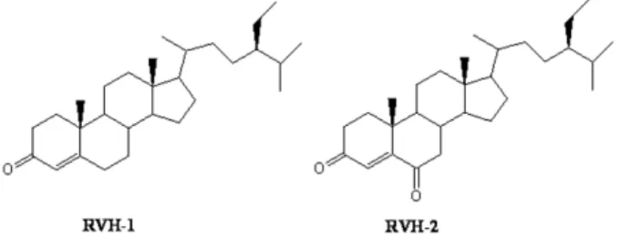
Fig. 1. Chemical structure of RVH-1 (stigma-4-ene-3-one) and RVH-2 (stigma-4-ene-3,6-dione).
sults in the death of pyramidal neurons and concurrent re- active gliosis in hippocampus [13,14].
In the present study, we examined the neuroprotective effects of RVH-1 and RVH-2, isolated from mushroom- de- toxified stem bark of Rhus verniciflua, on hippocampal CA3 neuronal damage induced by intracerebroventricular in- jection of KA.
METHODS Animals and reagents
Male ICR mice weighing 23∼25 g were obtained from Folas-International, Ltd. (Seoul, Korea). All of the animal experiments were conducted in accordance with the animal care guidelines of the National Institutes of Health (NIH) and Korean Academy of Medical Sciences (KAMS). Mice were housed five per cage in a room maintained at 22±2oC with an alternating 12/12 hr light/dark cycle. Food and wa- ter were available ad libitum. KA was obtained from Sigma Chemical Co. (St. Louis, MO, USA). KA was prepared as a stock solution at a concentration of 1 mg/ml in sterile 0.1 M phosphate-buffered saline (PBS, pH 7.4), and aliquots were stored at −20oC until use.
Compounds isolated from detoxified Rhus verniciflua RVH-1 (stigma-4-en-3-one) and RVH-2 (stigma-4-en-3,6- dione) were isolated and identified from mushroom- detoxi- fied stem bark of Rhus verniciflua [1]. These compounds (Fig. 1) were orally administered using an oral zonde daily for 7 consecutive days at a dose of 10 mg/kg. Saline was administered to control mice. More than three mice were used for each group. KA was intracerebroventricularly ad- ministered 1 hr after the last administration of these com- pounds.
Intracerebroventricular (icv) injection of KA
The administration of KA (0.1 μg/5 μl) was performed according to the procedure established by Laursen and Belknap [15]. Briefly, KA was injected at the bregma point with a 50-μl Hamilton microsyringe fitted with a 26-gauge needle that was inserted to a depth of 2.4 mm. Mice were sacrificed 24 hr after KA administration.
Immunohistochemistry
All mice were sacrificed 24 hr after KA injection. Mice
were transcardially perfused and post-fixed for 4 hr in 4%
paraformaldehyde. Brains were cryoprotected in 30% su- crose, sectioned coronally (40 μm) on a freezing microtome, and collected in cryoprotectant for storage at −20oC. For cresyl violet staining, the sections were mounted in gela- tin-coated slides and allowed to air-dry overnight. The mounted sections were submerged in 0.1% cresyl violet sol- ution for 5 min. The sections were rinsed in 70% ethanol and dehydrated in a graded series of ethanol, immersed in xylene, mounted in Permount (Fisher Scientific, NJ, USA) and cover slipped. The free-floating immunohistochemis- try of the brain of the brain sections was processed as pre- viously described [16]. Sections that were collected from cryoprotectant were washed with PBS, pre-incubated for 30 min in 0.1 M PBS with 1% bovine serum albumin and 0.2%
Triton X-100, and incubated for 2 days at 4oC with the fol- lowing primary antibodies: OX-6 (1:1,000; BD Pharmingen) or NeuN (1:1,000; Santa Cruz, Santa Cruz, CA, USA).
After 2 days of incubation with primary antibody, the anti- gens were detected with 3,3-diaminobenzidine tetrahydro- chloride using Vectastain Elite ABC kits (Vector, Burlin- game, CA, USA). Sections were mounted, air-dried, dehy- drated through graded ethanol, cleared in histoclear, and cover slipped using Permount.
In situ labeling of DNA fragmentation
The analysis of cells exhibiting DNA fragmentation, which are suggestive of apoptosis as previously described [17], was performed according to the manufacturer’s instructions us- ing terminal deoxynucleotidyl transferase with per- oxide-12-UTP nick-end labeling (TUNEL) (Roche Molecular Biochemicals, Indianapolis, IN, USA). Brain sections for TUNEL staining were prepared according to the same pro- cedure as above for immunohistochemistry. The percentage of TUNEL-positive cells was assessed by analysis of digi- tized images from 5 or more microscopic fields of TUNEL-stained cells from TIFF files (Adobe Photoshop).
Statistical analysis
Data were analyzed using Mann-Whitney’s U test and SPSS software 12K (SPSS statistics, Chicago, IL, USA) for independent samples to compare mice treated with KA and saline, and mice treated with KA and RVH-1 or RVH-2.
A value of p<0.05 was accepted as statistically significant.
Results are expressed as mean±SEM values.
RESULTS
Both RVH-1 and RVH-2 attenuated KA-induced neuronal cell death
Intracerebroventricular injection of KA resulted in ex- tensive neuronal cell death in the CA3 region of mouse hip- pocampus, whereas neuronal cell death was negligible in vehicle-, RVH-1-, or RVH-2-treated mice (Fig. 2). Although both RVH-1 and RVH-2 attenuated KA-induced neuronal cell death, they did not completely prevent neuronal cell death at a RVH concentration of 10 mg/kg. RVH-2 appeared to be more protective than RVH-1. However, dose-depend- ent inhibition of KA-induced neuronal cell death was not observed (data not shown).
Fig. 2. Representative neuroprotective effects of RVH-1 and RVH-2 on KA-induced neuronal cell death in CA3 region of mouse hippocampus. Hippocampal cell death was examined with cresyl violet staining. Intracerebroventricular (icv) injection of KA showed marked loss of neurons in the CA3 region of hippocampus (KA).
However, RVH-1 or RVH-2 treatment prior to KA injection showed attenuation of neuronal cell loss compared to KA alone. RVH-2 appeared to be more protective than RVH-1, albeit not significantly so. Neuronal cell death was not observed in vehicle (Cont)-, RVH- 1-only-, or RVH-2-only-treated mice. More than three mice were used for each group. Scale bar: 100 μm.
A B
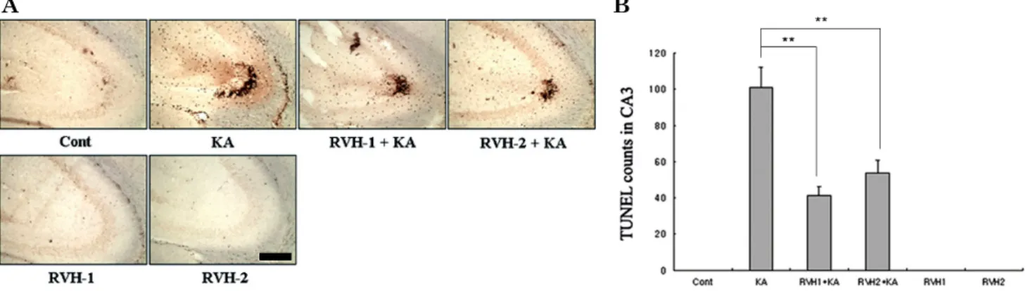
Fig. 3. Representative (A) and quantitative (B) analysis of neuronal cell death with Terminal deoxytransferase-mediated dUTP-nick end labeling (TUNEL) assay. A considerable number of TUNEL-positive neurons appeared with KA treatment within the CA3 region after 24 hr. Pre-administration of RVH-1 or RVH-2 significantly reduced the number of TUNEL-positive cells compared to the KA-only group. RVH-2 pretreatment showed fewer TUNEL-positive cells than RVH-1. A single treatment of RVH-1 or RVH-2 had no noticeable effects on cell survival. More than three mice were used for each group. Quantitative data represent three independent experiments and are expressed as mean±SEM. **p<0.01 indicates statistically significant difference from the KA-only treated group. Scale bar: 100 μm.
Both RVH-1 and RVH-2 attenuated KA-induced apo- ptotic neuronal cell death
Representative and quantitative analysis of apoptotic neuronal cell death was determined using TUNEL staining (Fig. 3). In accordance with the data from cresyl violet staining (Fig. 2), TUNEL-positive neurons in the CA3 re- gion were observed abundantly in KA-injected mice. Mice pretreated with RVH-1 or RVH-2 showed attenuated neuro- nal cell damage (Fig. 3). There was no apparent difference between RVH-1 and RVH-2. The number of TUNEL-pos- itive neurons was negligible in vehicle-treated mice (oral administration of saline as pretreatment and icv injection
of PBS instead of KA), RVH-1-only-treated mice (oral ad- ministration of RVH-1 or RVH-2 for 7 days daily as pre- treatment and icv injection of PBS instead of KA), and RVH-2-only-treated mice.
Protection of pyramidal neurons by RVH-1 and RVH-2 was confirmed with neuronal immunostaining in the CA3 region of hippocampus
To further elucidate the neuroprotective effects of RVH-1 and RVH-2 on pyramidal neurons in the hippocampus, im- munostaining of NeuN, a neuronal marker, was carried out.
In accordance with the data from cresyl violet and TUNEL staining, both RVH-1 and RVH-2 attenuated KA-induced loss of pyramidal neurons in the CA3 region of mouse hip- pocampus (Fig. 4). Neither RVH-1 nor RVH-2 had a notice- able effect on the viability of pyramidal neurons.
Both RVH-1 and RVH-2 attenuated KA-induced micro- glial activation
Given the previous report that KA-induced neuronal cell death accompanies microglial activation in the hippo- campus [13], the suppressive effect of RVH-1 and RVH-2 on KA-induced microglial activation was examined using immunostaining with OX-6, a microglial activation marker.
The intracerebroventricular administration of KA resulted in a considerable amount of microglial activation in the hip- pocampus, especially in the CA3 region where neuronal cell death was most obvious (Fig. 5). However, seven daily ad- ministrations of RVH-1 and RVH-2 prior to KA markedly attenuated KA-induced microglial activation (Fig. 5).
DISCUSSION
In the present study, we demonstrated that RVH-1 and RVH-2 isolated from mushroom-detoxified stem bark of Rhus verniciflua, exhibited neuroprotective activity against KA-induced excitotoxic damage in the CA3 region of mouse hippocampus. RVH-1 and RVH-2 significantly attenuated KA-induced pyramidal neuronal cell death and also sup-
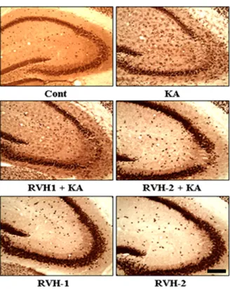
Fig. 4. Representative images of neuronal protection by RVH-1 and RVH-2 against KA-induced neuronal cell death in the CA3 region of hippocampus. In order to further elucidate the neuroprotective properties of RVH-1 and RVH-2, NeuN immunoreactivity, which specifically stains pyramidal neurons in the hippocampus, was examined. Pretreatment of RVH-1 or RVH-2 attenuated KA-induced death of pyramidal neurons in the CA3 region of hippocampus.
RVH-2 showed more neuronal protection against KA-induced excitotoxicity compared to RVH-1. RVH-1 and RVH-2 showed no noticeable change in neuronal viability. More than three mice were used for each group. Scale bar: 100 μm.
Fig. 5. Suppressive effects of RVH-1 and RVH-2 on KA-induced microglial activation. Given the previous report that KA-mediated neuronal death accompanies microglial activation, expression of OX-6, a microglial activation marker, was examined with immuno- histochemical staining at 24 hr after KA or vehicle treatments. KA resulted in increased microglial activation (KA). However, KA-induced microglial activation was attenuated with RVH-1 or RVH-2 pretreatment. RVH-2 appears to be more suppressive than RVH-1.
RVH-1 or RVH-2 showed negligible effects on microglial activation.
Representative images were obtained from three independent experiments. More than three mice were used for each group. Scale bar: 100 μm.
pressed microglial activation in the hippocampus.
RVH-1 and RVH-2, which belong to the stigmasterol fam- ily, were previously isolated from various natural plants [6].
Although RVH-1 and RVH-2 have been reported to inhibit platelet-derived growth factor (PDGF)-driven proliferation of hepatic stellate cells [18], biological activity of these com- pounds has not been extensively studied. In the meantime, it has been reported that stigmasterol exhibits anti-in- flammatory effects in a primary culture model of osteo- arthritis chondrocytes by inhibiting the production of pro- inflammatory mediators [4]. The authors suggested that the anti-inflammatory properties of stigmasterol might be mediated through the inhibition of the NF-kB pathway.
Given the previous report that activation of the NF-kB pathway might contribute to neuronal death in a KA-in- duced excitotoxicity model [19], it is plausible that RVH-1 and RVH-2 might exert a protective effect through in- hibition of NF-kB signaling in a KA-induced excitotoxicity model.
Excitotoxicity may be involved in a variety of pathological conditions such as stroke, traumatic brain injury, and spi- nal cord injury [20-22]. Furthermore, accumulating evi- dence suggests that excitotoxicity contributes to the patho- genesis of neurodegenerative disorders such as Alzheimer’s
disease, Parkinson’s disease, and Huntington’s disease [23,24].
In accordance with a previous study [13], we found that icv administration of KA resulted in the death of neurons mainly in the CA3 region of mouse hippocampus and daily pretreatment of RVH-1 and RVH-2 for 7 day significantly reduced KA-induced death of pyramidal neurons in mouse hippocampus. A single pretreatment with these compounds showed no noticeable inhibition of KA-induced neuronal death (data not shown), suggesting that a single treatment with these compounds is not sufficient to be effective.
Although these compounds exerted protective effects against KA-induced excitotoxicity, it is not certain whether the neu- roprotection by RVH-1 and RVH-2 is through the sup- pression of the NF-kB pathway. Therefore, further studies are necessary to determine the mechanism by which these compounds protect KA-induced neuronal cell death.
KA-induced excitotoxicity also accompanies activation of microglia in the hippocampus [13]. Microglia, the major im- munocompetent cells in the CNS, are believed to play an important role in inflammatory processes in the brain [25].
Concomitant microglial activation during excitotoxicity might contribute to secondary damage by producing neuro- toxic factors such as proinflammatory cytokines and nitric oxide [25,26]. It has been reported that a pathogenic
amount of nitric oxide contributes to neuronal damage in many disease conditions including neurodegenerative dis- eases [27-29]. In the present study, RVH-1 and RVH-2 sig- nificantly attenuated KA-induced microglial activation in the mouse hippocampus. However, it is not clear whether these compounds directly suppressed the microglia or whether the microglial suppression is secondary to the neu- roprotection of these compounds.
In conclusion, the present study demonstrates that RVH-1 and RVH-2 possess neuroprotective properties in an animal model of KA-induced excitotoxicity. These compounds might be potentially valuable in the treatment of brain patholo- gies associated with excitotoxic neuronal damage such as epilepsy, stroke, and traumatic brain injury. However, fur- ther studies are necessary to clearly understand the mecha- nism by which these compounds protect neurons against excitotoxicity. The present study suggests the possibility that detoxification of Rhus verniciflua utilizing mushrooms could be a valuable approach to yield neuroprotective com- pounds from Rhus verniciflua.
ACKNOWLEDGEMENTS
This study was supported by Rural Development Admini- stration of Bio-Green 21 project (PJ007090).
REFERENCES
1. Choi HS. Biological Detoxification of Lacpuer Tree (Rhus verniciflua Stokes) Stem Bark by Mushroom Species. Food Science and Biotechnology. 2007;16:935-942.
2. Awad AB, Fink CS. Phytosterols as anticancer dietary com- ponents: evidence and mechanism of action. J Nutr. 2000;130:
2127-2130.
3. Martins SL, Silva HF, Novaes MR, Ito MK. Therapeutic effects of phytosterols and phytostanols in cholesterolemia. Arch Latinoam Nutr. 2004;54:257-263.
4. Gabay O, Sanchez C, Salvat C, Chevy F, Breton M, Nourissat G, Wolf C, Jacques C, Berenbaum F. Stigmasterol: a phytosterol with potential anti-osteoarthritic properties. Osteoarthritis Cartilage. 2010;18:106-116.
5. Chung IM. Cytotoxic Chemical Constituents from the Mush- room of Pholiota adiposa. Food Science and Biotechnology.
2005;14:255-258.
6. Gupta S. Isolation and characterization of a dihydroxysterol from LAWSONIA INERMIS. Natural Product Letters. 1994;4:
195-201.
7. Greca MD. Stigmasterols from Typha latifolia. J Nat Prod.
1990;53:1430.
8. Chang YC, Chang FR, Khalil AT, Hsieh PW, Wu YC. Cytotoxic Benzophenanthridine and Benzylisiquinoline Alkaloids from Argemone mexicana. Z Naturforsch C. 2003;58:521-526.
9. Itokawa H. Several oxidized sterols isolated from callus tissue of Stephania cepharantha. Chem Pharm Bull. 1973;21:1386- 1387.
10. Choi DW, Rothman SM. The role of glutamate neurotoxicity in hypoxic-ischemic neuronal death. Annu Rev Neurosci. 1990;
13:171-182.
11. Doble A. The role of excitotoxicity in neurodegenerative disease:
implications for therapy. Pharmacol Ther. 1999;81:163-221.
12. Izquierdo LA, Barros DM, Ardenghi PG, Pereira P, Rodrigues C, Choi H, Medina JH, Izquierdo I. Different hippocampal molecular requirements for short- and long-term retrieval of one-trial avoidance learning. Behav Brain Res. 2000;111:93-98.
13. Byun JS, Lee SH, Jeon SH, Kwon YS, Lee HJ, Kim SS, Kim YM, Kim MJ, Chun W. Kainic Acid-induced Neuronal Death is Attenuated by Aminoguanidine but Aggravated by L-NAME in Mouse Hippocampus. Korean J Physiol Pharmacol. 2009;13:
265-271.
14. Giusti P, Lipartiti M, Franceschini D, Schiavo N, Floreani M, Manev H. Neuroprotection by melatonin from kainate-induced excitotoxicity in rats. FASEB J. 1996;10:891-896.
15. Laursen SE, Belknap JK. Intracerebroventricular injections in mice. Some methodological refinements. J Pharmacol Methods.
1986;16:355-357.
16. Baker H, Farbman AI. Olfactory afferent regulation of the dopa- mine phenotype in the fetal rat olfactory system. Neuroscience.
1993;52:115-134.
17. Henshall DC, Bonislawski DP, Skradski SL, Araki T, Lan JQ, Schindler CK, Meller R, Simon RP. Formation of the Apaf-1/
cytochrome c complex precedes activation of caspase-9 during seizure-induced neuronal death. Cell Death Differ. 2001;8:
1169-1181.
18. Badria FA, Dawidar AA, Houssen WE, Shier WT. In vitro study of flavonoids, fatty acids, and steroids on proliferation of rat hepatic stellate cells. Z Naturforsch C. 2005;60:139-142.
19. Nakai M, Qin ZH, Chen JF, Wang Y, Chase TN. Kainic acid-induced apoptosis in rat striatum is associated with nuclear factor-kappaB activation. J Neurochem. 2000;74:647-658.
20. Choi DW. Glutamate neurotoxicity and diseases of the nervous system. Neuron. 1988;1:623-634.
21. Lee JM, Zipfel GJ, Choi DW. The changing landscape of ischaemic brain injury mechanisms. Nature. 1999;399:A7-14.
22. McNamara JO. Emerging insights into the genesis of epilepsy.
Nature. 1999;399:A15-22.
23. Coyle JT, Puttfarcken P. Oxidative stress, glutamate, and neurodegenerative disorders. Science. 1993;262:689-695.
24. Salinska E, Danysz W, Lazarewicz JW. The role of excitotoxi- city in neurodegeneration. Folia Neuropathol. 2005;43:322-339.
25. Suzumura A, Takeuchi H, Zhang G, Kuno R, Mizuno T. Roles of glia-derived cytokines on neuronal degeneration and regeneration. Ann N Y Acad Sci. 2006;1088:219-229.
26. Milatovic D, Gupta RC, Dettbarn WD. Involvement of nitric oxide in kainic acid-induced excitotoxicity in rat brain. Brain Res. 2002;957:330-337.
27. Dehmer T, Lindenau J, Haid S, Dichgans J, Schulz JB.
Deficincye of inducible nitric oxide synthase protects against MPTP toxicity in vivo. J Neurochem. 2000;74:2213-2216.
28. Kim YM, Bombeck CA, Billiar TR. Nitric oxide as a bifunctional regulator of apoptosis. Circ Res. 1999;84:253-256.
29. Sugimoto K, Iadecola C. Effects of aminoguanidine on cerebral ischemia in mice: comparison between mice with and without inducible nitric oxide synthase gene. Neurosci Lett. 2002;331:
25-28.