Copyrightⓒ 2009, The Korean Academy of Oral Biology
1
Journal of Oral Biology
Participation of nitric oxide pathways in interleukin 1β-induced mechani- cal allodynia in the orofacial area of rats
Young M. Kang1, Min K. Lee1, Gwi Y. Yang1, Yong C. Bae2, and Dong K., Ahn1*
1Department of Oral Physiology and 2Oral Anatomy, School of Dentistry, Kyungpook National University, Daegu, Korea (received February 27, 2009 ; revised March 9, 2009 ; accepted March 13, 2009)
The purpose of the present study was to examine the role of peripheral nitric oxide (NO) pathways in the onset of interleukin (IL)-1β-induced mechanical allodynia in the orofacial area. Experiments were carried out on male Sprague-Dawley rats weighing 230-280 gm and surgical procedures were performed under pentobarbital sodium (40 mg/kg, i.p.). Under anesthesia, a polyethylene tube (PE10) was implanted into the subcutaneous area of one vibrissa pad, which enabled the injection of IL-1β or other chemicals. We subcutaneously injected 50µL of IL-1β into a vibrissa pad through the implanted polyethylene tube with a 100µL Hamilton syringe. After the administration of 0.01, 0.1, 1, or 10 pg of IL-1β, withdrawal behavioral responses were examined. The subcutaneous injection of saline had no effects on the air-puff thresholds. Following the sub- cutaneous injection of 0.01, 0.1, 1, or 10 pg of IL-1β, the threshold of air puffs decreased significantly to 12± 3, 7 ± 2, 5± 1, or 5 ± 1 psi, respectively, in a dose dependent manner.
Pretreatment with L-NAME, a nitric oxide synthase (NOS) inhibitor, blocked IL-1β-induced mechanical allodynia.
However, neither D-NAME, an inactive isomer of L- NAME, nor vehicle affected the IL-1β-induced mechanical allodynia. Subcutaneous injection of IL-1β increased the number of c-fos-like immunoreactive neurons, whereas pretreatment with L-NAME decreased this number, in the trigeminal caudal nucleus. These results suggest that pro- inflammatory cytokines and NO are important con- tributors to the pathogenesis of persistent and exaggerated IL-1β-induced pain states. Based on these observations,
peripheral application of NOS inhibitors may be of therapeutic value in treating pain disorders in the clinic.
Key wordk: NO in IL-1β-induced mechanical allodynia
Introduction
NO, produced from L-arginine by NOS, plays important roles in a wide variety of physiological and patho- physiological processes such as neurotransmission, regulation of vascular tone, and mediation of immune responses (Snyder, 1992; Moncada and Higgs, 1993; Prast and Philippu, 2001). At the site of inflammation, the released inflammatory agents may sensitize primary afferent fibers and cause plasma protein extravasation.
Inflammatory mediators, which produce the formation and release of endothelin-derived relaxing factor/ NO at the site of the injury, activate nociceptors inducing vasodilatation (Dickenson et al., 1992; Evans, 1995). Human and animal studies support a role for NO in mediating peripheral nociceptive transmission and inflammation. Subcutaneous injection of NO evoked pain in a dose dependent manner by direct exciting nociceptors in a human study (Holthusen and Arndt, 1994). In the animal study, intraplantar injection of NG-nitro-L-arginine methyl ester hydrochloride (L- NAME), a NOS inhibitor, produced dose-related anti- nociception assessed by use of three different experimental procedures including formalin-induced paw licking behavior, acetic acid induced abdominal constriction and the hot plate test (Moore et al., 1991). Evidence to support a role of NO in mediating the development of peripheral inflammation comes from the work of Honore et al. (1995), who observed that systemic injection of L-NAME reduced the edema formation induced by carrageenan injection in
*Corresponding author: Dong-Kuk, Ahn, DDS, PhD, Department of Oral Physiology, School of Dentistry, Kyungpook National University, 188-1 Sam Deok 2 ga, Chung-gu, Daegu (700-412), Korea. Tel.: +82-53-660-6840; Fax.: +8282-53-421-4077; E-mail:
the paw as well as c-fos expression in the spinal cord. NO can also be produced following activation of several pain- related chemicals including bradykinin (Dickenson et al., 1992), histamine (Beyak and Vanner, 1995) and N-methyl- D-aspartate (NMDA) receptors (Garthwaite et al., 1989).
These results suggest that NO pathway is involved in the development of inflammation and pain.
A recent study has demonstrated peripheral cytokine- induced mechanical allodynia in the orofacial area (Ahn et al., 2004, 2005). The air-puff thresholds that produced withdrawal behavioral responses decreased significantly in a dose-dependent manner after injection of IL-1β (Ahn et al., 2004, 2005). Pretreatment with an IL-1 receptor antagonist abolished the decrease in the air-puff thresholds.
These results suggest that peripheral cytokine can produce mechanical allodynia in the orofacial area. Moreover, there is growing evidences that cytokines and NO play an important role in central and peripheral modulation of nociception (Riedel and Neeck, 2001; Bredt and Snyder, 1989; Snyder, 1992). Expression of NO or release of pro- inflammatory cytokines has a high correlation with acceding pain intensity in chronic pain patients (Koch et al., 2007). The effect of the pro-inflammatory cytokine such as IL-1β on the inducible NOS-NO cascade in nociceptive signal transduction was examined in the intact rat spinal cord (Sung et al., 2004). Recently it has been shown that patients suffering from chronic orofacial pain experience significantly increased plasma levels of NO metabolites when compared with controls (Gratt and Anbar, 2005).
Although peripheral NO pathways are involved in the orofacial pain, direct behavioral evidence for participation of NO pathways in the IL-1β-induced mechanical allodynia in the orofacial area is unclear. The purpose of the present study was to examine a role of peripheral NO pathways in the IL-1β-induced mechanical allodynia in the orofacial area. To achieve this purpose, the present study investigated effects of L-NAME, a nitric oxide synthase (NOS) inhibitor, on IL-1β-induced mechanical allodynia and examined the effects of L-NAME on the expression of c-fos like immu- noreactive neurons in the trigeminal spinal nucleus in rats.
Materials and Methods
Animals
Experiments were carried out on male Sprague-Dawley rats weighing 230-280 gm and surgical procedures were performed under pentobarbital sodium (40 mg/kg, i.p.). All procedures involving the use of animals were approved by the Institutional Care and Use Committee of the School of Dentistry, Kyungpook National University and carried out in accordance with the ethical guidelines for the investigation of experimental pain in conscious animals of IASP. All behavioral responses were measured by an experimenter who was blind to the treatment group.
Surgery
Under anesthesia, a polyethylene tube (PE10) was implanted into the subcutaneous area of one vibrissa pad, which enabled the injection of IL-1β or drugs in freely moving rats. The polyethylene tubes were subcutaneously led to the top of the skull and secured in place by means of a stainless steel screw and dental acrylic resin on the skull.
Animals were housed one per cage and were maintained under constant temperature and lighting conditions with a 12- hour light/dark cycle. Food and water were freely available. Animals had a recovery time of at least 72 hours from surgery.
Evaluation of mechanical allodynia
For behavioral observation, rats were placed in customized observation cages with holes opened in the top so that the head could move through it for the application of air puffs. The cage was placed in a darkened and noise-free room and the animals habituated for at least 30 min. We injected 50µL of IL-1β into a vibrissa pad subcutaneously through the implanted polyethylene tube with 100µL Hamilton syringe. The cannula had a dead space volume of 7µL, and 10 µL saline was injected to flush the cannula following each drug microinjection. After the administr- ation of 0.01, 0.1, 1, or 10 pg of IL-1β, we examined withdrawal behavioral responses produced by 10 successive trials of constant pressure of air puffs (4 sec duration, 10 sec interval) applied to the IL-1β injection site. The intensity of air puffs was controlled by a pneumatic pump module (BH2 system, Harvard apparatus). Air puffs were applied through a 26 gauge metal tube (length, 10 cm) located 1 cm away from skin. Allodynia was defined as an intensity of air puffs when rats attempted to escape or aggressive behavioral responses including biting the metal tube. The 50%
threshold of air puffs was calculated for each rat. The cut-off pressure of air-puff was 25 psi. Trials were terminated if the withdrawal responses did not occur within 25 psi. The normal animals usually did not respond below 25 psi pressure.
Evaluation of nitric oxide pathways on IL-1β-induced mechanical allodynia
The air-puff thresholds were measured at 10, 30, 60, 120, or 180 min after 50µl of IL-1β was applied to the vibrissa pad subcutaneously through the implanted tube. These responses, suggestive of noxious sensory input, are evoked by cutaneous stimuli and are only elicited by stimulation of discrete and localized cutaneous regions near the cytokine injection site. The participation of peripheral NO pathways in the IL-1β-induced mechanical allodynia was investigated. A NG-nitro-L-arginine methyl ester (L- NAME, 100µg, 30 µl), a NOS inhibitor, or NG-nitro-D- arginine methyl ester (D-NAME, 100µg, 30 µl), an inactive isomer of NAME, was administered through the implanted tube 10 min prior to injection of 10 pg IL-1β. In the control
group, saline was injected subcutaneously.
Immunohistochemical detection of c-Fos protein in trigeminal caudal nucleus
The anesthetized animals were divided into three groups:
saline-treated group, IL-1β group, and L-NAME-pretreated group. After air-puffs stimulation (10 sec duration with 10 sec interval) for 3 minutes, animals were deeply anesthetized with pentobarbital sodium (40 mg /kg i.p.) in 3 hr after the air-puff stimulation. The anesthetized rats were transcardically perfused with 100 mL saline followed by 400 ml of 4% paraformaldehyde in 0.1 M phosphate buffer (pH 7.2). After fixation, the lower brain stem (from the obex to 5 mm caudal to the obex) was removed, postfixed for 4 hr in the same fixative, and cryoprotected overnight in 30%
sucrose at 4oC. The frozen transverse sections (30µm thick) were cut with a cryostat. After pretreatment with 3%
H2O2 to inhibit endogenous peroxidase, the free-floating sections were kept in blocking serum (0.1% Triton X-100 and 2% bovine serum albumin, 5% normal goat serum, 5%
fetal bovine serum in 0.01 M PBS) for 1 hr at room temperature. The sections were incubated for 48 hrs at 4 °C with rabbit polyclonal anti-Fos antibody (Oncogene Science, 1:1000) and were sequentially incubated in biotinylated goat anti-rabbit immunoglobulins (IgG) and avidin–biotin peroxidase complex (ABC-Staining System, Vector Labs., USA). The immune reaction was visualized using 3,3’-diaminobenzidine (DAB) (Sigma, USA). The control was performed by omitting the primary antibody.
Statistical analysis
Differences between groups were compared using analysis of repeated measures ANOVAs followed by LSD post hoc analysis. Comparisons between the two means were performed by a Student’s t-test. In all statistical comparisons, p<0.05 was used as the criterion for statistical significance. All data is presented as mean± SEM.
Results
The present results showed a subcutaneous injection of IL- 1β into the orofacial area produced mechanical allodynia.
The changes in the air-puff thresholds after a subcutaneous injection of IL-1β are illustrated in Fig. 1. The naive animals did not respond to below 25 psi pressure. The subcutaneous injection of saline had no effects on the air-puff thresholds.
After the subcutaneous injection of 0.01, 0.1, 1, or 10 pg of IL-1β, the air-puff thresholds decreased significantly to 12 ± 3, 7 ± 2, 5 ± 1, or 5 ± 1 psi respectively, with a dose- dependent manner (p<0.05). The decrease in the air-puff thresholds appeared 10 mins after an injection of IL-1β and persisted for over 3 hrs (p<0.05).
Fig. 2 illustrates effects of pretreatment with L-NAME or D-NAME on IL-1β-induced mechanical allodynia. The
subcutaneous injection of 10 pg of IL-1β significantly decreased air-puffs thresholds (p<0.05). Pretreatment with L-NAME, a NOS inhibitor, significantly blocked IL-1β- induced mechanical allodynia. However, neither pretreatment with D-NAME, an inactive isomer of NAME, nor saline (vehicle) affected IL-1β-induced mechanical allodynia.
The present study examined c-fos expression in the trigeminal spinal nucleus after air-puffs stimulation (10 sec Fig. 1. Changes in the threshold of air puffs producing withdrawal behavioral responses after IL-1β was injected subcutaneously into a vibrissa pad. Subcutaneous injection of IL-1β produced mechani- cal allodynia. All doses of IL-1β produced a significant decrease in the threshold of air puffs ipsilateral to the IL-1β injection site com- pared with the saline-treated group (p<0.05). The mechanical allo- dynia persisted for over 3 hours after IL-1β injection. The cut-off pressure of air puff intensity was 25 psi. Trials were terminated if the responses did not occur below 25 psi of air pressure. There were 9 animals in each group.
Fig. 2. Changes in the IL-1β-induced air-puffs threshold after L- NAME was injected subcutaneously into a vibrissa pad 10 min prior to the IL-1β injection. Pretreatment with L-NAME, a NOS inhibitor, blocked IL-1β-induced mechanical allodynia. However, neither D-NAME, an inactive isomer of NAME, nor vehicle affected IL-1β-induced mechanical allodynia. There were 9 ani- mals in each group. * p < 0.05 vs. saline-treated group.
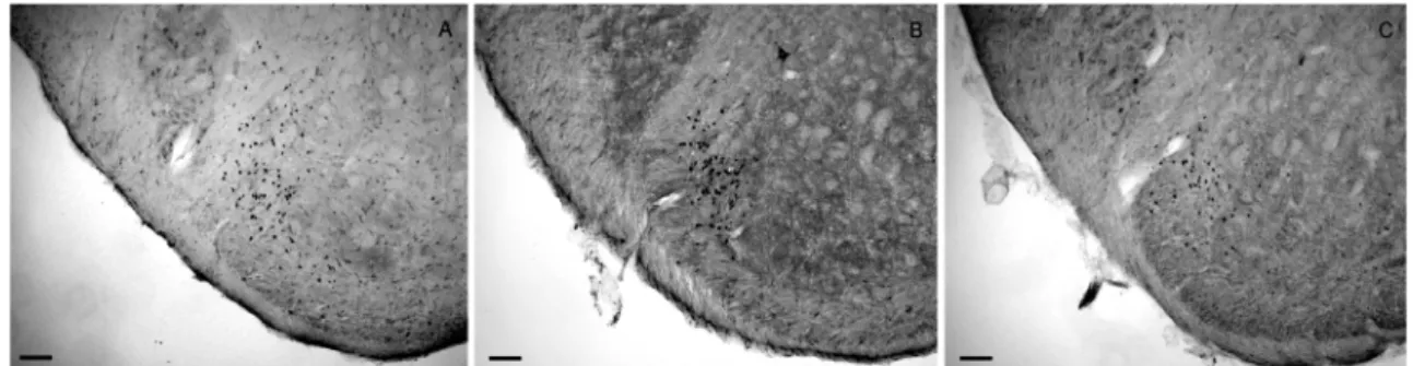
duration with 10 sec interval) for 3 minutes. The typical microphotographics of c-fos like immunoreactive neurons are illustrated in Fig. 3. The most c-fos like immunoreactive neurons were detected lamina III and IV. IL-1β increased the number of c-fos like immunoreactive neurons and pretreatment with L-NAME blocked increases in the number of c-fos like immunoreactive neurons produced by subcutaneous injection of IL-1β. The c-fos like immunoreactive neurons were counted subsequent 5 sections and the average number of c-fos like immunoreactive neurons are illustrated in Fig. 4. The air- puffs stimulation produced 33 ± 6 of c-fos like immunoreactive neurons in the saline-treated rats. IL-1β significantly increased c-fos like immunoreactive neurons to 75 ± 7 neurons per section, as compared with saline- treated rats. Pretreatment with L-NAME, NOS inhibitor, significantly blocked increases in the number of the c-fos like immunoreactive neurons to 46 ± 8 neurons per section.
Discussion
The present study demonstrated that the subcutaneous injection of L-NAME, NOS inhibitor, reduced IL-1β- induced mechanical allodynia in the orofacial area of rats.
The particaipation of NO pathways in the nociceptive signal transduction of pro-inflammatory cytokine in the intact rat spinal cord has been examined in a previous study (Sung et al., 2004). Intrathecal administration of IL-1β produced thermal hyperalgesia and the expression of inducible nitric oxide synthase (iNOS) protein, and release of NO (Sung et al., 2004). Wieseler-Frank et al. (2007) also demonstrated that release of pro-inflammatory cytokine, including NOS, in the meninges has a potential role in the modulation of pain. Intrathecal administration of gp120 increased gene expression of pro-inflammatory signals, including tumor necrosis factor-alpha (TNF-α), IL-1β, IL-6, and iNOS in meninges. The present study also demonstrated that peripheral administration of L-NAME, but not D-NAME, blocked decreased air-puff thresholds produced by IL-1β injection. These results, taken together with previous data,
suggest that pro-inflammatory cytokines and NO are important contributors to the pathogenesis of persistent and exaggerated pain states as humoral signaling molecules.
Tissue injury or the presence of foreign material initiates a series of pathophysiological events that may manifest as inflammatory pain. The initiating factor can produce the release of several pain mediators that control the threshold and activation of nociceptors. It has been suggested that many nociceptors associated with inflammatory pain are dormant, and are activated by cyclo-oxygenase(COX) metabolites and sympathomimetic amines in hyperalgesia.
In this state, pain receptors may be activated by previously ineffective stimuli. The release of TNF-α initiates the release of IL-1 and IL-8, which in turn liberate COX metabolites and sympathomimetic amines, respectively. It is assumed that sensitization of nociceptors is due to increased concentrations of cAMP/Ca++ in the sensory neurons. The effect of increased cAMP concentrations may be counteracted by stimulation of the arginine/nitric oxide/
cGMP pathway (Ferreira, 1993).
Moreover, local inflammatory mediators may play an Fig. 3. Photomicrographs of c-fos like immunoreactive neurons in the ipsilateral trigeminal spinal nucleus. Air puff stimulation (10 sec dura- tion with 10 sec interval) was applied for 3 min in the saline-treated rat (left panel), IL-1β-treated rat (middle panel), and L-NAME-pre-treated rat (right panel). Scale bars = 100µm.
Fig. 4. Effects of pretreatment with L-NAME on IL-1β-induced c- fos like immunoreactive neurons. *p < 0.05 vs. saline-treated group.
# p < 0.05 vs. IL-1β-treated group.
important role in the development of neuropathic pain following peripheral nerve injury. One important participant in the inflammatory response of injured peripheral nerve may be NO. Sciatic nerve injury associated with behavioral evidence of neuropathic pain had substantial rises in local blood flow. The NOS inhibitor, L-NAME, but not D- NAME, reversed the hyperaemia in a dose-dependent fashion. Aminoguanidine, a NOS inhibitor, also reversed nerve hyperaemia distal to the constriction. These results provide evidence for critical role of local NOS and NO in the chronic constriction injury model of neuropathic pain.
These results suggest that NO has local physiological actions that include vasodilatation of microvessels and that may be important in the development of pain sensitivity (Levy and Zochodne, 1998).
Combined injections of L-NAME with formalin into the plantar aspect of one hindpaw of normal rats elicited a dose- dependent suppression of c-fos expression as compared to that induced by formalin alone. However, combined injections of L-arginine with formalin elicited considerable enhancement of c-fos expression when the dosages of L- arginine were less than 20µM, while it elicited marked suppression of c-fos expression when the dosages were in the range from 50 to 100µM (Wang et al., 1999). These results suggest that although NO pathway may participate in the pain modulation, the action of NO is still in controversy.
Recently participation of peripheral NO pathways in antinociception of COX inhibitors has been introduced (Ventura-Martinez et al., 2004). Oral administration of indomethacin, a nonselective COX inhibitor, significantly decreased the nociceptive response elicited by uric acid injected into the knee joint of the right hind limb.
Indomethacin-induced antinociception was reduced by local pretreatment with L-NAME, a NOS inhibitor. These results suggest that the antinociceptive effect of indomethacin involved, at least in part, the NO-cyclic GMP pathway at a peripheral level (Ventura-Martinez et al., 2004).
The present study demonstrated that subcutaneous injection of IL-1β produced mechanical allodynia in the orofacial area. Inflammation is associated with a complex pattern of local and systemic changes including inflammatory cell migration, cytokines release, edema, erythema, release of acute phase proteins, fever, pain and hyperalgesia. The precise molecular events responsible for sensory changes at the site of inflammation and surrounding tissue are not yet fully understood, but changes in the transduction sensitivity and activation of chemosensitive nociceptors by inflammatory mediators are involved (Treede et al., 1992). IL-1β, a cytokine, released from activated macrophages and monocytes during infection, plays an important role in acute inflammatory responses (Dinarello, 1998). Since the first reports showed cytokines having a role in mediating inflammatory hyperalgesia (Ferreira et al., 1988; Schweizer et al., 1998) several studies
have demonstrated cytokines to be involved in pain modulations. Several studies have demonstrated peripheral cytokines to be involved in hyperalgesia. Peripheral IL-1β may play an important role in the cutaneous hyperalgesia by activating polymodal receptors to mechanical and thermal stimulation in rats (Fukuoka et al., 1994). Cytokines mediated inflammatory hyperalgesia were also limited by an IL-1β receptor antagonist (Cunha et al., 2000). Several cell types produced IL-1β including blood monocytes, tissues macrophages, blood neutrophils during inflammation (Dinarello, 1991; Libby et al., 1986). The final hyperalgesic mediator might be a prostaglandin or sympathomimetic amine and the release of these agents is usually preceded by the release of the other mediator.
Besides this kind of mediators, the present study investigated the participation of peripheral NO pathways in IL-1β-induced mechanical allodynia.
An air puff or air jet stimulation was used as an evaluation of mechanical allodynia in the orofacial area. The air puff stimulation was applied to the IL-1β injection site and then the air-puff threshold was measured when rats produced withdrawal behavioral responses. An air puffs test is a convenient method for producing and qualifying mechanical allodynia compared to conventional methods for evaluating pain in the orofacial region of rats. The air puff stimulation was also used as a useful pain evaluating method in previous studies. Air puffs or air jet stimulation produced facilitation of single unit activity at spinal cord in anesthetized rats (Sherman et al., 1997; Yaksh, 1989) and air puffs startle stimulation was used to evaluate pain related hormonal changes in the rats (Anand et al., 1999). In the present study, the normal animals usually did not respond to air puffs within the proposed range. The subcutaneous injection of IL-1β produced orofacial mechanical allodynia in the experiment conditions.
In summary, the subcutaneous injection of IL-1β produced mechanical allodynia in the orofacial area.
Pretreatment with L-NAME, a NOS inhibitor, but not D- NAME, an inactive isomer of NAME, blocked IL-1β- induced mechanical allodynia. IL-1β significantly increased the number of c-fos like immunoreactive neurons and pretreatment with L-NAME, a NOS inhibitor, significantly blocked increases in the number of the c-fos like immunoreactive neurons. These results suggest that pro-inflammatory cytokines and NO are important contributors to the pathogenesis of persistent and exaggerated pain states. Based on these observations, peripheral application of NOS inhibitors may be of therapeutic value in treating pain disorders in the clinic.
Acknowledgement
This work was supported by the Korea Research Foundation Grant funded by the Korean Government
(MOEHRD, Basic Research Promotion Fund)(KRF-2008- 314-E00213).
References
Ahn DK, Jung CY, Lee HJ, Choi HS, Ju JS, Bae YC.
Peripheral glutamate receptors participate in interleukin- 1beta-induced mechanical allodynia in the orofacial area of rats. Neurosci Lett. 2004;357: 203-6.
Ahn DK, Kim KH, Jung CY, Choi HS, Lim EJ, Youn DH, Bae YC. Role of peripheral group I and II metabotropic glutamate receptors in IL-1beta-induced mechanical allodynia in the orofacial area of conscious rats. Pain 2005;118:53-60.
Anand KJ, Coskun V, Thrivikraman KV, Nemeroff CB, Plotsky PM. Long term behavioral effects of repetitive pain in neonatal rat pups. Physiol Behav. 1999;66:627-37.
Beyak M, Vanner S. Histamine H1 and H3 vasodilator mechanisms in the guinea pig ileum. Gastroenterology 1995;108:712-8.
Bredt DS, Snyder SH. Nitric oxide mediates glutamate-linked enhancement of cGMP levels in the cerebellum. Proc Natl Acad Sci. USA 1989;86:9030–3.
Cunha JM, Cunha FQ, Poole S, Ferreira SH. Cytokine- mediated inflammatory hyperalgesia limited by interleukin- 1 receptor antagonist. Br J Pharmacol. 2000;103:1418-24.
Dickenson A, Haley J, Schachter M, Chapman V.
Electrophysiological approaches to the study of bradykinin and nitric oxide in inflammatory pain. Agents Actions Suppl. 1992;38:358-65.
Dinarello CA. Biology of interleukin 1. FASEB J. 1998;2:108-15.
Dinarello CA. Interleukin-1 and interleukin-1 antagonism.
Blood 1991;8:1627-52.
Evans CH. Nitric oxide: what role does it play in inflammation and tissue destruction? Agents Actions Suppl. 1995;47:107- 16.
Ferreira SH, Lorenzetti BB, Bristow AF, Poole S. Interleukin- 1 beta as a potent hyperalgesic agent antagonized by a tripeptide analogue. Nature 1988;334:698-700.
Ferreira SH. The role of interleukins and nitric oxide in the mediation of inflammatory pain and its control by peripheral analgesics. Drugs 1993;46(Suppl 1):1-9.
Fukuoka H, Kawatani M, Hisamitsu T, Takeshige C.
Cutaneous hyperalgesia induced by peripheral injection of interleukin-1 β in the rat. Brain Res. 1994;657:133-40.
Garthwaite J, Garthwaite G, Palmer RM, Moncada S. NMDA receptor activation induces nitric oxide synthesis from arginine in rat brain slices. Eur J Pharmacol. 1989;172:413-6.
Gratt BM, Anbar M. A pilot study of nitric oxide blood levels in patients with chronic orofacial pain. Oral Surg Oral Med Oral Pathol Oral Radiol Endod. 2005;100:441-8.
Holthusen H, Arndt JO. Nitric oxide evokes pain in humans on intracutaneous injection. Neurosci Lett. 1994;165:71-4.
Honore P, Chapman V, Buritova J, Besson JM. Reduction of carrageenan oedema and the associated c-Fos expression in the rat lumbar spinal cord by nitric oxide synthase inhibitor.
Br J Pharmacol. 1995;114:77-84.
Koch A, Zacharowski K, Boehm O, Stevens M, Lipfert P, von Giesen HJ, Wolf A, Freynhagen R. Nitric oxide and pro- inflammatory cytokines correlate with pain intensity in chronic pain patients. Inflamm Res. 2007;56:32-7.
Levy D, Zochodne DW. Local nitric oxide synthase activity in a model of neuropathic pain. Eur J Neurosci. 1998;10:1846- 55.
Libby P, Ordovas JM, Auger KR, Robbins AH, Birinyi LK, Dinarello CA. Endotoxin and tumour necrosis factor induce interleukin-1 gene expression in human vascular endothelial cells. Am J Pathol. 1986;124:179-85.
Moncada S, Higgs A. The l-arginine-nitric oxide pathway. N Engl J Med. 1993;329:2002-12.
Moore PK, Oluyomi AO, Babbedge RC, Wallace P, Hart SL.
L-NG-nitro arginine methyl ester exhibits antinociceptive activity in the mouse. Br J Pharmacol. 1991;102:198-202.
Prast H, Philippu A. Nitric oxide as modulator of neuronal function. Prog Neurobiol. 2001;64:51-68.
Riedel W, Neeck G. Nociception, pain, and antinociception:
current concepts. Z Rheumatol. 2001;60:404–15.
Schweizer A, Feige U, Fontana A, Muller K, Dinarello CA.
Interleukin-1 enhances pain reflexes. Mediation through increased prostaglandin E2 levels. Agents Actions 1998;25:246-51.
Sherman SE, Luo L, Dostrovsky JO. Spinal strychinin alters responses properties of nociceptive-specific neurons in rat medial thalamus. J Neurophysiol. 1997;78:628-637.
Snyder SH. Nitric oxide: first in a new class of neurotransmitters. Science 1992;257:494-6.
Sung CS, Wen ZH, Chang WK, Ho ST, Tsai SK, Chang YC, Wong CS. Intrathecal interleukin-1beta administration induces thermal hyperalgesia by activating inducible nitric oxide synthase expression in the rat spinal cord. Brain Res.
2004;1015:145-53.
Treede RD, Meyer RA, Raja SN, Campbell JN. Peripheral and central mechanisms of cutaneous hyperalgesia. Prog Neurobiol. 1992;38:397-421.
Ventura-Martinez R, Deciga-Campos M, Diaz-Reval MI, Gonzalez-Trujano ME, Lopez-Munoz FJ. Peripheral involvement of the nitric oxide-cGMP pathway in the indomethacin-induced antinociception in rat. Eur J Pharmacol. 2004;503:43-8.
Wang H, Nie H, Zhang RX, Qiao JT. Peripheral nitric oxide contributes to both formalin- and NMDA-induced activation of nociceptors: An immunocytochemical study in rats. J Neurosci Res. 1999;57:824-9.
Wieseler-Frank J, Jekich BM, Mahoney JH, Bland ST, Maier SF, Watkins LR. A novel immune-to-CNS communication pathway: cells of the meninges surrounding the spinal cord CSF space produce proinflammatory cytokines in response to an inflammatory stimulus. Brain Behav Immun.
2007;21:711-8.
Yaksh TL. Behavioral and autonomic correlates of the tactile evoked allodynia produced by spinal glycine inhibition.
Effects of modulatory receptor systems and excitatory amino acid antagonists. Pain 1989;37:111-23.