363 http://dx.doi.org/10.4196/kjpp.2011.15.6.363
ABBREVIATIONS: ERK, extracellular signal-regulated kinase; GPX, glutathione peroxidase; IL-1β, interleukin-1β; iNOS, inducible nitric oxide synthase; L-NIL, N6-(1-iminoethyl)-L-lysine; MAPK, mitogen-activated protein kinase; MSU, monosodium urate; SOD, superoxide dismutase; TNF-α, tumor necrosis factor-α.
Received September 30, 2011, Revised December 15, 2011, Accepted December 16, 2011
Corresponding to: So-Young Park, Department of Physiology, Yeung- nam University College of Medicine, 170, Hyunchung-ro, Nam-gu, Daegu 705-717, Korea. (Tel) 82-53-620-4334, (Fax) 82-53-651-3651, (E-mail) sypark@med. yu.ac.kr
CCThis is an Open Access article distributed under the terms of the Creative Commons Attribution Non-Commercial License (http://creativecommons.org/licenses/
by-nc/3.0) which permits unrestricted non-commercial use, distribution, and reproduction in any medium, provided the original work is properly cited.
Inhibition of Inducible Nitric Oxide Synthase Attenuates Monosodium Urate-induced Inflammation in Mice
Tae-Jin Ju1,2, Jin-Myoung Dan3, Young-Je Cho4, and So-Young Park1,2
1Department of Physiology, 2Aging-associated Vascular Disease Research Center, College of Medicine, Yeungnam University, Daegu 705-717,
3Department of Orthopedic Surgery, Gumi CHA University Hospital, Gumi 730-728, 4School of Food Science & Biotechnology, Food &
Bio-Industry Research Institute, Kyungpook National University, Daegu 702-701, Korea
The present study elucidated the effect of the selective inducible nitric oxide synthase (iNOS) inhibitor N6-(1-iminoethyl)-L-lysine (L-NIL) on monosodium urate (MSU) crystal-induced inflammation and edema in mice feet. L-NIL (5 or 10 mg/kg/day) was administered intraperitoneally 4 h before injection of MSU (4 mg) into the soles of mice hindlimb feet. Twenty-four hours after MSU injection, foot thickness was increased by 160% and L-NIL pretreatment reduced food pad swelling in a dose dependent manner. Pretreatment of 10 mg/kg/day L-NIL significantly suppressed the foot pad swelling by MSU. Plasma level of nitric oxide (NO) metabolites and gene expression and protein level of iNOS in feet were increased by MSU, which was suppressed by L-NIL pretreatment. Similar pattern of change was observed in nitrotyrosine level. MSU increased the gene expression of tumor necrosis factor (TNF)-α and interleukin (IL)-1β and L-NIL pretreatment suppressed MSU-induced cytokines expression. The mRNA levels of superoxide dismutase and glutathione peroxidase1 were increased by MSU and L-NIL pretreatment normalized the gene expression. Phosphorylation of extracellular signal-regulated kinase 1/2 and p38 was increased by MSU, which was suppressed by L-NIL pretreatment. The mRNA levels of iNOS, TNF-α, and IL-1β were increased by MSU in human dermal fibroblasts, C2C12 myoblasts, and human fetal osteoblasts in vitro, which was attenuated by L-NIL in a dose dependent manner. This study shows that L-NIL inhibits MSU-induced inflammation and edema in mice feet suggesting that iNOS might be involved in MSU-induced inflammation.
Key Words: Uric acid, Gout, iNOS
INTRODUCTION
Gout is caused by hyperuricemia through altered purine metabolism. Although asymptomatic hyperuricemia is com- mon and the majority of patients never develop gout [1], gout is a common cause of arthritis affecting more than 1%
of the adult population [2]. When the local concentration of uric acids exceeds the limit of solubility, monosodium ur- ate (MSU) crystals are generated and precipitate in the joints, kidneys, and soft tissues, causing inflammation and leading to gout [3]. MSU crystals stimulate local connective tissue cells, monocyte-macrophages, and neutrophils to pro- duce a variety of inflammatory cytokines including tumor necrosis factor (TNF)-α, interleukin (IL)-8, IL-1β, IL-6 and monocyte chemotactic factor, which collectively induce
acute inflammation [4,5].
Nitric oxide (NO) is a small gas molecule synthesized by three isoforms of NO synthase (NOS) [6]. Although NO from two constitutive isoforms (endothelial NOS and neuro- nal NOS) is critical in a wide variety of physiological func- tions [6], overexpression of inducible NOS (iNOS) is in- volved in a variety of pathological conditions including in- flammation [7,8]. Although the major metabolites of NO are nitrite and nitrate [9], NO can be transformed to peroxyni- trite under oxidative stress, which produces nitrated pro- teins including nitrotyrosine [10]. Nitrotyrosine is a marker of peroxynitrate production and nitrative stress [11,12].
Expression of iNOS is also increased in MSU-stimulated chondrocytes and in the synovial tissue of gouty arthritis patients, suggesting a potential role of iNOS in the patho- genesis of arthritis [13,14]. However, no direct evidence of the involvement of iNOS in gout has been reported. In this study, the role of iNOS in gouty arthritis was elucidated by examining whether a selective iNOS inhibitor sup- pressed MSU-induced inflammation in a mouse foot model.
METHODS Animals
Seven-week-old male C57BL/6 mice were housed in a room operating with a 12:12 h light/dark cycle. All the mice were fed a standard chow diet with free access to water.
For the preparation of MSU crystals, 4 g of uric acid was dissolved at 60oC in 800 ml of 0.5 M NaOH (pH 8) and cooled overnight at 4oC. After discarding the supernatant, precipitated crystals were collected, washed and dried. The needle-like crystals were dissolved in saline (0.04 g/500μl) and 4 mg/50μl of the solution was injected into soles of hindlimb feet of the mice [15]. This concentration is high enough to induce inflammation since the solubility of uric acid in plasma is 6.8 mg/dl [16]. The iNOS selective in- hibitor N6-(1-iminoethyl)-L-lysine (L-NIL; Cayman Chemi- cal, Ann Arbor, MI, USA)[17,18] was injected into mice in- traperitoneally (10 mg/kg) 4 h before MSU injection.
Twenty four hours after MSU injection, edema of feet was measured using digital calipers (Mitutoyo Corporation, Kawasaki-shi, Kanagawa, Japan) in mice anesthetized with an intraperitoneal injection of tiletamine and zolezepam (25 mg/kg) and zylazine (10 mg/kg). Blood was collected from the retro-orbital plexus using micro-hematocrit capillary tubes coated with heparin. The blood was centrifuged and plasma was stored at −80oC for further analysis. The feet were excised and stored at −80oC for further analysis. This study was conducted in accordance with the guidelines for the care and use of laboratory animals provided by Yeungnam University, and all experimental protocols were approved by the Ethics Committee of Yeungnam Uni- versity.
Cell culture
Human dermal fibroblast (HDF) was purchase from Lonza (Walkersville, MS, USA) and mouse myoblast cell line, C2C12, and human fetal osteoblastic cell line, hFOB1.19, were purchased from the American Type Cul- ture Collection (Manassas, VA, USA). HDF and C2C12 were culture in Dulbecco’s Modified Eagle Media (DMEM;
GIBCO, Grand Island, NY, USA) at 37oC in a 5%
CO2-humidifier incubator. hFOB1.19 cells were cultured in a 1:1 mixture of DMEM and F12 (GIBCO) without phenol red at 36.5oC in an atmosphere containing 5% CO2. The cells were plated at 1×10⁶ to each of 6 wells and treated with 2 mg MSU for 2 h. L-NIL (0.25 or 0.5 mg/ml) was added into the cells 30 min before MSU treatment. RNA extraction was performed for the quantitative real time pol- ymerase chain reaction (qRT-PCR).
qRT-PCR
qRT-PCT was performed as previously described [19].
Briefly, 25 mg tissue samples were homogenized in TRI re- agent (Sigma-Aldrich, St. Louis, MO, USA) and RNA was reverse transcribed to cDNA using a reverse transcription kit (Applied Biosystems, Foster City, CA, USA). Quantita- tive real-time PCR was performed using the Real-Time PCR 7500 System and Power SYBR Green PCR Master Mix (Applied Biosystems) according to the manufacturer's instructions. Expression levels of β-actin were used for sample normalization. Each reaction mixture was in- cubated at 95oC for 10 min followed by 45 cycles of 95oC
for 15 s, 55oC for 20 s and 72oC for 35 s. Sequences of pri- mers were based on the National Center for Biotechnology Information nucleotide database and were designed using the Primer Express Program (Applied Biosystems). The pri- mer sequences were: mouse β-actin (121 bp: forward, 5'-TGG ACA GTG AGG CAA GGA TAG-3'; reverse, 5'-TAC TGC CCT GGC TCC TAG CA-3'), mouse iNOS (71 bp: for- ward, 5'-CTC CTG CCT CAT GCC ATT-3'; reverse, 5'-TGT TCC TCT ATT TTT GCC TCT TTA-3'), mouse TNF-α (71 bp: forward, 5'-CTA TCT CCA GGT TCT CTT CAA-3'; re- verse, 5'-GCA GAG AGG AGG TTG ACT TTC), mouse IL-1β (71 bp: forward, 5'-GCC CAT CCT CTG TGA CTC-A-3'; re- verse, 5'-AGT GCA GCT GTC TAA TGG GA-3'), mouse su- peroxide dismutase (SOD; 71 bp: forward, 5'-CTG CTC TAA TCA GGA CCC ATT-3'; reverse, 5'-GTG CTC CCA CAC GTC AAT C-3'), mouse glutathione peroxidase 1 (GPx1; 71 bp: forward, 5'-GAA GTG CGA AGT GAA TGG TG-3'; re- verse, 5'-TGG GTG TTG GCA AGG C-3'), human β-actin (72 bp: forward, 5'-ACC GCA TCG TCA CCA AC-3'; reverse, 5'-CCA CAC GCA GCT CAT TGT A-3'), human iNOS (192 bp:forward, 5'-TTA TGA CTC CCA AAA GTT TGA CCA-3';
reverse, 5'-CCG TCA GTT GGT AGG TTA CTG TTG-3'), human TNF-α (72 bp: forward, 5'-AGA GGG CCT GTA CCT CAT CTA-3'; reverse, 5'-AGC AGC ACA TGG GTC GAG-3'), and IL-1β (133 bp: forward, 5'-TCC AGG GAC AGG ATA TGG AG-3'; reverse, 5'-TCT TTC AAC ACG CAG GAC AG-3').
Western blotting
Foot samples were used for measurement of the protein level of phosphorylated extracellular signal-related kinase (pERK; Cell Signaling Technologies, Danvers, MA, USA), ERK (Cell Signaling Technologies), p38 (Cell Signaling Technologies), p-p38 (Cell Signaling Technologies), iNOS (Santa Cruz Biotechnology, Santa Cruz, CA, USA), nitro- tyrosine (Upstate Biotechnology, Lake Placid, NY, USA), and glyceraldehyde 3-phosphate dehydrogenase (GAPDH;
Santa Cruz Biotechnology). Western blotting was per- formed as previously described [12]. Briefly, 25 mg tissue samples were homogenized in lysis buffer (Invitrogen, Carlsbad, CA, USA) and extracted protein was separated by 10% sodium dodecyl sulfate-polyacrylamide gel electro- phoresis (SDS-PAGE). Resolved proteins were transferred to a polyvinylidene fluoride membrane (Millipore, Billerica, MA, USA). After blocking with 5% skim mile, the mem- brane was incubated overnight at 4oC with primary anti- body and then specific antibody binding was detected using sheep anti-rabbit IgG horseradish peroxidase or goat an- ti-mouse IgG horseradish peroxidase (Bio-Rad, Hercules, CA, USA) for 1 h at room temperature, except for nitro- tyrosine, which was detected using mouse anti-mouse IgG horseradish peroxidase (Bio-Rad). The binding was vi- sualized using an enhanced chemiluminescence detection regent (Millipore).
Nitrite and nitrate
Plasma concentrations of the NO-derived end products nitrite and nitrate were measured by a TotalNOAssay Kit (R&D Systems, Minneapolis, MN, USA). To minimize inter- ference with plasma protein, the sample was ultra-filtered through a 10 kDa cut-off filter (Millipore) prior to the assay.
Fig. 1. Thickness of feet in mice injected with monosodium-urate (MSU) into hindlimb feet sole. N6-(1-iminoethyl)-L-lysine (L-NIL;
5 and 10 mg/kg) was injected intraperitoneally 4 h before MSU (4 mg) injection. Data is presented as mean±SE. Experimental cases are seven in each group. *p<0.05 vs. Saline, #p<0.05 vs.
MSU.
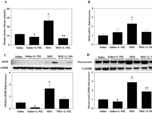
Fig. 2. Nitric oxide metabolites in plasma (A) and the expression of inducible nitric oxide synthase (iNOS;
B, C) and nitrotyrosine (D) in the feet of mice injected with monoso- dium-urate (MSU) into hindlimb sole. N6-(1-iminoethyl)-L-lysine (L- NIL; 10 mg/kg) was injected intra- peritoneally 4 h before MSU (4 mg) injection. Data is presented as mean
±SE. Experimental cases are seven in each group. *p<0.05 vs. Saline, #p
<0.05 vs. MSU.
Statistical analysis
Data are expressed as mean ± standard error (SE). The difference among groups was analyzed by ANOVA with a post-hoc analysis by Duncan’s multiple test. Statistical sig- nificance was at p<0.05.
RESULTS Feet edema
MSU injection into the soles induced redness, swelling, and heat in mice feet (data not shown) and significantly
increased the thickness of feet by 160% at 24 h compared with the saline injected feet. Pretreatment of 5 and 10 mg/kg L-NIL suppressed MSU-induced edema by 12% and 40%, respectively, while L-NIL alone had no effect on the thickness of feet (Fig. 1). Since 10 mg/kg/day L-NIL sig- nificantly reduced MSU-induced foot pad swelling, this con- centration of L-NIL was used in this experiment.
iNOS expression
Plasma levels of the NO metabolites nitrite and nitrate was reduced by L-NIL administration compared with control. MSU injection increased the NO metabolites in plasma, which was reversed by L-NIL pretreatment (Fig.
2A). While gene expression of iNOS was not affected by L-NIL, the mRNA level of iNOS in feet was significantly increased by MSU. L-NIL pretreatment suppressed MSU- induced gene expression of iNOS (Fig. 2B). Protein level of iNOS in feet showed the same pattern of changes as with iNOS gene expression, except that L-NIL treatment alone significantly reduced the level of iNOS protein (Fig. 2C).
Nitrotyrosine level in feet was significantly increased by MSU injection, which was partially suppressed by L-NIL pretreatment. Like the iNOS protein level, nitrotyrosine was significantly reduced by L-NIL treatment (Fig. 2D).
Gene expression of inflammatory cytokines and anti- oxidant enzyme
Gene expression of TNF-α was also increased by MSU, which was significantly reduced by L-NIL pretreatment.
However, MSU-induced TNF-α expression was not com- pletely normalized by L-NIL pretreatment compared with saline-injected control mice. L-NIL treatment did not alter the mRNA level of TNF-α. While L-NIL had no effect on the gene expression of IL-β, the mRNA level of IL-1β was significantly increased by MSU. L-NIL pretreatment sup-
Fig. 3. The mRNA level of inflam- matory cytokines and antioxidant enzymes in the foot of mice injected with monosodium-urate (MSU) into hindlimb sole. N6-(1-iminoethyl)-L- lysine (L-NIL; 10 mg/kg) was in- jected intraperitoneally 4 h before MSU (4 mg) injection. Interleukin (IL)-1β (A); tumor necrosis factor (TNF)-α (B); glutathione peroxidase (GPx) 1 (C); superoxide dismutase (SOD) (D). Data is presented as mean±SE. Experimental cases are seven in each group. *p<0.05 vs.
Saline, #p<0.05 vs. MSU.
Fig. 4. The phosphorylation of extra- cellular signal-related kinase (ERK) 1/2 (A) and p38 (B) in the foot of mice injected with monosodium-urate (MSU) into hindlimb sole. N6-(1-imi- noethyl)- L-lysine (L-NIL; 10 mg/kg) was injected intraperitoneally 4 h before MSU (4 mg) injection. Data is presented as mean±SE. Experimen- tal cases are seven in each group. *p
<0.05 vs. Saline, #p<0.05 vs. MSU.
pressed MSU-induced gene expression of IL-1β. MSU also elevated the mRNA level of GPX1 and SOD and L-NIL pre- treatment suppressed MSU-induced the gene expression of these antioxidants enzymes (Fig. 3).
Mitogen-activated protein kinase (MAPK) expression Phosphorylation of ERK1/2 and p38 was increased by MSU, which was reversed by L-NIL pretreatment. L-NIL alone did not alter the phosphorylation of ERK1/2 and p38 (Fig. 4). Phosphorylation of c-Jun-N-terminal kinase (JNK) was not affected by either MSU or L-NIL (data not shown).
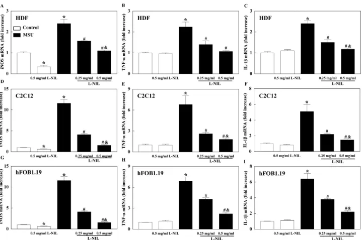
Gene expression of iNOS and cytokines in cell lines Gene expression of iNOS was increased in HDF, C2C12, and hFOB1.19 by MSU and L-NIL pretreatment attenu- ated iNOS mRNA expression in a dose dependent manner.
Gene expression of iNOS was reduced by L-NIL treatment alone. MSU also increased mRNA expression of TNF-α and
IL-1β in HDF, C2C12, and hFOB1.19, which were sig- nificantly attenuated by L-NIL in a dose dependent manner (Fig. 5).
DISCUSSION
The present study demonstrates that MSU induces ede- ma and increases inflammatory cytokine expression in feet that is accompanied with increased iNOS expression. The selective iNOS inhibitor L-NIL suppresses the edema and cytokine expression. These results suggest that iNOS is in- volved in the development of gout.
The association between iNOS and rheumatoid arthritis or/and osteoarthritis has been extensively investigated and a large body of evidence suggests the important role of iNOS in these arthritis. The expression of iNOS is in- creased in the synovium, chondrocytes, vascular smooth muscle and inflammatory cells of rheumatoid arthritis and osteoarthritis subjects [20-24]. Moreover, suppression of
Fig. 5. The mRNA level of iNOS, tumor necrosis factor (TNF)-α, and interleukin (IL)-1β in human dermal fibroblast (HDF; A∼C), C2C12 myoblast, and (D∼F) human fetal osteoblast (hFOB)1.19 (G∼I). The cells were treated with 2 mg/ml monosodium-urate (MSU) for 2 h.
N6-(1-iminoethyl)-L-lysine (L-NIL) was treated into cells 30 min before MSU treatment. Data is presented as mean±SE of three separate experiments. *p<0.05 vs. Control, #p<0.05 vs. MSU, &p<0.05 vs. 0.25 mg/ml L-NIL.
iNOS expression in rheumatoid arthritis and osteoarthritis attenuates cell apoptosis, cytokine production and arthritis [20,25-27].
Despite extensive investigations as to the role of iNOS in rheumatoid arthritis and osteoarthritis, the relationship between iNOS and gouty arthritis has not been well- defined. Previous research has indicated that MSU in- creases iNOS expression in cells such as macrophages and chondrocytes in vitro [13,14,28]. Similar to this finding, we presently showed that MSU increased iNOS expression in fibroblast, myoblast, and osteoblast in vitro. Furthermore, we also observed that iNOS expression was increased in mice feet by MSU treatment, which is the first study show- ing increased iNOS expression by MSU treatment in vivo.
The elevated levels of NO metabolites in plasma indirectly support increased iNOS expression by MSU. Additionally, the suppression of MSU-induced iNOS expression by L-NIL resulted in attenuated cytokine expression and edema.
Consistent with our results, iNOS expression is enhanced in synovial tissues of gouty arthritis patients [13]. Together with this previous data, the current results support the no- tion that increased iNOS expression plays a causative role in the inflammation induced by MSU.
The mechanism by which MSU induces iNOS expression is unclear presently, but it is possible that MSU increases iNOS expression through MAPK pathways. MSU increases
the MAPK subfamily member JNK, p38, and ERK1/2 [13,29,30], which are involved in a variety of MSU-induced pathological pathways [31]. ERK1/2 is involved in MSU- mediated transcriptional activation of IL-8 that functions as a neutrophil chemoattractant factor [29]. Inhibition of ERK1/2 or p38 reduces MSU-induced monocyte chemo- attractant protein-1 in vascular smooth muscle cells [32].
MSU-induced iNOS expression is also mediated by p38 and ERK1/2 in chondrocytes and macrophages [13,14,28]. MSU activates p38 through the phosphorylation of proline-rich tyrosine kinase 2/focal adhesion kinase/protein paxillin, which increases iNOS expression and NO production [14].
Inhibition of ERK1/2 by specific inhibitor suppresses MSU- induced iNOS expression [13,28]. Consistent with these previous results, we presently observed that MSU in- creased the phosphorylation of ERK1/2 and p38. Addition- ally, the suppression of MSU-induced iNOS expression by L-NIL was accompanied with decreased the phosphor- ylation of both ERK1/2 and p38. Taken together, these re- sults suggest the involvement of ERK/12 or/and p38 in MSU-induced iNOS expression. Although JNK is also in- creased in human monocyte cells line by MSU [33], JNK was not presently increased by MSU. Difference of species and experimental conditions could be the possible reasons.
We assume that increased iNOS expression could acti- vate the production of inflammatory cytokines, which sub-
sequently play a key role in gouty arthritis. Our notion is supported by the observation that suppression of MSU-in- duced iNOS expression by L-NIL presently attenuated cyto- kine expression and edema. A Previous study also demon- strated that the NO donor S-nitroso-acetyl penicillamine in- creases inflammatory cytokines such as TNF-α [34]. Gouty arthritis and administration of MSU crystal results in the production of a variety of inflammatory cytokines [35-37]
and increased expressions of anti-inflammatory cytokines such as transforming growth factor β1, IL-1 receptor an- tagonist, IL-10, and soluble TNF receptor are correlated with spontaneous resolution of gouty arthritis [38].
Gouty arthritis is associated with oxidative stress [39]
and the suppression of oxidative stress can reduce the symptoms [40]. Oxidative stress in gout may involve MSU- induced iNOS expression since elevated NO production from iNOS induces oxidative stress [12]. Presently, MSU increased the gene expression of anti-oxidant enzymes, which was normalized by L-NIL treatment. Antioxidant en- zyme may be increased to protect the tissue from oxidative damage and attenuated oxidative stress by L-NIL normal- izes the enzyme levels. Increased nitrotyrosine level by MSU was attenuated by L-NIL treatment presently, which also supports our theory.
In summary, MSU induces iNOS expression that is asso- ciated with increased expression of inflammatory cytokines, oxidative stress, and edema. ERK1/2 or/and p38 may medi- ates MSU-induced iNOS expression. Suppression of iNOS could be a new therapeutic target for gout.
ACKNOWLEDGEMENTS
This work was supported by the Ministry of Knowledge Economy grant (70007082) and by the Korea Science and Engineering Foundation grant (2010-0007389) funded by the Korea government.
REFERENCES
1. Keith MP, Gilliland WR. Updates in the management of gout.
Am J Med. 2007;120:221-224.
2. Sabina EP, Rasool M, Mathew L, Ezilrani P, Indu H. 6-Shogaol inhibits monosodium urate crystal-induced inflammation--an in vivo and in vitro study. Food Chem Toxicol. 2010;48:229-235.
3. Eggebeen AT. Gout: an update. Am Fam Physician. 2007;76:
801-808.
4. Di Giovine FS, Malawista SE, Nuki G, Duff GW. Interleukin 1 (IL 1) as a mediator of crystal arthritis. Stimulation of T cell and synovial fibroblast mitogenesis by urate crystal-induced IL 1. J Immunol. 1987;138:3213-3218.
5. Sabina EP, Chandal S, Rasool MK. Inhibition of monosodium urate crystal-induced inflammation by withaferin A. J Pharm Pharm Sci. 2008;11:46-55.
6. Michel T, Feron O. Nitric oxide synthases: which, where, how, and why? J Clin Invest. 1997;100:2146-2152.
7. Noronha BT, Li JM, Wheatcroft SB, Shah AM, Kearney MT.
Inducible nitric oxide synthase has divergent effects on vascular and metabolic function in obesity. Diabetes. 2005;54:
1082-1089.
8. Tsuchiya K, Sakai H, Suzuki N, Iwashima F, Yoshimoto T, Shichiri M, Hirata Y. Chronic blockade of nitric oxide synthesis reduces adiposity and improves insulin resistance in high fat-induced obese mice. Endocrinology. 2007;148:4548-4556.
9. Kelm M. Nitric oxide metabolism and breakdown. Biochim Biophys Acta. 1999;1411:273-289.
10. Torres SH, De Sanctis JB, de L Briceño M, Hernández N, Finol HJ. Inflammation and nitric oxide production in skeletal muscle of type 2 diabetic patients. J Endocrinol. 2004;181:419- 11. Beckman JS, Koppenol WH. Nitric oxide, superoxide, and per-427.
oxynitrite: the good, the bad, and ugly. Am J Physiol. 1996;271:
C1424-1437.
12. Cha HN, Kim YW, Kim JY, Kim YD, Song IH, Min KN, Park SY. Lack of inducible nitric oxide synthase does not prevent aging-associated insulin resistance. Exp Gerontol. 2010;45:711- 718.
13. Chen L, Hsieh MS, Ho HC, Liu YH, Chou DT, Tsai SH. Stim- ulation of inducible nitric oxide synthase by monosodium urate crystals in macrophages and expression of iNOS in gouty arthritis. Nitric Oxide. 2004;11:228-236.
14. Liu R, Lioté F, Rose DM, Merz D, Terkeltaub R. Proline-rich tyrosine kinase 2 and Src kinase signaling transduce mono- sodium urate crystal-induced nitric oxide production and mat- rix metalloproteinase 3 expression in chondrocytes. Arthritis Rheum. 2004;50:247-258.
15. Sabina EP, Rasool M. An in vivo and in vitro potential of Indian ayurvedic herbal formulation Triphala on experimental gouty arthritis in mice. Vascul Pharmacol. 2008;48:14-20.
16. Becker MA, Schumacher HR Jr, Wortmann RL, MacDonald PA, Eustace D, Palo WA, Streit J, Joseph-Ridge N. Febuxostat compared with allopurinol in patients with hyperuricemia and gout. N Engl J Med. 2005;353:2450-2461.
17. Connor JR, Manning PT, Settle SL, Moore WM, Jerome GM, Webber RK, Tjoeng FS, Currie MG. Suppression of adjuvant- induced arthritis by selective inhibition of inducible nitric oxide synthase. Eur J Pharmacol. 1995;273:15-24.
18. Chicoine LG, Tzeng E, Bryan R, Saenz S, Paffett ML, Jones J, Lyons CR, Resta TC, Nelin LD, Walker BR. Intratracheal adenoviral-mediated delivery of iNOS decreases pulmonary vasoconstrictor responses in rats. J Appl Physiol. 2004;97:1814- 1822.
19. Cha HN, Hong GR, Kim YW, Kim JY, Dan JM, Park SY.
Deficiency of iNOS Does Not Prevent Isoproterenol-induced Cardiac Hypertrophy in Mice. Korean J Physiol Pharmacol.
2009;13:153-159.
20. van’t Hof RJ, Hocking L, Wright PK, Ralston SH. Nitric oxide is a mediator of apoptosis in the rheumatoid joint. Rheuma- tology (Oxford). 2000;39:1004-1008.
21. Mäki-Petäjä KM, Cheriyan J, Booth AD, Hall FC, Brown J, Wallace SM, Ashby MJ, McEniery CM, Wilkinson IB. Inducible nitric oxide synthase activity is increased in patients with rheumatoid arthritis and contributes to endothelial dysfunc- tion. Int J Cardiol. 2008;129:399-405.
22. Grabowski PS, Wright PK, Van’t Hof RJ, Helfrich MH, Ohshima H, Ralston SH. Immunolocalization of inducible nitric oxide synthase in synovium and cartilage in rheumatoid arthritis and osteoarthritis. Br J Rheumatol. 1997;36:651-655.
23. Heale CE, Fåhraeus-Van Ree GE, Rahman P, Richardson VJ.
Progressive and concordant expression of PKC-eta and iNOS phenotypes in monocytes from patients with rheumatoid arthritis: association with disease severity. J Histochem Cyto- chem. 2007;55:495-503.
24. Borderie D, Hilliquin P, Hernvann A, Kahan A, Menkes CJ, Ekindjian OG. Nitric oxide synthase is expressed in the lymphomononuclear cells of synovial fluid in patients with rheumatoid arthritis. J Rheumatol. 1999;26:2083-2088.
25. Vuolteenaho K, Moilanen T, Hämäläinen M, Moilanen E.
Regulation of nitric oxide production in osteoarthritic and rheumatoid cartilage. Role of endogenous IL-1 inhibitors.
Scand J Rheumatol. 2003;32:19-24.
26. Järvinen K, Vuolteenaho K, Nieminen R, Moilanen T, Knowles RG, Moilanen E. Selective iNOS inhibitor 1400W enhances anti-catabolic IL-10 and reduces destructive MMP-10 in OA cartilage. Survey of the effects of 1400W on inflammatory mediators produced by OA cartilage as detected by protein antibody array. Clin Exp Rheumatol. 2008;26:275-282.
27. Cuzzocrea S, Chatterjee PK, Mazzon E, McDonald MC, Dugo
L, Di Paola R, Serraino I, Britti D, Caputi AP, Thiemermann C. Beneficial effects of GW274150, a novel, potent and selective inhibitor of iNOS activity, in a rodent model of collagen-induced arthritis. Eur J Pharmacol. 2002;453:119-129.
28. Jaramillo M, Naccache PH, Olivier M. Monosodium urate crystals synergize with IFN-gamma to generate macrophage nitric oxide: involvement of extracellular signal-regulated kinase 1/2 and NF-kappa B. J Immunol. 2004;172:5734-5742.
29. Liu R, O'Connell M, Johnson K, Pritzker K, Mackman N, Terkeltaub R. Extracellular signal-regulated kinase 1/extra- cellular signal-regulated kinase 2 mitogen-activated protein kinase signaling and activation of activator protein 1 and nuclear factor kappaB transcription factors play central roles in interleukin-8 expression stimulated by monosodium urate monohydrate and calcium pyrophosphate crystals in monocytic cells. Arthritis Rheum. 2000;43:1145-1155.
30. Liu W, Kato M, Itoigawa M, Murakami H, Yajima M, Wu J, Ishikawa N, Nakashima I. Distinct involvement of NF-kappaB and p38 mitogen-activated protein kinase pathways in serum deprivation-mediated stimulation of inducible nitric oxide synthase and its inhibition by 4-hydroxynonenal. J Cell Bio- chem. 2001;83:271-280.
31. Liu-Bryan R, Lioté F. Monosodium urate and calcium pyro- phosphate dihydrate (CPPD) crystals, inflammation, and cell- ular signaling. Joint Bone Spine. 2005;72:295-302.
32. Kanellis J, Watanabe S, Li JH, Kang DH, Li P, Nakagawa T, Wamsley A, Sheikh-Hamad D, Lan HY, Feng L, Johnson RJ.
Uric acid stimulates monocyte chemoattractant protein-1 pro- duction in vascular smooth muscle cells via mitogen-activated protein kinase and cyclooxygenase-2. Hypertension. 2003;41:
1287-1293.
33. Inokuchi T, Ka T, Yamamoto A, Moriwaki Y, Takahashi S,
Tsutsumi Z, Tamada D, Yamamoto T. Effects of ethanol on monosodium urate crystal-induced inflammation. Cytokine.
2008;42:198-204.
34. McInnes IB, Leung BP, Field M, Wei XQ, Huang FP, Sturrock RD, Kinninmonth A, Weidner J, Mumford R, Liew FY. Pro- duction of nitric oxide in the synovial membrane of rheumatoid and osteoarthritis patients. J Exp Med. 1996;184:1519-1524.
35. di Giovine FS, Malawista SE, Thornton E, Duff GW. Urate crystals stimulate production of tumor necrosis factor alpha from human blood monocytes and synovial cells. Cytokine mRNA and protein kinetics, and cellular distribution. J Clin Invest. 1991;87:1375-1381.
36. Chapman PT, Yarwood H, Harrison AA, Stocker CJ, Jamar F, Gundel RH, Peters AM, Haskard DO. Endothelial activation in monosodium urate monohydrate crystal-induced inflamma- tion: in vitro and in vivo studies on the roles of tumor necrosis factor alpha and interleukin-1. Arthritis Rheum. 1997;40:955- 37. Rider TG, Jordan KM. The modern management of gout. 965.
Rheumatology (Oxford). 2010;49:5-14.
38. Chen YH, Hsieh SC, Chen WY, Li KJ, Wu CH, Wu PC, Tsai CY, Yu CL. Spontaneous resolution of acute gouty arthritis is associated with rapid induction of the anti-inflammatory factors TGFβ1, IL-10 and soluble TNF receptors and the intracellular cytokine negative regulators CIS and SOCS3. Ann Rheum Dis. 2011;70:1655-1663.
39. Ghio AJ, Kennedy TP, Rao G, Cooke CL, Miller MJ, Hoidal JR. Complexation of iron cation by sodium urate crystals and gouty inflammation. Arch Biochem Biophys. 1994;313:215-221.
40. Facchini FS. Near-iron deficiency-induced remission of gouty arthritis. Rheumatology (Oxford). 2003;42:1550-1555.