257 DOI: 10.4196/kjpp.2010.14.5.257
ABBREVIATIONS: KA, kainic acid; GFAP, glial fibrillary acidic protein; IR, immunoreactivity; IL-1β, interleukin-1beta; TNF-α, tumor necrosis factor-alpha; IL-6, interleukin-6; COX-2, cyclooxy- genase-2.
Received May 11, 2010, Revised September 3, 2010, Accepted September 10, 2010
Corresponding to: Hong-Won Suh, Department of Pharmacology, College of Medicine, Hallym University, 1, Okchun-dong, Chuncheon 200-702, Korea. (Tel) 82-33-248-2614, (Fax) 82-33-248-2612, (E-mail) [email protected]
*Equally contributed to first author.
Neuroprotective Effect of Visnagin on Kainic Acid-induced Neuronal Cell Death in the Mice Hippocampus
Min-Soo Kwon2,*, Jin-Koo Lee1,*, Soo-Hyun Park1, Yun-Beom Sim1, Jun-Sub Jung1, Moo-Ho Won3, Seon-Mi Kim1, and Hong-Won Suh1
1Department of Pharmacology, Institute of Natural Medicine, College of Medicine, Hallym University, Chuncheon 200-702, 2Department of Aerospace Medical Research, Aerospace Medical Center, ROKAF (Republic of Korea Air Force), Cheongwon 363-842, 3Department of Anatomy and Neurobiology, and Institute of Neurodegeneration and Neuroregeneration, College of Medicine, Hallym University, Chuncheon 200-702, Korea
Visnagin (4-methoxy-7-methyl-5H-furo[3,2-g][1]-benzopyran-5-one), which is an active principle extracted from the fruits of Ammi visnaga, has been used as a treatment for low blood-pressure and blocked blood vessel contraction by inhibition of calcium influx into blood cells. However, the neuroprotective effect of visnagin was not clearly known until now. Thus, we investigated whether visnagin has a neuroprotective effect against kainic acid (KA)-induced neuronal cell death. In the cresyl violet staining, pre-treatment or post-treatment visnagin (100 mg/kg, p.o. or i.p.) showed a neuro- protective effect on KA (0.1 μg) toxicity. KA-induced gliosis and proinflammatory marker (IL-1β, TNF- α, IL-6, and COX-2) inductions were also suppressed by visnagin administration. These results suggest that visnagin has a neuroprotective effect in terms of suppressing KA-induced pathogenesis in the brain, and that these neuroprotective effects are associated with its anti-inflammatory effects.
Key Words: Neuroprotection, Kainic acid, Hippocampus, Visnagin, Cytokines
INTRODUCTION
Kainic acid (KA), which is an analog of the excitatory amino acid L-glutamate, elicits neuronal cell death followed by severe status epilepticus in the pyramidal layer of the hippocampal CA3 region [1] when KA is administered in- tracerebroventricularly (i.c.v.). It may be due to excessive activation of neurons by excitatory neurotransmitters (e.g.
glutamate), which are massively released as a consequence of energy depletion and which result in excitotoxic neuron death [2,3]. Recent studies have demonstrated that the KA-induced neuronal death is associated with the activa- tion of microglia and astrocytes in the hippocampus, and that these processes are induced by enhanced reactive oxy- gen species (ROS) production and cytokine expressions [4,5]. The microglia also can be detected using the comple- ment receptor type 3 (OX-42) IR. The complement receptor type 3 is important in the adherence of neutrophils and monocytes to stimulated endothelium, and also in the phag- ocytosis of complement coated particles that raises the last step of the microglial activation [6]. Expressions, hyper- trophy and proliferation of Glial fibrillary acidic protein (GFAP) specifically is found in astroglia, a cell type which
is highly responsive to neurologic insults [7]. After brain injury, astrocytes undergo a number of cellular syntheses and release of a variety of growth factors and immunomod- ulatory cytokines [7]. In addition, recent studies have dem- onstrated that inflammatory and apoptotic processes con- tribute to the later stages of the damage induced by various brain injuries, and that these detrimentally affect neuro- logic outcome [5,8]. Among them, it has been reported well that pro-inflammatory mediators such as IL-1β, TNF-α, IL-6, and COX-2 can lead to deteriorative effect in the brain, and KA can trigger an aberrant inflammatory cyto- kine response by microglial cells and accelerated disease progression [4].
Visnagin (4-methoxy-7-methyl-5H-furo[3,2-g][1]-benzopyran- 5-one) is an active principle extracted from the fruits of Ammi visnaga [9]. The fruit or its isolated active compo- nents have been used for the treatment of angina pectoris due to their peripheral and coronary vasodilator activity [10,11]. In isolated aorta, visnagin, and other related active principles present in these fruits such as visnadin and khel- lin inhibited vascular smooth muscle contractility, probably by acting at multiple sites to decrease the availability of Ca2+ required for activation [12-14]. Several reports have demonstrated that visnagin descents blood pressure and blocks blood vessel contraction as inhibiting calcium influx into cell [15]. However, the neuroprotective effect of visna- gin on kainic acid (KA)-induced neuronal death has not
been demonstrated until now although visnagin has been studied for therapeutic use for over 10 years.
In the present study, we explored the neuroprotective ef- fects of visnagin in KA-induced neuronal cell death model.
The peroral (p.o.) or intraperitoneal (i.p.) administration of visnagin remarkably suppresses hippocampal cell death, in- dicating that visnagin has a neuroprotective effect against KA-induced neuronal cell death.
METHODS
These experiments were approved by the Hallym Univer- sity Animal Care and Use Committee. All procedures were conducted in accordance with the 'Guide for Care and Use of Laboratory Animals' published by the National Institutes of Health. Male ICR mice (MJ Co., Seoul, Korea) weighing 25∼28 g were used for all the experiments.
Experimental animals
Male ICR mice (MJ Co., Seoul, Korea) weighing 25∼28 g were used for all the experiments. Animals were housed 5 per cage in a room maintained at 22±0.5oC with an alter- nating 12 hr light-dark cycle. Food and water were avail- able ad libitum. The animals were allowed to adapt to the laboratory for at least 2 hr before testing and were only used once. To reduce variation, all experiments were per- formed during the light phase of the cycle (10:00∼17:00).
Drug treatment and i.c.v. kainic acid injection Visnagin was purchased from Acros organics. Visnagin was prepared following steps: (A) 1 g of decursinol was dis- solved in 0.5 ml of ethanol plus 0.5 ml of polyethylene glycol 400 (B) Separately; 100 mg of sodium carboxymethyl- cellulose was dissolved in 9 ml of distilled water. (C) Finally, Solution (A) and Solution (B) were vigorously mixed. These solutions excluding visnagin were used as ve- hicle control. The KA (Sigma, USA) was dissolved in a phos- phate buffer solution. The i.c.v. administrations of KA were performed following the procedure established by Laursen and Belknap [16]. Briefly, each mouse was injected at breg- ma with a 50 μl Hamilton microsyringe fitted with a 26-gauge needle that was inserted to a depth of 2.4 mm.
Cresyl violet staining method and histological analysis Animals were sacrificed for the brain sample by perfusion at 1 day after KA administration. All perfusion procedures were worked in the fume hood. For perfusion, all mice were first deeply anesthetized with sodium pentobarbital (100 mg/kg, i.p., Hanlim Pharm., Korea) and perfused intra- cardially with physiological saline followed with ice-cold phosphate-buffered 4% paraformaldehyde (pH 7.4). Whole brain was removed from the skull and postfixed in the same fixative for 4 hrs at 4oC. Then the brains were cryoprotected in 30% sucrose for 24 hrs at 4oC and sectioned coronally (45 μm) on a freezing microtome and collected in cryopro- tectant for storage at −20oC until processed. Prepared sec- tions were rinsed 3×10 min in PBS to remove cryopro- tectant. Sections were mounted on microscope slides (Fisher, USA) and dried on air. The slides were soaked in cresyl violet working solution (0.02% buffer solution; 0.2%
sodium acetate, 0.3% acetic acid) for 30 min. Then the sec-
tions were dehydrated through alcohol and xylene and cov- erslipped using Permount (Fisher, USA).
Histological analysis method in pyramidal layer of hippo- campal CA3 region was performed following under proce- dures [17]. The number of cresyl violet-positive neurons was counted by two blinded observers at the same time us- ing an image analyzing system equipped with a com- puter-based CCD camera (Olympus AX70, USA). The num- ber of cresyl violet-positive neurons in the CA3 region of the hippocampus was counted in 3 sections in reference to the mouse atlas [18] for each animal [19]. Starting from the first section (interaural 2.10 mm, bregma −1.70 mm), counts were taken from at least three coronal sections at 0.135 mm increments. Thus, we could always perform neu- ronal counting of the same brain region and minimize any counting bias. The number of cresyl violet-positive neurons was compared to that of the control group of the same brain area from all animals. All experiments were conducted in- dependently twice. The neuronal counting of the same group was combined for final analysis.
Isolation of total RNA
The entire process for isolation of total RNA was con- ducted three times independently. Finally, the animal num- ber used for each group was nine. Three animals of each group were dissected for real time PCR analysis. Total cel- lular RNA was extracted from dissected hippocampus tis- sue using a rapid guanidine thiocyanate-watersaturated phenol/chloroform extraction procedure and subsequent precipitation with acidic sodium acetate [20]. Total cellular RNA in the aqueous phase was precipitated with ice-cold isopropyl alcohol. Isolated RNA samples were subjected to spectrophotometric analysis at 260 nm and 280 nm. The separated organic layer was extracted twice with an equal volume of sterilized (Millipore, USA) water and proteins were precipitated by adding two volumes of absolute etha- nol to the water-extracted organic phase. The dried pellets were dissolved in a denaturing buffer (6 M guanidium chloride, 20 mM Tris-HCl [pH 8.0], and 1 mM EDTA).
Real-time PCR analysis
Expression of TNF-α, IL-1β, IL-6, COX-2 and GAPDH mRNA was evaluated by real-time PCR using QuantiTectTM SYBRⓇ Green PCR Kit (Qiagen, Germany). All PCRs were performed in total volume of 20 μl using the QuantiTectTM SYBRⓇ Green PCR Kit (Qiagen, Germany). Each reaction contained 1.5 μl of cDNA, 6.5 μl RNase-Free Water, each 1 μl of sense and antisense primer (20 μM) and 10 μl of 2X SYBRⓇ Green PCR Master Mix (containing QuantiTect SYBR Green PCR buffer, dNTPs, SYBR Green I dye, ROX dye, and HotStarTaq DNA polymerase). After an initial de- naturation step at 95oC for 30 s, temperature cycling with a total of 40 cycles was initiated. Each cycle consisted of a denaturation phase at 95oC for 30 s, an annealing phase at 60oC for 30 s and an elongation phase at 72oC for 30 s. Amplification was followed by melting curve analysis to verify the correctness of the amplification. A negative con- trol with water instead of cDNA was run within every PCR to assess specificity of the reaction. To verify the accuracy of the amplification, PCR products were further analyzed on ethidium bromide-stained 2% agarose gel. For data anal- ysis, Rotor-Gene 6000 Series Software 1.7 (Build 87) was used. Results are given as a ratio of the amount of TNF-α,
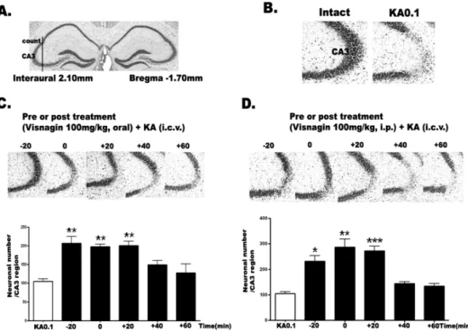
Fig. 1. The effect of visnagin ad- ministered orally or intraperitone- ally on KA-induced neuronal death in the hippocampus. The location of the hippocampal CA3 region per- forming cresyl violet-positive neu- ronal count is indicated (A) refer- ring Franklin [18]. I.c.v. kainic acid injecton induced neuronal cell death in the pyramidal cells in the CA3 region of hippocampus (B).
Mice were administered visnagin orally (C) or intraperitoneally (D) 20 min (−20) prior to KA (0.1 μg/
5 μl) injection or 0, 20 (+20), 40 (+40), 60 (+60) min after KA treatment (0.1 μg/5 μl). And then, the cresyl violet staining was per- formed at 1 day after KA. The ver- tical bars indicate the standard er- ror of mean. *p<0.05, **p<0.01,
***p<0.001 (KA 0.1 vs other groups). The mice number of each group was 10.
IL-1β, IL-6, COX-2 mRNA to that of GAPDH mRNA. The following primers were used: TNF-α (NM_013693) sense:
CAT CTT CTC AAA ATT CGA GTG ACA A, antisense: TGG GAG TAG ACA AGG TAC AAC CC; IL-1β (NM_008361) sense: TCT CGC AGC AGC ACA TCA, antisense: CAC ACA CCA GCA GGT TAT; IL-6 (NM_031168) sense: GAG GAT ACC ACT CCC AAC AGA CC, antisense: AAG TGC ATC ATC GTT GTT CAT ACA; COX-2 (AF344876) sense: TTC AAA AGA AGT GCT GGA AAA GGT, antisense: GAT CAT CTC TAC CTG AGT GTC TTT and GAPDH (NM_008084) sense: TCG TGG ATC TGA CGT GCC GCC TG, antisense:
CAC CAC CCT GTT GCT GTA GCC GTA T. The entire process from dissection to RT-PCR was conducted three times independently. Finally, the animal number used for each group was nine. Each real time PCR result (indepen- dently three times) was quantified with GraphPad Prism Version 4.0 for Windows (GraphPad Software, USA) for statistical analysis.
Immunohistochemistry
Sections were cut with a cryostat at a thickness of 45 μm.
Immuno-histochemical staining was performed with Elite ABC Kit (Vector Laboratories, USA). Sections were first rinsed with 0.1 M PBS three times for 10 min each, then pre-incubated in 0.1M PBS containing 1% BSA and 0.2%
Triton X-100 for 30 min. After rinsing twice with 0.1 M PBS containing 0.5% BSA for 10∼15 min each, sections were incubated with antibody against GFAP (1:50,000; Sigma, USA) and OX-42 (1:75,000; Accurate Chemical, USA) di- luted with 0.1 M PBS containing 0.5% BSA and 0.05% so- dium azide at 4oC. After overnight incubation, sections were rinsed and incubated with biotinylated secondary antibody 1:200 diluted with 0.1 M PBS containing 0.5% BSA for 1 hr at room temperature. After rinsing, the sections were incubated with ABC reagent 1:200 diluted with PBS for 1 hr at room temperature and then rinsed with PBS fol-
lowed with 0.1 M phosphate buffer (PB). Finally sections were incubated in SIGMA FAST DAB kit (Sigma, USA), until the desired stain intensity developed. We stand- ardized the lengths of DAB reaction time (10 min for all brain sections) to allow for uniform intensity of staining across the experimental groups. Sections were rinsed with 0.1 M PB, and then mounted to gelatin-coated slides, and dehydrated through alcohol and xylene. To quantify, we counted GFAP or OX-42 IR in each section. The animal number used for immuostaining was 5 per group.
Statistical analysis
Statistical analysis was carried out by one-way analysis of variance (ANOVA) with a Bonferroni post-hoc test using GraphPad Prism Version 4.0 for Windows (GraphPad Soft- ware, USA). p values less than 0.05 were considered to in- dicate statistical significance. All values were expressed as the mean±S.E.M.
RESULTS
The effect of visnagin administered orally or intra- peritoneally on KA-induced neuronal death in the hippocampus
To determine a dose of visnagin showing a neuroprotective effect, we examined the dose-dependent effect (50, 80, 100 mg/kg, p.o.) of visnagin against KA toxicity. Visnagin was pretreated orally 20 min prior to KA injection. We observed that visnagin has a significant neuroprotective effect at 80 and 100 mg/kg, but not at 50 mg/kg (data not shown). In this study, visnagin was treated at the dose of 100 mg/kg showing a significant neuroprotective effect against KA toxicity.
To examine the time course effect of visnagin on KA-in-
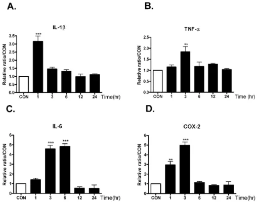
Fig. 2. The time course alteration of proinflammatory cytokines mRNA on KA injection in the hippocampus.
Alteration of IL-1β, TNF-α (B), IL-6 (C), and COX-2 (D) mRNA at 1, 3, 6, 12, 24 hrs after intracerebral (i.c.v.) KA (0.1 μg/5 μl) injectio was examined in the hippocampus. Control group (CON) was injected with i.c.v. PBS.
The mice number of each group was 3. Experiments were conducted inde- pendently three times, giving a total of nine per group in the final statistical analysis. **p<0.01(CON vs other groups), ***p<0.001 (CON vs other groups).
duced neuronal death, we administered visnagin orally (p.o.) or intraperitoneally (i.p.) 20 min (−20 min) prior to KA (0.1 μg/5 μl, i.c.v.) injection or 0, 20 (+20), 40 (+40), 60 (+60) min after KA injection (0.1 μg/5 μl, i.c.v.). As shown in Fig. 1A, KA induced neuronal cell death in the pyramidal layer of the hippocampal CA3 region. When vis- nagin was treated before or after KA injection orally or in- traperitneally, we observed that visnagin had neuroprotec- tive effect on KA-induced neuronal cell death. However, de- layed visnagin administration (+40 and +60 min) in both p.o. (Fig. 1B) and i.p. (Fig. 1C) did not show any neuro- protective effect on KA-induced neuronal death. The visna- gin administered p.o. or i.p. at −20, 0, +20 min had similar neuroprotective effect on KA-induced neuronal death in the hippocampus.
The course alteration of proinflammatory cytokines mRNA on KA injection in the hippocampus
Previous studies suggest that inflammatory and apoptotic processes contribute to the later stages of the damage in- duced by brain injuries, and that these detrimentally affect neurologic outcome [5,21,22]. Therefore, we investigate the course alteration of proinflammatory cytokines mRNA on KA injection in the hippocampus. The mRNA levels of vari- ous proinflammatory markers such as IL-1β, TNF-α, IL-6, and COX-2 in the hippocampus were examined at 1, 3, 6, 12, 24 hrs after KA administration (0.1 μg/5 μl, i.c.v.).
As shown in Fig. 2, intracerebroventricular injection with KA increased TNF-α and COX-2 mRNA levels, and the lev- els showed peak at 3 hrs after KA treatment. On the other hand, IL-1β mRNA level reached maximum level at 1hr after KA treatment, and IL-6 mRNA level showed peak lev- el at 3 and 6 hrs after KA treatment. The increased cyto- kines mRNA level decreased gradually after peak level.
The effect of visnagin on pro-inflammatory cytokines increased by KA in the hippocampus
To investigate whether the neuroprotective effect of vis- nagin is accompanied by suppressions of proinflammatory markers such as IL-1β, TNF-α, IL-6, and COX-2 in the hippocampus, we observed an effect of visnagin on pro-in- flammatory cytokines increased by KA in the hippocampus.
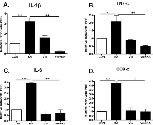
The time point when pro-inflammatory cytokines peaked was refereed by Fig 2 result. The induction of IL-1β, TNF- α, IL-6, and COX-2 mRNA hippocampus were inhibited by visnagin pretreatment (Fig. 3). However, visnagin itself did not affect pro-inflammatory cytokines expression.
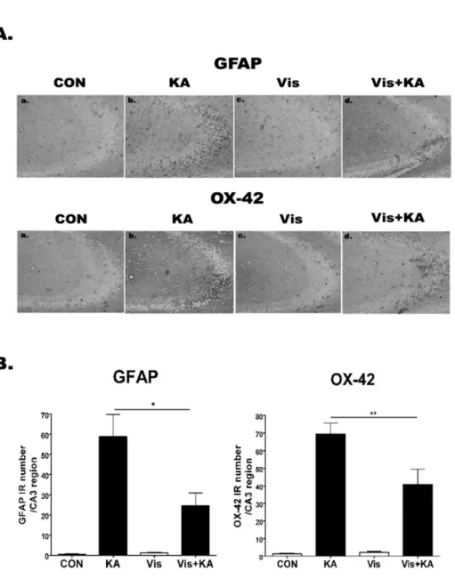
The effect of visnagin on the GFAP, OX-42 expression induced by KA in the hippocampal CA3 region To investigate whether visnagin affects GFAP or OX-42 expression induced by KA, the activations of microglia and astrocytes were analyzed using anti-OX-42 (microglia mark- er) and anti-GFAP (astrocyte marker) antibodies, respec- tively (Fig. 4). In PBS treated control mice, both microglias and astrocytes having ramified morphology were barely detected. However, the number of anti-OX-42 cells and an- ti-GFAP cells was remarkably elevated at 1 day after i.c.v.
KA treatment in the hippocampal CA3 region. In addition, visnagin administered orally at −20 min attenuated OX-42 and GFAP expression induced by KA.
DISCUSSION
There is an accumulating body of evidence which sug- gests that inflammation contributes to brain damage occur-
Fig. 3. The effect of visnagin on pro- inflammatory cytokines increased by KA in the hippocampus. Alteration of -1β, TNF-α (B), IL-6 (C), and COX-2 (D) at 1 (A), 3 hrs (B, D), or 6 hrs (C) after intracerebroventri- cular (i.c.v.) KA injection was exa- mined in the hippocampus. The in- creased IL-1β, TNF-α (B), IL-6 (C), and COX-2 (D) by KA was decreased by visnagin treated prior to 20 min.
The mice number of each group was 3. Experiments were conducted inde- pendently three times, giving a total of nine per group in the final statis- tical analysis. *p<0.05, **p<0.01,
***p<0.001 (CON vs KA), ++p<
0.01 (Vis vs Vis+KA). CON, vehicle (i.p.)+PBS (i.c.v.); KA, vehicle (i.p.)
+KA (i.c.v.); Vis, Visnagin (i.p.)+
PBS (i.c.v.).
ring after acute injury, and that it detrimentally affects neurological outcome [5,8]. In addition, under inflammatory conditions, free oxygen radicals, nitric oxide, and in- flammatory cytokines produced by activated microglial cells seem to cause neuronal damage. Because the hippocampus is densely populated with microglia and is one of the most sensitive and malleable regions of the brain [23], it is specu- lated that excessive production of inflammatory cytokines in the hippocampus would be associated with KA toxicity.
In the present study, we observed that pre- or post-visnagin administered orally (p.o.) or intraperitoneally (i.p.) in- hibited KA-induced neuronal cell death in the hippocampal CA3 region. Visnagin not only inhibited microglial and as- troglial activation but also attenuated the inflammatory marker expressions concomitantly, suggesting that visna- gin exerts its neuroprotective effects via an anti-inflam- matory mechanism in KA model.
Visnagin has been shown to relax KCl-and noradrenaline induced contractions in guinea-pig aortic strips to a similar extent [13] and this vasorelaxant effect was explained by the inhibition of Ca2+ entry into vascular smooth muscle cells. During excitotoxicity, glutamate concentrations in- crease in the synapse leads to intracellular Ca2+ concen- tration increase, activating a cell death pathway [2,3,24].
Thus, it is speculated that visnagin may have a neuro- protective effect against KA toxicity by inhibiting Ca2+
influx. Although inhibition of visnagin on Ca2+ entry into hippocampal neuron was not confirmed in the present study, it may be one mechanism on the neuroprotective ef- fect of visnagin. In regard to its anti-inflammatory effect, we also observed whether visnagin attenuated inflammatory markers against KA toxicity in the hippocampus. It appears that khellin extracts have some antimicrobial activity; this might be attributable to both the khellin and visnagin con- stituents, which both seem to have antifungal, antibacterial, and antiviral activity [25]. In addition, several reports have
demonstrated that drugs or compounds having an anti-in- flammatory effect can show a neuroprotective effect [4,26,27].
Thus, we examined the effect of visnagin on proinflamma- tory mediators expression induced by KA.
Recently, it has been demonstrated that proinflammatory cytokines have established potent pro-excitatory actions [28-31]. Collectively these reports show that proinflam- matory cytokines simultaneously facilitate excitatory gluta- matergic pathways while concurrently reducing inhibitory GABAergic transmission. Thus it is speculated that the combinatory synergic effect of these cytokines increased by KA and known facilitatory action of KA on excitatory neuro- transmission may lead to neuronal cell death in the hippocampus. Thus, the inhibitory effect of visnagin on cy- tokines expression showing synergic effect on neuronal cell death by KA may explain the neuroprotective effect of visnagin. It has been reported that COX-2 inhibitors may protect the brain against neurodegenerative diseases [32].
In addition, COX-2 specific inhibitors have a neuroprotec- tive effect in models of focal [33] and global brain ischemia [34]. Furthermore, reactive oxygen species are generated by COX-2 activity. Oxidative stress has been demanded to induce neurodegeneration in a variety of disease states [35].
Prostaglandins, which are the product of cyclooxygenase metabolism, can produce injury by inflammatory and vas- cular mechanisms, as well as directly lead to apoptosis in some cell types [36]. Furthermore, it has been reported that antioxidative effects were also observed in the case of visna- gin [37]. Thus, it is speculated that COX-2 inhibition and the antioxidative effect of visnagin may be another mecha- nism explaining the neuroprotective effect on KA toxicity.
Several earlier reports showed microglia expressing many inflammatory mediators after inflammatory injury [38,39].
Recently, it has been reported well that activated microglia is a significant source of redundant extracellular glutamate that induces excitotoxic neuronal death [40]. In addition,
Fig. 4. The effect of visnagin on the GFAP, OX-42 expression induced by KA in hippocampal CA3 region. OX-42 and GFAP immunoreactivities (A) in the hippocampus were examined at 24 hr after i.c.v. injection of KA (0.1 μg/5 μl). Animals (N=5 per each group) were pretreated orally with either PBS or visnagin (100 mg/kg) 20 min prior to KA (0.1 μg /5 μl). To quantify, we counted GFAP or OX-42 IR in each section (B). CON (a):
vehicle (i.p.)+PBS (i.c.v.), KA (b):
vehicle (i.p.)+KA (i.c.v.), Vis (c):
Visnagin (i.p.)+PBS (i.c.v.), Vis+
KA (d): Visnagin (i.p.)+KA (i.c.v.).
Antibody against OX-42 and GFAP was used at 1:10,000 dilution for immunostaining. *p<0.05 (KA vs Vis+KA), **p<0.01 (KA vs Vis+KA).
there is a report that excitatory amino acids released by microglia are suggested to compose the major determinant of neurotoxicity rather than reactive oxygen intermediates and cytokines [41]. Furthermore, Liang et al. [42] have demonstrated that astrocytes prevented excito-neurotoxicity by the reduction of exogenous glutamate whereas microglia did not, and conversely, activated microglia released an ex- cess of glutamate that induced excitotoxic neuronal death.
Taken together, it is speculated that activated glial cells induced by KA may be involved in increasing extracellular glutamate concentration finally as regulating glutamate re- lease each other, or induce synergic effect on KA toxicity.
Although we can not confirm that visnagin plays important roles as glutamate scavenger or glutamate receptor blocker, it seems that visnagin contributes to partly attenuate glu- tamate release as inhibiting gliosis.
We also observed that visnagin co-treatment (at 0 hr, p.o.) with KA injection produced a similar protective effect with Pre- or post-treatment of visnagin in the CA3 region.
This result suggests that in addition to the anti-inflam- matory effects, visnagin may also be effective during the acute damage process. First, direct inhibition of Ca2+ influx
into neurons may be one possibility as mentioned above [13]
although the underlying mechanism remains to be explored in detail. Second, the antioxydative effect of visnagin may be another candidate on co-treatment effect [37]. However, further study should be conducted to elucidate these hypotheses.
In conclusion, based on all this information, we speculate that the neuroprotective effects of visnagin in vivo are the results of multiple mechanisms, and that one of these may be associated with the suppression of inflammatory pro- cesses.
ACKNOWLEDGEMENTS
This study was supported by a grant of the Korea Healthcare technology R&D Project, Ministry of Health, Welfare & Family Affairs, Republic of Korea (A081028), Priority Research Centers Program through the National Research Foundation of Korea (NRF, 2009-0094072) and Grant (2009K001254) from Brain Research Center of the 21st Century Frontier Research Program funded by the
Ministry of Education, Science and Technology.
REFERENCES
1. Sperk G. Kainic acid seizures in the rat. Prog Neurobiol. 1994;
42:1-32.
2. Beal MF. Mechanisms of excitotoxicity in neurologic diseases.
Faseb J. 1992;6:3338-3344.
3. Rothman SM, Olney JW. Glutamate and the pathophysiology of hypoxic--ischemic brain damage. Ann Neurol. 1986;19:105-111.
4. Jin Y, Lim CM, Kim SW, Park JY, Seo JS, Han PL, Yoon SH, Lee JK. Fluoxetine attenuates kainic acid-induced neuronal cell death in the mouse hippocampus. Brain Research. 2009;1281:
108-116.
5. Penkowa M, Florit S, Giralt M, Quintana A, Molinero A, Carrasco J, Hidalgo J. Metallothionein reduces central nervous system inflammation, neurodegeneration, and cell death following kainic acid-induced epileptic seizures. J Neurosci Res. 2005;79:
522-534.
6. Kreutzberg GW. Microglia: a sensor for pathological events in the CNS. Trends Neurosci. 1996;19:312-318.
7. Ridet JL, Malhotra SK, Privat A, Gage FH. Reactive astrocytes:
cellular and molecular cues to biological function. Trends Neurosci. 1997;20:570-577.
8. Kim JB, Yu YM, Kim SW, Lee JK. Anti-inflammatory mechanism is involved in ethyl pyruvate-mediated efficacious neuroprotection in the postischemic brain. Brain Research.
2005;1060:188-192.
9. Smith E, Pucci LA, Bywater WG. Crystalline Visnagan. Science.
1952;115:520-521.
10. Anrep GV, Barsoum GS, Kenawy MR, Misrahy G. Ammi Visnaga in the treatment of the anginal syndrome. Br Heart J. 1946;8:171-177.
11. Anrep GV, Barsoum GS, Kenawy MR. The pharmacological actions of the crystalline principles of Ammi Visnaga Linn. J Pharm Pharmacol. 1949;1:164-176.
12. Duarte J, Perez-Vizcaino F, Torres AI, Zarzuelo A, Jimenez J, Tamargo J. Vasodilator effects of visnagin in isolated rat vascular smooth muscle. Eur J Pharmacol. 1995;286:115-122.
13. Rauwald HW, Brehm O, Odenthal KP. The involvement of a Ca2+ channel blocking mode of action in the pharmacology of Ammi visnaga fruits. Planta Medica. 1994;60:101-105.
14. Ubeda A, Tejerina T, Tamargo J, Villar A. Effects of khellin on contractile responses and 45Ca2+ movements in rat isolated aorta. J Pharm Pharmacol. 1991;43:46-48.
15. Duarte J, Torres AI, Zarzuelo A. Cardiovascular effects of visnagin on rats. Planta Medica. 2000;66:35-39.
16. Laursen SE, Belknap JK. Intracerebroventricular injections in mice. Some methodological refinements. J Pharmacol Methods.
1986;16:355-357.
17. Sapolsky RM, Krey LC, McEwen BS. Prolonged glucocorticoid exposure reduces hippocampal neuron number: implications for aging. J Neurosci. 1985;5:1222-1227.
18. Franklin KBJ, Paxinos G. The mouse brain in stereotaxic coordinates. 3rd ed. San Diego: Academic Press; 1997.
19. Kwon MS, Seo YJ, Choi SM, Choi HW, Jung JS, Park SH, Suh HW. The differential effects of single or repeated restraint stress on kainic acid-induced neuronal death in the hippo- campal CA3 region: the role of glucocorticoid and various signal molecules. J Neurochem. 2007;103:1530-1541.
20. Chomczynski P, Sacchi N. Single-step method of RNA isolation by acid guanidinium thiocyanate-phenol-chloroform extraction.
Anal Biochem. 1987;162:156-159.
21. Kim SW, Yu YM, Piao CS, Kim JB, Lee JK. Inhibition of delayed induction of p38 mitogen-activated protein kinase attenuates kainic acid-induced neuronal loss in the hippo- campus. Brain Research. 2004;1007:188-191.
22. Weiss JH, Sensi SL, Koh JY. Zn(2+): a novel ionic mediator of neural injury in brain disease. Trends Pharmacol Sci.
2000;21:395-401.
23. McEwen BS. Physiology and neurobiology of stress and adap- tation: central role of the brain. Physiol Rev. 2007;87:873-904.
24. White BC, Sullivan JM, DeGracia DJ, O'Neil BJ, Neumar RW, Grossman LI, Rafols JA, Krause GS. Brain ischemia and reperfusion: molecular mechanisms of neuronal injury. J Neurol Sci. 2000;179:1-33.
25. Hudson J, Towers GHN. Phytomedicines as antivirals. Drugs Future. 1999;24:295-300.
26. Cho IH, Kim SW, Kim JB, Kim TK, Lee KW, Han PL, Lee JK. Ethyl pyruvate attenuates kainic acid-induced neuronal cell death in the mouse hippocampus. J Neurosci Res. 2006;84:
1505-1511.
27. Yoo KY, Hwang IK, Kim JD, Kang IJ, Park J, Yi JS, Kim JK, Bae YS, Won MH. Antiinflammatory effect of the ethanol extract of Berberis koreana in a gerbil model of cerebral ischemia/reperfusion. Phytother Res. 2008;22:1527-1532.
28. Beattie EC, Stellwagen D, Morishita W, Bresnahan JC, Ha BK, Von Zastrow M, Beattie MS, Malenka RC. Control of synaptic strength by glial TNFalpha. Science. 2002;295:2282-2285.
29. Stellwagen D, Beattie EC, Seo JY, Malenka RC. Differential regulation of AMPA receptor and GABA receptor trafficking by tumor necrosis factor-alpha. J Neurosci. 2005;25:3219-3228.
30. Viviani B, Bartesaghi S, Gardoni F, Vezzani A, Behrens MM, Bartfai T, Binaglia M, Corsini E, Di Luca M, Galli CL, Marinovich M. Interleukin-1beta enhances NMDA receptor- mediated intracellular calcium increase through activation of the Src family of kinases. J Neurosci. 2003;23:8692-8700.
31. Wang S, Cheng Q, Malik S, Yang J. Interleukin-1beta inhibits gamma-aminobutyric acid type A (GABA(A)) receptor current in cultured hippocampal neurons. J Pharmacol Exp Ther.
2000;292:497-504.
32. McGeer PL, Schulzer M, McGeer EG. Arthritis and anti- inflammatory agents as possible protective factors for Alzhei- mer's disease: a review of 17 epidemiologic studies. Neurology.
1996;47:425-432.
33. Nogawa S, Zhang F, Ross ME, Iadecola C. Cyclo-oxygenase-2 gene expression in neurons contributes to ischemic brain damage. J Neurosci. 1997;17:2746-2755.
34. Nakayama M, Uchimura K, Zhu RL, Nagayama T, Rose ME, Stetler RA, Isakson PC, Chen J, Graham SH. Cyclooxygenase-2 inhibition prevents delayed death of CA1 hippocampal neurons following global ischemia. Proc Natl Acad Sci U S A. 1998;95:
10954-10959.
35. Coyle JT, Puttfarcken P. Oxidative stress, glutamate, and neurodegenerative disorders. Science. 1993;262:689-695.
36. Funk CD. Prostaglandins and leukotrienes: advances in eicosa- noid biology. Science. 2001;294:1871-1875.
37. Aboul-Enein HY, Kladna A, Kruk I, Lichszteld K, Michalska T. Effect of psoralens on Fenton-like reaction generating reactive oxygen species. Biopolymers. 2003;72:59-68.
38. Buttini M, Appel K, Sauter A, Gebicke-Haerter PJ, Boddeke HW. Expression of tumor necrosis factor alpha after focal cerebral ischaemia in the rat. Neuroscience. 1996;71:1-16.
39. Chao CC, Hu S, Molitor TW, Shaskan EG, Peterson PK.
Activated microglia mediate neuronal cell injury via a nitric oxide mechanism. J Immunol. 1992;149:2736-2741.
40. Barger SW, Basile AS. Activation of microglia by secreted amyloid precursor protein evokes release of glutamate by cystine exchange and attenuates synaptic function. J Neurochem.
2001;76:846-854.
41. Piani D, Spranger M, Frei K, Schaffner A, Fontana A. Macro- phage-induced cytotoxicity of N-methyl-D-aspartate receptor positive neurons involves excitatory amino acids rather than reactive oxygen intermediates and cytokines. Eur J Immunol.
1992;22:2429-2436.
42. Liang J, Takeuchi H, Doi Y, Kawanokuchi J, Sonobe Y, Jin S, Yawata I, Li H, Yasuoka S, Mizuno T, Suzumura A.
Excitatory amino acid transporter expression by astrocytes is neuroprotective against microglial excitotoxicity. Brain Research. 2008;1210:11-19.