• 검색 결과가 없습니다.

신생아 수신증:

N/A
N/A
Protected

Academic year: 2021

Share "신생아 수신증:"

Copied!
5
0
0

로드 중.... (전체 텍스트 보기)

전체 글

(1)

59

REVIEW ARTICLE

신생아 수신증: 진료지침을 위한 제안

Neonatal Hydronephrosis: Proposal for Korean Guideline Sungchan Park, Minki Baek

1

From the D epartm ent of U rology, U niversity of U lsan College of M edicine, U lsan,

1

Sungkyunkw an U niversity School of M edicine, Seoul, Korea

박성찬ㆍ백민기1

울산대학교, 1성균관대학교 의과대학 비뇨기과학교실

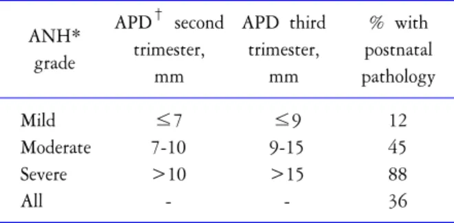
Hydronephrosis is defined as a dilation of the renal pelvis and calyces.

The widespread use of prenatal ultrasonography has resulted in an in- creased recognition of fetal hydronephrosis. Hydronephrosis can result from anatomic or functional processes interrupting the flow of urine, or from other congenital urologic anomalies such as vesicoureteral reflux;

however, this condition can also result from benign processes, such as physiologic dilation. The challenge in the management of neonatal hydro- nephrosis is to decide which child can be observed, which one can be managed medically, and which one requires surgical intervention.

Despite the fact that hydronephrosis is a very frequent condition in the neonatal population, there is a surprising lack of consensus regarding di- agnosis and management of this common condition. This article reviews the diagnosis and treatment of neonatal hydronephrosis.

Key Words: Hydronephrosis, Children, Kidney

(Received: November 23, 2010, Accepted: November 25, 2010)

교신저자 백민기

성균관대학교 삼성서울병원 비뇨기과

서울시 강남구 일원동 50, 135-710

Tel: 02-3410-3553 Fax: 02-3410-3027 E-mail: minki.baek

@samsung.com

서 론

수신증은 어떠한 구체적인 질환을 지칭하기 보다 는 신우와 신배가 확장되어 있는 현상을 지칭하는 용 어이다. 신생아에서 수신증이 있다고 하여 반드시 요 로폐색이나 방광요관역류와 같은 요로계 이상이 존 재하는 것은 아니며, 증상이 없고 시간이 지남에 따라 별다른 치료 없이 저절로 호전되는 경우도 있다.

1,2

신 생아 수신증은 대부분 산전 수신증으로 발견되거나 요로감염으로 병원을 방문하여 시행한 검사에서 발 견된다. 최근 사회 경제적 발전과 더불어 산전 진찰의 증가에 따른 산전 수신증의 발견이 흔해지면서 신생 아 수신증의 진료에 대한 중요성이 증가하였다.

3

신생 아 수신증 환아에서는 치료가 필요할 것인지를 조기 에 진단하고, 필요한 경우 적절한 시기에 치료를 시행 함으로써 신손상을 예방하는 것이 중요하다. 실제 임 상 영역에서 신생아 수신증 환아를 진료하는데 있어

서 어떤 검사를 시행할 것인지, 검사의 시행 시기와 간격, 예방 항생제 사용 여부, 수술 적응증 등을 결정 하는 데에서 많은 선택의 문제에 직면하게 된다.

4,5

하 지만 신생아 수신증의 진단과 치료에 대해서는 아직 까지 논란이 되는 부분이 많은 실정이며, 우리나라 비 뇨기과 전문의들을 대상으로 한 설문 조사에서도 신 생아 수신증의 진단과 치료에 대하여 다양한 의견이 존재함을 알 수 있었다.

6

저자는 본 글에서 신생아 수 신증의 진단과 치료 및 신생아 수신증의 흔한 원인인 신우요관이행부폐색증 (ureteropelvic junction obstruc- tion)의 치료에 대하여 언급하겠다.

신생아 수신증의 진단 1. 산전 수신증의 정의와 단계

산전 수신증은 재태 기간에 따라 정의와 단계를 달

리한다. 일반적으로 재태 16-18주부터 산전 초음파에

서 태아 신장을 관찰할 수 있는 것으로 알려져 있다.

(2)

60 Korean Journal of Pediatric Urology Vol. 2, 59-63, 2010

Table 1. Antenatal hydronephrosis grade according to anterior-posterior diameter of renal pelvis

ANH*

grade

APD

second trimester,

mm

APD third trimester,

mm

% with postnatal pathology

Mild ≤7 ≤9 12

Moderate 7-10 9-15 45 Severe >10 >15 88

All - - 36

*ANH: antenatal hydronephrosis,

APD: anterior-poste- rior diameter

Table 2. Hydronephrosis grades for Society of Fetal Urology Grade 0: No or minimal collecting system visible; consi-

dered normal

Grade 1: Just the renal pelvis visible with an axial diameter

<5-7 mm; usually considered normal

Grade 2: Axial renal pelvis diameter 5/7-10 mm; some calices with normal forniceal shape visible Grade 3: Marked dilatation of the renal calices and pelvis

>10 mm with reduced forniceal and papillary differentiation without parenchymal narrowing Grade 4: Gross dilatation of the collecting system with

narrowing of the parenchyma

(Grade 5: Used in some places additionally, to communicate an extreme hydronephrosis with only a thin, membrane-like residual renal parenchymal rim) 산전 수신증의 정도는 산전 초음파에서 태아 신장 종

축의 전후 길이 (anterior-posterior diameter; APD)를 기 준으로 구분하며, 일반적으로 사용되고 있는 산전 수 신증의 단계는 Table 1과 같다.

7,8

2. 신생아 수신증의 원인 질환

수신증은 여러 질환에 의해 발현된다. 신생아 수신 증의 원인이 되는 질환으로서는, 신우요관이행부협착 증, 요관방광이행부폐색증 (ureteroveiscal junction ob- struction), 방광요관역류 (vesicoureteral reflux), 후부요도 판막 (posterior urethral valve) 등이 있다.

7

신우요관이행 부협착증은 신생아수신증의 가장 흔한 원인이 되는 질환으로서, 신우 (renal pelvis)와 근위부 요관 (proximal ureter) 사이가 좁아져 소변의 흐름이 원활하지 못하여 집뇨계 (collecting system)가 확장된 상태를 말한다.

9

이 는 대략적으로 신생아 500명 중 1명에서 발생하며, 여 아에 비하여 남아에서 2배 많다. 요관방광이행부폐색 증은 원위부 요관 (distal ureter)에서 방광 (bladder)으로 들어가는 부위가 좁아진 상태를 말한다. 폐색으로 인 한 요관 확장을 동반하게 되므로 폐색성 거대요관 (obstructive megaureter)의 한 원인이기도 하는데, 남아 에서 흔하며 좌측에 흔히 발생한다.

10

3. 신생아 수신증의 초기 진단

1) 초음파검사 (ultrasonography): 초음파는 비침습적 이고 간단하게 시행할 수 있는 반면 요로계 이상을 비교적 명확하게 알 수 있도록 해주므로 신생아 수신 증의 원인 질환을 진단하는데 가장 기본이 되는 검사 이다. 초음파검사를 통해서 신우와 신배의 확장 정도, 신장의 크기, 신실질의 두께 및 반향 (echogenicity), 요

관 확장 여부, 방광의 모양과 두께 등을 관찰할 수 있 다.

초음파검사의 적용 범위로는, 산전 수신증이 있었 던 모든 환아에서는 출생 후 초음파검사를 시행할 것 을 추천한다 (Grade A recommendation). 검사의 시행시 기는, 산전에 심한 양측 수신증이 있었거나, 단일 신 장에 수신증이 있었던 경우, 산전에 양수의 양이 적었 던 경우 (oligohydramnios)에는 출생 후 바로 초음파 검 사를 시행할 것을 추천한다.

7

일반적으로 심하지 않은 산전 수신증의 경우, 생후 48-72시간 동안에는 신생아 에서 소변 생성이 적게 이루어져 수신증 정도가 더 감소되어 나타날 수 있으므로 이 시기에는 초음파 검 사를 시행하지 않는 것이 좋으며, 생후 7-30일에 시행 할 것을 추천한다.

7,11

심한 일측성 수신증의 경우에는 생 후 가능한 빨리 퇴원 전 초음파검사를 시행하여 귀가 전에 상태를 파악하는 것이 권장된다. 이때 생리 적 탈수증에 의한 위음성 (false negative)을 고려하여야 한다.

초음파검사로 수신증의 단계를 구분할 수 있다. 신

생아 수신증은 신우와 신배의 확장 정도와 신실질 두

께를 기준으로 단계를 구분하며 일반적으로 사용되

는 기준은 Society of Fetal Urology (SFU)에서 제안한 것

으로서 Table 2와 같다.

12,13

낮은 단계 수신증 (1, 2단

계)은 시간이 지날수록 자연 호전 가능성이 크지만,

높은 단계 (3, 4단계) 수신증은 신우요관이행부폐색증

과 같은 요로 폐색 가능성이 높은 것으로 알려져 있

다.

11

신생아 수신증이 있는 경우 방광요관역류가 동

(3)

Fig. 1. Evidence level and recommendation grade according to diagnostic flow-chart and test of neonatal hydronephrosis VUR: vesicoureteral reflux, US: ultrasound, VCUG: voiding cystourethrography, MAG3: mercaptuacetyltriglycine3 renal scan, DTPA: diethylenetriamene pentaacetate renal scan, SFU: Society of Fetal Urology.

반 가능성이 있으나 (8.5-15%), 수신증 단계와 방광요 관역류의 동반 가능성과는 상관 관계가 없는 것으로 알려져 있다.

7,14

2) 배뇨중방광요도조영술 (voiding cystourethrography):

배뇨중방광요도조영술은 요도에 관을 삽입하여 시행 하여야 하는 비교적 침습적인 검사이지만, 방광요관 역류의 존재 여부와 단계를 평가하는데 필수적인 검 사이다. 또한 이를 통하여 방광과 요도의 모양을 평가 할 수 있으며 요도 판막 (urethral valve), 요관류 (urete- rocele), 방광 게실 (bladder diverticulum) 등의 질환의 진 단에도 도움을 준다.

후부요도판막과 같이 방광출구폐색이 의심되는 질 환에서는 출생 후 퇴원 전 배뇨중방광요도조영술 시 행을 추천한다.

11

또한 양측의 심한 수신증, 방광 원위 부 폐색이 의심되는 경우에도 응급 배뇨중방광요도 조영술 시행을 추천한다. 요관의 확장, 중복 요관이 의심되는 경우, 초음파검사에서 방광의 에코와 모양 이 비정상적인 경우에는 응급은 아니더라도 가능한 한 배뇨중방광요도조영술을 시행할 것을 추천한다.

11

초음파검사에서 SFU 1, 2 단계 수신증이 있는 경우 배 뇨중방광요도조영술을 시행해야 하는지에 대해서는 아직 논란이 많다 (Grade D recommendation). 방광요관

역류의 유병률이 일반적인 소아에서는 1% 정도이지 만 수신증이 있는 경우 15% 정도라고 알려져 있으나, 낮은 단계 수신증에서 발견된 방광요관역류는 대부 분 낮은 단계이므로 그 임상적 중요성에 대해서는 의 문이 있다.

14

초음파검사에서 높은 단계 (4단계) 수신 증이 지속되는 경우 배뇨중방광요도조영술을 시행할 것을 추천한다 (Grade C recommendation).

7

이러한 경우 방광요관역류가 동반된다면 대부분 높은 단계인 경 우가 많다.

3) 이뇨성 신스캔 (diuretic renal scan): 최근 가장 널 리 받아들여지고 있는 요로 폐색의 정의는 ‘소변의 흐 름이 방해를 받는 상태로서, 치료가 늦어진 경우 신장 의 손상이 초래되는 경우’이다. DTPA와 MAG3와 같 은 이뇨성 신스캔은 요로 폐색의 진단과 더불어 상대 신기능을 평가하는데 유용한 검사이지만, 하나의 검 사만으로 요로 폐색 유무를 결정하는 것은 매우 어려 운 문제이다. 또한 영아 (infant)에서는 이뇨성 신스캔 결과만으로 요로 폐색을 결정하는 데에는 주의를 필 요로 한다.

11,15

적용 범위로는, SFU 3, 4단계 수신증이 있는 환아가

배뇨중방광요도조영술 검사에서 방광요관역류가 발

견되지 않았다면 생후 4-6주에 이뇨성 신스캔 시행을

(4)

62 Korean Journal of Pediatric Urology Vol. 2, 59-63, 2010

고려해야 한다.

11

이 시기 전에는 사구체여과율 (glo- merular filtration rate; GFR)이 낮으므로 이뇨성 신스캔 을 하더라도 정확하지 않다. 하지만 심한 수신증 환아 에서는 생후 4주 이전이라도 이뇨성 신스캔을 시행할 수 있다.

4) 그 외 검사방법: 수신증의 원인 중 이차적으로 신

우요관이행부 폐색을 유발하는 것으로는 교차하는 혈관 (crossing, aberrant vessel)이 요관을 누르는 경우가 흔하다. 이러한 진단을 위하여는 컬러도플러 초음파 (color Doppler ultrasonography)나 전산화 단층촬영 (computed tomography; CT), 자기공명영상 (magnetic resonance imaging; MRI)이 필요할 수가 있다.

16

신우요 관이행부 폐색의 원인을 찾아 더 나은 치료방법을 선 택하고 더 나은 결과를 얻기 위해서 혈관전산화 단층 촬영 (CT angiography)이 도움이 될 수 있다.

17

4. 신생아 수신증의 추적 및 추가 진단 1) SFU 1, 2단계 수신증 환아

(1) 배뇨중방광요도조영술을 시행하지 않은 경우;

광요관역류의 동반 가능성은 존재한다. 전신성 요로 감염의 징후가 있거나 보호자가 명확한 진단을 원하 는 경우 배뇨중방광요도조영술을 시행한다.

18

6개월 후 추가 초음파 검사를 권유하기도 한다 (Grade C recommendation).

19

(2) 배뇨중방광요도조영술에서 방광요관역류가 동반

된 경우; 방광요관역류 진료 지침에 준하여 치료한다.

(3) 배뇨중방광요도조영술에서 방광요관역류가 동반

되지 않은 경우; 이 수신증은 임상적으로 중요하지 않

을 가능성이 높다. 6개월 후 추가 초음파 검사를 권유 하기도 한다 (Grade C recommendation).

2) SFU 3, 4단계 수신증 환아

(1) 배뇨중방광요도조영술에서 방광요관역류가 동반

된 경우; 방광요관역류 진료 지침에 준하여 치료한다.

(2) 배뇨중방광요도조영술에서 방광요관역류가 동반

되지 않은 경우; 주기적으로 초음파 검사를 시행하여

수신증의 변화를 관찰한다. 이뇨성 신스캔을 시행하 여 요로 폐색 여부를 알아보는 것도 추천한다 (Grade C recommendation). 초음파검사에서 수신증이 심하고 이뇨성신주사에서 명백한 폐색소견이 있을 때는 출 생 후 3, 6개월에 추적 초음파검사와 이뇨성 신스캔을 시행하며 검사시기는 수신증의 정도와 폐색의 정도 에 따라 달라질 수 있다.

신생아 수신증의 치료 1. 예방 항생제

산전 수신증이 있는 환아가 그렇지 않은 환아와 비 교하여 요로감염 발생률이 유의하게 높다고 알려져 있다.

20

하지만 산전 수신증이 발견된 환아에서 산후 에 완전한 진단이 이루어지기까지 예방적으로 항생 제를 투여하는 것에 대하여는 논란이 많다 (Grade D recommendation). 일반적으로 방광요관역류 환아에서 예방 항생제가 요로감염을 예방할 것이라고 생각하 지만, 아직까지 구체적인 증거가 부족한 실정이다. 신 생아 (neonate)에서 주로 사용되는 예방 항생제는 amoxicillin (50 mg/일)이다. Trimethoprim-sultamatholxa- zole이나 nitrofurantoin도 흔히 사용되는 예방 항생제이 지만 2-3개월 이하 신생아에서는 가능하다면 사용하 지 않는 것이 좋다.

2. 신우요관이행부폐색증의 치료

증상이 없는 경우 주기적인 추적이 필요하다. 다만 가능하다면 동일한 조건에서 추적하는 것이 좋다. 증 상이 있는 경우 (반복적인 측복부 통증 또는 요로감염 등) 수술 적응증이 된다. 또한, 폐색이 있는 신장의 상 대 신기능 (relative renal function)이 40% 이하인 경우, 동일한 조건으로 시행하여 추적한 이뇨성 신스캔에 서 신장 기능이 5-10% 이상 감소한 경우, 초음파에서 신장 종축의 전후 길이가 증가한 경우, SFU 수신증 기 준 3단계 이상, 신실질이 더 얇아지는 등 수신증의 진 행이 명확히 관찰될 때 수술 적응증이 된다.

21

결 론

최근에 널리 보편화된 산전 초음파에 의해서 신우 요관이행부나 요관방광이행부 폐색에 의한 수신증의 진단이 늘고 있다. 꼼꼼하면서 반복적인 산후 관리가 신장기능의 저하를 초래하는, 수술적인 치료를 필요 로 하는 수신증의 폐색의 원인을 찾는데 꼭 필요하다.

수술적인 방법은 대부분 표준화되어 있고 좋은 임상 결과를 보인다.

REFERENCES

1. Lim DJ, Park JY, Kim JH, Paick SH, Oh SJ, Choi H.

(5)

Clinical characteristics and outcome of hydronephrosis detected by prenatal ultrasonography. J Korean Med Sci 2003;18:859-62

2. Choi SM, Jung JH, Hyun JS, Chung KH. Clinical course of prenatally-detected hydronephrosis: focus on uretero- pelvic junction obstruction. Korean J Urol 2008;49:1125- 30

3. Gunn TR, Mora JD, Pease P. Antenatal diagnosis of urinary tract abnormalities by ultrasonography after 28 weeks’ gestation: incidence and outcome. Am J Obstet Gynecol 1995;172:479-86

4. Kang YS, Han SW, Choi SK. The ultrasonographic follow up results and the prognosis of prenatal hydronephrosis.

Korean J Urol 1995;36:1100-5

5. Song PH, Cho CK, Park TC. Clinical outcome of prenatal hydronephrosis. Korean J Urol 2003;44:556-60

6. Baek M, Sohn DW, Oh KJ, Lee T, Chang HS, Han SW, et al. Nationwide questionnaire survey on practice patterns for pediatric hydronephrosis of Korean urologists at secon- dary or university hospitals. Korean J Urol 2009;50:1231- 47

7. Lee RS, Cendron M, Kinnamon DD, Nguyen HT. Ante- natal hydronephrosis as a predictor of postnatal outcome:

a meta-analysis. Pediatr 2006;118:586-93

8. Psooy K, Pike J. Investigation and management of antenatally detected hydronephrosis. Can Urol Assoc J 2009;3:69-72

9. Lebowitz RL, Griscom NT. Neonatal hydronephrosis: 146 cases. Radiol Clin North Am 1977;15:49-59

10. Brown T, Mandell J, Lebowitz RL. Neonatal hydrone- phrosis in the era of sonography. Am J Roentgenol 1987;

148:959-63

11. Belarmino JM, Kogan BA. Management of neonatal hydronephrosis. Early Hum Dev 2006;82:9-14

12. Fernbach SK, Maizels M, Conway JJ. Ultrasound grading of hydronephrosis: introduction to the system used by the Society of Fetal Urology. Pediatr Radiol 1993;23:478-80 13. Riccabona M, Avni FE, Blickman JG, Dacher JN, Darge

K, Lobo ML et al. Imaging recommendations in paediatric ruoradiology: minutes of the ESPR workgroup session on urinary tract infection, fetal hydronephrosis, urinary tract ultrasonography and voiding cystourethrography, Barce- lona, Spain, June 2007. Pediatr Radiol 2008;38:138-45 14. Gloor JM, Ramsey PS, Ogburn PL Jr, Danilenko-Dixon

DR, DiMarco CS, Ramin KD. The association of isolated mild fetal hydronephrosis with postnatal vesicoureteral reflux. J Matern Fetal Neonatal Med 2002;12:196-200 15. Koff SA, Binkovitz L, Coley B, Jayanthi VR. Renal pelvis

volume during diuresis in children with hydronephrosis:

implications for diagnosing obstruction with diuretic reno- graphy. J Urol 2005;174:303-7

16. Mitterberger M, Pinggera GM, Neururer R, Peschel R, Colleselli D, Aigner F, et al. Comparison of contrast-en- hanced color Doppler imaging (CDI), computed tomogra- phy (CT), and magnetic resonance imaging (MRI) for the detection of crossing vessels in patients with ureteropelvic junction obstruction (UPJO). Eur Urol 2008;53:1254-60 17. Rabah D, Soderdahl DW, McAdams PD, Knowles YK,

Vingan HL, Shaves SC, et al. Ureteropelvic junction obstruction: does CT angiography allow better selection of therapeutic modalities and better outcome? J Endourol 2004;18:427-30

18. Hallerstein S, Nickell E. Prophylactic antibiotics in children at risk for urinary tract infection. Pediatr Nephrol 2002;

17:506-10

19. Aksu N, Yavascan O, Kangin M, Kara OD, Aydin Y, Erdoğan H, et al. Postnatal management of infants with antenatally detected hydronephrosis. Pediatr Nephrol 2005;

20:1253-9

20. Harding LJ, Malone PSJ, Wellesley DG. Antenatal mini- mal hydronephrosis: is its followup an unnecessary cause of concern? Prenat Diagn 1999;19:701-5

21. Riedmiller H, Androulakakis P, Beurton D, Kocvara R,

Gerharz E. EAU guidelines on paediatric urology. Eur Urol

2001;40:589-99

수치

Table  2.  Hydronephrosis  grades  for  Society  of  Fetal  Urology  Grade 0: No  or  minimal  collecting  system  visible;
Fig.  1.  Evidence  level  and  recommendation  grade  according  to  diagnostic  flow-chart  and  test  of  neonatal  hydronephrosis VUR:  vesicoureteral  reflux,  US:  ultrasound,  VCUG:  voiding  cystourethrography,  MAG3:  mercaptuacetyltriglycine3  re

참조

관련 문서

Antibiotic prophylaxis for the prevention of recurrent urinary tract infection in children with low grade vesicoureteral reflux: results from a

Antibiotic Prophylaxis for the prevention of recurrent urinary tract infection in children with low grade vesicoureteral reflux: results from a prospective

Our case demonstrated that high grade vesicoureteral reflux patient with reduced renal function, bladder and bowel dysfunction at presentation is more likely to progress to

The patients’ medical records were assessed concerning gender, presence of vesicoureteral reflux (VUR), hydronephrosis, type of spinal dysraphism, level of spinal dysraphism,

Incidence and Classification of Vesicoureteral Reflux According to the Level of Spinal Cord Injury.. Level

Antibiotic prophylaxis for the prevention of recurrent urinary tract infection in children with low grade vesicoureteral reflux: re- sults from a prospective

Purpose: The purpose of this study is to evaluate the incidence and risk factors of febrile urinary tract infection (UTI) after endoscopic therapy for vesicoureteral reflux

While endoscopic subureteral injection of bulking agents has become a first-line therapy for the treatment of vesicoureteral reflux (VUR), mainly due to its high success rates