49 접수일: 2010년 3월 12일, 게재결정: 2010년 3월 19일
*교신저자: 김찬종, 501-757, 전남대학교 의과대학 소아과학교실, Phone: 062-220-6646, FAX: 062-220-6103, E-mail: [email protected] Chonnam Medical Journal
Vol. 46, No. 1, pp. 49∼55 DOI: 10.4068/cmj.2010.46.1.49
요로감염 환아의 방광 요관 역류에 대한 선별검사로서 초음파와
99m
Technetium Dimercaptosuccinic Acid (DMSA) Scan의 효용성
전남대학교 의과대학 소아과학교실
양은미ㆍ김상정ㆍ김찬종*ㆍ우영종
Clinical Usefulness of Ultrasonography and
99mTechnetium
Dimercaptosuccinic Acid Scan for Predicting the Vesicoureteral Reflux in Children with Urinary Tract Infection
Eun Mi Yang, Sang Jeong Kim, Chan Jong Kim* and Young Jong Woo Department of Pediatrics, Chonnam National University Medical School, Gwangju, Korea
The purpose of this study was to evaluate the predictive value of ultrasonography (US) and 99mtechnetium dimercaptosuccinic acid (DMSA) scan for vesicoureteral reflux (VUR) in children with urinary tract infection (UTI). This study was a retrospective review of 114 children who were diagnosed as UTI from January 2004 to December 2007. A total of 114 patients underwent a US, DMSA scan and VCUG. The patients were divided into three groups according to the results of VCUG. The findings of the US and DMSA scan were compared with VCUG results. Of the 114 patients, there were 79 (69.3%) without VUR, 12 (10.5%) with low-grade VUR (grade I, II) and 23 (20.2%) with high-grade VUR (more than grade III). The US predicted 15 of 35 VUR with a sensitivity of 42.9% and a specificity of 70.9%. A DMSA scan predicted 26 of 35 VUR with a sensitivity of 74.3%. If either the US or DMSA scan was abnormal, this condition predicted 29 of 35 VUR with a sensitivity of 82.9%, negative predictive value (NPV) of 82.9%. VUR was associated with abnormal DMSA scan findings and abnormal findings of either US or DMSA scan. If either the US or DMSA renal scan was abnormal in children with a urinary tract infection, this was predictable factor for VUR. And this condition was more accurate in high-grade VUR. As screening examination for VUR, US and DMSA scan are useful and should be performed together. If both tests are normal in children with a urinary tract infection, there may be little or no clinically significant VUR.
Key Words: Urinary tract infections; Technetium Tc 99m dimercaptosuccinic acid; Vesico-ureteral reflux;
Ultrasonography
서 론
요로감염은 소아에서 흔히 발생하는 감염으로 여아에서는 3∼5%, 남아에서는 1%의 발병률을 보인다.1 방광 요관 역류 는 소아의 요로감염과 신반흔을 일으키는 원인 중의 하나로 요로감염이 있는 환아의 약 25∼50%에서 발생하며, 역류가 있는 환아 중 30∼49%는 신반흔을 가진 것으로 보고되고 있 다.2-4 여러 연구에서 방광 요관 역류 환아에서 발생한 신반흔 은 향후 신장 기능의 저하, 고혈압, 임신성 고혈압 및 임신 중독증 등 합병증의 증가와 연관되어 있다고 알려져 있다.5-9 따라서 첫 요로감염의 경우에 영상학적 검사로 신장 초음파,
99mtechnetium dimercaptosuccinic acid (DMSA) scan, 배 뇨성 방광 요도 조영술 등의 검사가 추천된다.10 초음파는 신 장의 크기, 요로 폐쇄 유무와 정도, 신장 또는 신장 주위 농양, 요관 확장, 방광 기형 등을 진단하고 DMSA scan은 신 반흔, 신 위축 등을 진단하며 급성기에 시행 시 급성 신우 신염을 진단 할 수 있으며 배뇨성 방광 요도 조영술은 방광 요관 역 류, 후부요도 판막의 진단에 필요하다.11 그러나 배뇨성 방광 요도 조영술은 환아들에게 통증을 유발할 수 있으며 방사선 노출 및 요로감염부터 방광천공까지 다양한 합병증을 초래할 수 있다.12,13 따라서 배뇨성 방광 요도 조영술의 선별적인 시 행을 주장하는 여러 연구가 있으며 침습적인 배뇨성 방광 요 도 조영술을 대체하기 위한 다양한 검사방법이 고안되고 있 다.14-19
이에 저자는 초음파 및 DMSA scan과 같이 비침습적인 검사로써 방광 요관 역류를 예측할 수 있는지 알아보고, 배 뇨성 방광 요도 조영술의 빈도를 줄일 수 있는 지를 조사해 보고자 하였다.
대상 및 방법
2004년 1월부터 2007년 12월까지 전남대학교병원에서 요 로감염으로 진단 받은 환아 중에서 초음파, DMSA scan, 배 뇨성 방광 요도 조영술을 모두 시행한 114명을 대상으로 후 향적으로 분석하였다. 요로감염의 진단은 38oC 이상의 발열 이 있는 환아 중 소변검사에서 고배율 시야에서 5개(5/HPF, high power field) 이상의 농뇨가 있으면서 소변 배양 검사에 서 단일 균주가 mL당 105 집락 형성 단위(colony forming units) 이상 배양되었을 경우 또는 항생제 사용 후 배양 검사 를 받은 경우는 DMSA scan 상 신우신염이 있는 경우로 하
였다. 소변은 2세 이상의 환아 중 요의를 표현할 수 있는 경우 중간뇨를, 요의를 표현하지 못하는 2세 이상의 환아 및 2세 미만의 모든 환아에 대해서는 무균 채뇨백을 이용하여 채취하 였으며, 베타딘으로 요도 주위를 닦은 뒤 채뇨백을 부착하는 방법으로 시행하였다. 다른 감염이 동반된 환아와 신경학적 방광 기능이상이 있는 환아들은 연구에서 제외하였다.
초음파는 내원 3일 이내에 시행하였으며 수신증, 요관의 확장, 신 실질 부위의 위축이나 증가, 요로결석, 방광벽의 비 후, 마제신, 중복 요관이나 다낭성 신질환 등의 소견이 있는 군을 양성으로 하였다. DMSA scan은 내원일로부터 평균 5 일 이내에 시행하였으며 급성 신우신염이나 신반흔을 시사하 는 신피질 결손이 있거나 동위원소 섭취율이 저하된 경우, 신 위축 소견이 있는 경우, 마제신이나 중복요관이 있는 경우를 양성으로, 이러한 소견이 없는 군을 음성으로 하는 두 군으로 분류하였다. 배뇨성 방광 요도 조영술을 시행 후 방광 요관 역류의 존재 유무에 따라 있는 군과 없는 군으로 분류하였으 며, 세부등급은 국제소아역류연구회의 방광 요관 역류 등급 분류(International Reflux Study Committee, 1981)에 따 라 나누었고 grade I-II를 경증 역류, grade III-V를 중증 역 류로 분류하였다.
환아의 성별, 나이, 원인 균주, 혈중 백혈구 수치와 혈장 CRP 수치 및 초음파와 DMSA scan의 이상소견과 방광 요관 역류와 연관성에 대해 조사하였다. 또한 방광 요관 역류에 대해 초음파, DMSA scan의 민감도(sensitivity), 특이도(specificity) 및 양 성예측도(positive predictive value)와 음성예측도(negative predictive value)를 조사하였다. 통계처리는 통계 전문 상용 소프트웨어인 SPSS for windows version 12 (SPSS Inc., Chicago, USA)을 이용하였으며 방광 요관 역류의 등급에 따른 각 인자의 연관성은 일원배치 분산분석(one-way ANOVA test)을, 영상학적 검사와 방광 요관 역류와의 관계는 Fisher의 직접 확률계산법(Fisher's exact test)을 이용하여 p값이 0.05 미만인 경우를 통계적으로 유의한 것으로 정의하였다.
결 과
1. 대상 환아의 특성
대상 환아의 나이는 평균 16.94±27.73개월이며 남아가 66명 (57.9%), 여아는 48명(42.1%)이었다. 평균 혈중 백혈구 수치 는 13,230±6,832/mm3, 혈장 CRP 수치는 3.42±4.44 mg/dL였 다. 초음파에서 이상소견을 보인 환아는 38명(33.3%), DMSA
Table 1. Patients characteristics
Characteristics (n=114) No. of patients Sex (%)
Male Female Mean age (month) Mean WBC counts (/mm3) Mean serum CRP level (mg/dL) Ultrasonography result (%) Normal
Abnormal
DMSA scan result (%) Normal
Abnormal
Voiding cystourethrography result (%) Absence of VUR
VUR grade I-II VUR grade III-V Isolated organism (n=79) Escherichia coli (%) Enterococcus feacalis (%) Klebsiella pneumoniae (%) The others (%)
66 (57.9) 48 (42.1) 16.94±27.73 13,230±6,832 3.42±4.44
76 (66.7) 38 (33.3)
46 (40.4) 68 (59.6)
79 (69.3) 12 (10.5) 23 (20.2)
50 (63.3) 9 (10.1) 8 (11.4) 12 (15.2) DMSA, 99mtechnetium dimercaptosuccinic acid; VUR, vesicoureteral reflux.
Table 2. Relationship between clinical characteristics and vesicoureteral reflux
Variable Absence
(n=79)
Low-grade*
(n=12)
High-grade†
(n=23) p value
Sex (male) Age (month) WBC counts (/mm3) CRP level (mg/dL) Isolated organism (E.coli)
45 (56.9%) 14.91±26.66 13,361±7,152 3.29±4.45 34 (43.0%)
6 (50%) 23.83±36.22 14,070±6,493 5.76±6.23 8 (66.7%)
15 (65.2%) 20.348±26.86 12,327±6,136 2.67±3.01
8 (34.8%)
0.66 0.47 0.79 0.26 0.45
*Low-grade, grade I-II; †High-grade, grade III-V.
Table 3. Associations of each ultrasonographic finding with vesi- coureteral reflux and high-grade vesicoureteral reflux
US finding Number
(%) VUR p
value
High-grade VUR*
p value Hydronephrosis
APN
Hydroureterosis MCDK
Renal hypoplasia Urolithiasis Duplicated ureter Horseshoe kidney Bladder wall thickening
18 (47.4) 9 (23.7) 3 (7.9) 2 (5.3) 2 (5.3) 1 (2.6) 1 (2.6) 1 (2.6) 1 (2.6)
6 4 0 1 2 0 1 1 0
0.471 0.266 0.553 0.513 0.089
- - - -
5 4 0 0 1 0 1 1 0
0.251 0.070 0.519 0.647 0.353
- - - - US, ultrasonography; VUR, vesicoureteral reflux; APN, acute pyelonephritis;
MCDK, multicystic dysplastic kidney.
*High-grade VUR, grade III-V.
Table 4. Associations of each 99mtechnetium dimercaptosuccinic acid scan finding with vesicoureteral reflux and high-grade vesicoureteral reflux
DMSA finding Number
(%) VUR p
value
High-grade VUR*
p value APN
Renal hypoplasia MCDK
Horseshoe kidney Duplicated ureter
63 (92.6) 2 (2.9) 1 (1.5) 1 (1.5) 1 (1.5)
22 2 0 1 1
0.134 0.089
- - -
18 1 0 1 1
0.004† 0.353
- - - DMSA, 99mtechnetium dimercaptosuccinic acid scan; VUR, vesicoureteral reflux; APN, acute pyelonephritis; MCDK, multicystic dysplastic kidney.
*High-grade VUR, grade III-V; †Odds ratio 4.70 (95% CI, 1.48∼15.0).
scan에서 이상소견을 보인 환아는 68명(59.6%)이었고 방광요 관역류가 없는 군은 79명(69.3%)이었으며 경증 역류는 12명 (10.5%), 중증 역류는 23명(20.2%)이었다. 소변배양검사에서 79명의 환아에서 균주가 동정되었으며 E. coli가 50명(63.3%) 으로 가장 많았고, Enterococus species 9명(11.4%), Klebsiella pneumoniae 8명(10.1%)순이었고, 이외 Staphylococcus species 4명(5.0%), Morganella morganii 3명(3.8%), Enterobacter cloacae 2명(2.5%), Pseudomonas aeruginosa 1명(1.3%) 등 의 균들이 배양되었다(Table 1).
2. 방광 요관 역류와 임상적 특성 및 검사실 소견과의 관계 배뇨성 방광 요도 조영술에 따라 역류가 없는 군은 79명 (69.3%), 경증 역류가 12명(10.5%), 중증 역류가 있는 군 이 23명(20.2%)이었으며 역류의 유무와 정도에 따른 성별,
나이, 혈중 백혈구 수치, 혈장 CRP 수치, 원인 균주는 의의 있는 차이를 보이지 않았다(Table 2).
3. 초음파와 DMSA scan 소견
초음파에서 양성을 보인 경우는 대상 환아 114명 중 38 명으로 수신증 18명(47.4%), 급성 신우신염 9명(23.7%), 요관 확장 3명(7.9%) 등의 순이었고 각각의 소견만으로 방 광 요관 역류와 중증 방광 요관 역류와 연관성은 없었다 (Table 3). DMSA scan에서 양성으로 확인된 환아는 68명
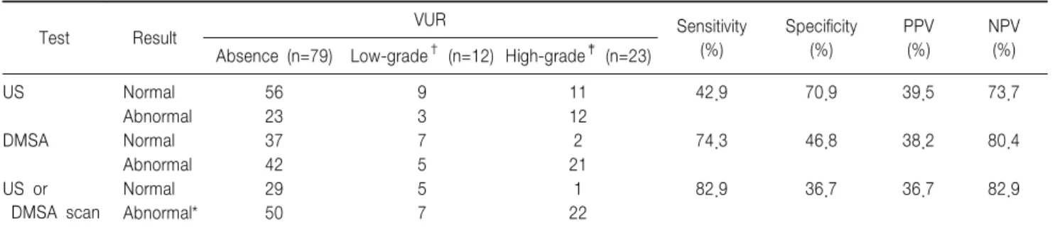
Table 5. Usefulness of US, DMSA scan and a both test for predicting VUR
Test Result VUR Sensitivity
(%)
Specificity (%)
PPV (%)
NPV Absence (n=79) Low-grade† (n=12) High-grade‡ (n=23) (%)
US Normal 56 9 11 42.9 70.9 39.5 73.7
Abnormal 23 3 12
DMSA Normal 37 7 2 74.3 46.8 38.2 80.4
Abnormal 42 5 21
US or DMSA scan
Normal 29 5 1 82.9 36.7 36.7 82.9
Abnormal* 50 7 22
US, renal ultrasonography; DMSA scan, 99mtechnetium dimercaptosuccinic acid scan; PPV, positive predictive value; NPV, negative predictive value;
*Abnormal, abnormal findings on either US or DMSA scan; †Low-grade, grade I-II; ‡High-grade, grade III-V.
Table 6. Relationship between abnormalities on image studies and the presence of vesicoureteral reflux
Test Odds ratio 95% CI p value
US DMSA
US or DMSA scan
1.82 2.54 2.80
0.79∼4.18 1.06∼6.12 1.04∼7.55
0.196 0.040 0.047 US, renal ultrasonography; DMSA, 99mtechnetium dimercaptosuccinic acid scan.
Table 7. Usefulness of US, DMSA scan and a both-test strategy for predicting high-grade vesicoureteral reflux
Test Result
No high- grade
VUR (n=91)
High- grade VUR*
(n=23) Sensi-
tivity (%)
Speci- ficity
(%) PPV
(%) NPV
(%)
US Normal 65 11 52.2 71.4 31.6 85.5
Abnormal 26 12
DMSA Normal 44 2 91.3 48.4 30.9 95.7
Abnormal 47 21
US or DMSA scan
Normal 34 1 95.7 37.4 27.8 97.1
Abnormal 57 22
US, renal ultrasonography; DMSA scan, 99mtechnetium dimercapto- succinic acid scan; PPV, positive predictive value; NPV, negative predictive value; *High-grade, grade III-V.
Table 8. Relationship between abnormalities on image studies and the presence of vesicoureteral reflux
Test Odds ratio 95% CI p value
US DMSA
US or DMSA scan
2.72 9.83 13.12
1.07∼6.95 2.18∼44.39 1.69∼101.8
0.047 0.001 0.002 US, renal ultrasonography; DMSA, 99mtechnetium dimercaptosuccinic acid scan.
으로 급성 신우신염이 63명(92.6%)으로 가장 많았으며 이 는 중증 방광 요관 역류와 의의 있는 연관성이 있었다(OR
=4.70, 95% CI: 1.48∼15.0, p=0.004) (Table 4).
4. 방광 요관 역류와 초음파, DMSA scan과의 관계 요로 감염 환아의 방광 요관 역류의 진단에서 초음파의 민 감도는 42.9%, 특이도는 70.9%이며 양성 예측도는 39.5%, 음성 예측도는 73.7%이었다. DMSA scan의 민감도와 특이 도는 74.3%, 46.8%이고 양성 예측도 및 음성 예측도는 38.2%, 80.4%이었다. 두 검사, 즉 초음파 혹은 DMSA scan 중 하나라도 양성소견이 있는 경우의 민감도와 음성 예측도 는 각각 82.9%, 82.9%으로 상승하였다(Table 5). 방광 요관 역류에 대한 비교 위험도를 조사한 결과 DMSA scan (OR= 2.54, 95% CI: 1.06∼6.12, p=0.040)과 두 검사 중 하나라도 양성 소견이 있는 경우(OR=2.80, 95% CI: 1.04
∼7.55, p=0.047)가 의의 있는 연관이 있었다(Table 6).
5. 중증 방광 요관 역류와 초음파, DMSA scan과의 관계 요로 감염 환아의 중증 방광 요관 역류의 진단에서 초음파 의 민감도는 52.2%, 특이도는 71.4%이며 양성 예측도는 31.6%, 음성 예측도는 85.5%이었다. DMSA scan의 민감도 와 특이도는 91.3%, 48.4%이고 양성 예측도 및 음성 예측도
는 30.9%, 95.7%이었다. 두 검사 중 하나라도 양성 소견이 있는 경우의 민감도는 95.7%, 음성예측도는 97.1%으로 상 승하였다(Table 7). 중증 방광 요관 역류에 대한 비교 위험 도를 조사한 결과 초음파(OR=2.72, 95% CI: 1.07∼6.95, p=0.047), DMSA scan (OR=9.83, 95% CI: 2.18∼44.39, p=0.001) 그리고 두 검사 중 하나라도 양성 소견이 있는 경 우(OR=13.12, 95% CI: 1.69∼101.8, p=0.002) 모두에서 의의 있는 연관이 있었다(Table 8).
고 찰
배뇨성 방광 요도 조영술은 요로감염이 발생한 환아에서 방광 요관 역류 유무를 확인하기 위해 보편적으로 시행되는 검사이다. 최근의 몇몇 연구에서 경증 방광 요관 역류(grade I-II)는 신손상과 의의있는 연관성이 없다는 연구결과가 있 으며,20 자연 소실율도 높은 것을 알려져 있으나 중증 방광 요관 역류는 영구적 신손상과 연관이 있으며 방광 요관 역 류로 인한 신장손상은 단백뇨, 고혈압, 만성신부전 등으로 이행될 수 있다고 알려져 있다.21-23 그러나 방광 요관 역류 가 있는 환아 중 상당수에서 신반흔이 나타나지 않고 검사 에 따른 고통과 방사선 조사의 위험성 때문에 DMSA scan 에서 신손상이 있는 환아에게만 선별적으로 배뇨성 방광 요 도 조영술을 시행하자는 주장이 있다.14,24,25 이러한 배경에 서 불필요한 배뇨성 방광 요도 조영술의 시행을 줄이기 위 해 방광 요관 역류를 예측할 수 있는 다양한 인자들에 대해 많은 연구가 시행되었다.
방광 요관 역류와 성별 및 연령과의 관련성에 대해서는 상반된 결과들이 발표되고 있다. Polito 등26의 보고에 따르 면 방광 요관 역류의 예측에 있어서 환아의 성별은 의의가 없었으며 Oostenbrink 등15은 저연령, 남아, 가족력, 혈장 CRP 수치, 초음파 결과가 첫 요로감염 환아들의 방광 요관 역류의 유무를 가늠할 수 있다고 하였으며 Lee 등16은 연령 및 성별은 역류와 연관성이 없었고 발열, 농뇨, 적혈구침강 속도, 혈장 CRP 수치 등이 역류의 유무와 연관이 있다고 하였다. 본 연구에서는 연령 및 성별이나 실험실검사, 배양 검사에서 동정된 균주 등과 역류의 유무, 역류의 등급과 연 관성이 없었다.
Ilyas 등27은 DMSA scan에서 급성 신우신염이나 신반흔 이 관찰되는 환아 중 방광 요관 역류가 모두에서 동반되는 것 은 아니나 중증 방광 요관 역류가 동반될수록 DMSA scan상 에서 의의 있게 이상소견이 발견되었으며 Preda 등17이 시행 한 전향적 연구에 따르면 DMSA scan에서 양성의 결과를 보 인 환아에서 1명을 제외한 26명 모두가 중증 방광 요관 역류 가 동반되어 있었다. 이외 여러 연구에서 DMSA scan과 VUR의 관련성이 언급되어 DMSA scan을 통한 VUR의 예 측가능성이 시사되었다.28-31 본 결과에서도 DMSA scan은 방광 요관 역류와 의의 있는 연관이 있었다. 그러나 방광 요관 역류의 진단에서 DMSA scan의 민감도는 연구에 따라 50∼
88%까지 많은 차이가 있으며14,16,18,32
본 연구에서 DMSA scan의 민감도는 74.3%으로 조사되어 DMSA scan 소견만
으로 방광 요관 역류를 예측하는 데는 무리가 있을 것으로 생 각된다.
초음파는 간단하고 이용하기 쉬우며 비침습적이고 수신증이 나 요관확장 등의 폐색성 병변이나 신 또는 신 주위 농양, 방광 기형 등을 비교적 빠르고 정확하게 평가할 수 있어 소아 요로감 염 환아 또는 의심되는 경우에도 일률적으로 시행한다.33 그러 나 신장초음파로 측정한 소아의 신길이(renal length)와 신부 피(renal volume)는 검사자에 따라 2년 정도의 정상 성장만큼 (about 2 years' normal growth)의 차이를 보이는데 이는 장내 가스 때문에 신장의 장축을 측정하기가 어렵고 수분공급 상태 의 변화에 따른 신부피의 변화가 생기기 때문이다.34,35 따라서 소아 신장초음파의 경우 초음파 시행자의 기술과 경험에 따른 결과의 차이가 있을 수 있다.36 더욱이 Tsai 등37은 방광 요관 역류의 경우 동적이고(dynamic) 비폐색성인 특성 때문에 초음 파에서 단지 경도의 수신증 또는 정상소견을 나타낼 수 있고 실제로 신생아 수신증의 상당수는 신우요관이행부 폐색이나 생리적 수신증이므로, 신생아 방광 요관 역류 발견을 위한 신장 초음파의 결과가 민감도, 특이도, 양성 예측치, 음성 예측치 등 모두에서 낮아 그 결과를 신뢰할 수 없다고 하였다. 본 연구 결과에서도 초음파는 방광 요관 역류의 유무와 통계적으로 의 의 있는 연관이 없었으며 낮은 민감도와 특이도를 나타내었다.
초음파와 DMSA scan을 함께 고려하였을 때, 즉 한 검사 에서라도 양성소견을 나타내었을 때 통계적으로 방광 요관 역 류와 의의 있는 연관성이 있었으며 민감도와 음성예측도도 상 승하였다. 이는 고등급의 방광 요관 역류와의 관계에서도 같 은 결과를 나타내었고 비교 위험도가 DMSA scan은 9.83, 두 검사를 고려하였을 때 13.12로 높게 나타나 중증 방광 요 관 역류에 있어서 더욱 영향력 있는 예측인자로 생각된다. 따 라서 초음파와 DMSA scan 모두에서 정상일 때 신손상을 유 발할 수 있는 중증 방광 요관 역류가 발생할 가능성은 낮아 배뇨성 방광 요도 조영술을 시행하지 않아도 될 것으로 생각 한다. Tseng 등14도 DMSA scan의 중증 역류에 대한 민감도 와 음성 예측도를 모두 100%로 보고하였으며 Preda 등17의 연구에서도 III등급 이상의 역류에 대한 DMSA scan의 민감 도와 음성 예측도를 각각 96%, 99%라고 하였다. Lee 등19은 중증 역류에 대해서 초음파와 DMSA scan 모두를 고려하였 을 때 민감도는 83.2%, 음성 예측도는 91.5%이며 초음파나 DMSA scan 각각의 민감도와 음성 예측도에 비해 그 결과가 상승하여 요로감염 환아에서 두 검사가 같이 시행되어야 하고 두 검사 중 하나라도 이상이 있을 때 배뇨성 방광 요도 조영술을 시행할 것을 제안하였다. 또한 Lee 등19은 초음파와 DMSA
scan에서 정상이었으나 중증 방광 요관 역류가 있었던 환아 는 236명 중 20명(8.47%)였으나 이 중 10명은 추적검사에서 역류가 소실되었고 6명은 역류가 저등급으로 호전되었으며 4 명만이 고등급의 역류가 지속되었다고 보고하였다. 본 연구에 서도 초음파와 DMSA scan에서 정상소견이었으나 중증 방 광 요관 역류가 있었던 환아가 1명(2.86%)이 있었으나 향후 추적검사에서 저등급으로 호전되었다.
DMSA scan의 이상소견은 방광 요관 역류의 예측인자이 며 특히 중증 방광 요관 역류에서 비교 위험도가 높게 나타 났다. 초음파와 DMSA scan을 함께 고려하였을 때, 즉 한 검사에서라도 이상소견이 있는 경우에는 방광 요관 역류의 대한 민감도와 음성 예측도가 상승하였으며 중증 방광 요관 역류에 대해서 음성 예측도는 97.1%로 높게 나타났다. 따라 서 요로감염 환아에서 방광 요관 역류에 대한 선별검사는 초음파와 DMSA scan이 동시에 시행되어야 효과적이며 두 검사가 모두 정상소견일 경우 중증 방광 요관 역류가 동반 될 가능성은 낮았다.
본 연구는 후향적인 연구라는 제한점과 초음파, DMSA scan, 배뇨성 방광 요도 조영술을 모두 시행한 환아를 대상 으로 하여 초음파나 DMSA scan에서 이상 소견이 없어 배 뇨성 방광 요도 조영술이 시행되지 않았던 경우가 제외되어 대상 환아수가 적었다는 한계점이 있다. 향후 다기관의 전향 적인 무작위 통제 연구를 통해 요로 감염 환아의 진단시에 시행되는 침습적인 배뇨성 방광 요도 조영술에 대한 합리적 인 적응증을 만들어 불필요한 배뇨성 방광 요도 조영술을 줄이도록 해야 할 것으로 생각한다.
References
1. Jack S. Elder. Urinary tract infection. Vesicoureteral reflux. In: Kliegman RM, Behrman RE, Jenson HB, Stanton BF, eds. Nelson textbook of pediatrics. 18th ed. Philadelphia: Saunders, 2007:2223-33.
2. Bellinger MF, Duckett JW. Vesicoureteral reflux: a comparison of non-surgical and surgical management. Contrib Nephrol 1984;39:81-93.
3. Weiss R, Tamminen-Möbius T, Koskimies O, Olbing H, Smellie JM, Hirche H, et al. Characteristics at entry of children with severe primary vesicoureteral reflux recruited for a multicenter, international therapeutic trial comparing medical and surgical management. The International Reflux Study in Children. J Urol 1992;148:1644-9.
4. Smellie JM, Ransley PG, Normand IC, Prescod N, Edwards D. Development of new renal scars: a collaborative study. Br Med J (Clin Res Ed) 1985;290:1957-60.
5. Jacobson SH, Hansson S, Jakobsson B. Vesico-ureteric reflux: occurrence and long-term risks. Acta Paediatr 1999;88:22-30.
6. Jakobsson B, Berg U, Svensson L. Renal scarring after acute pyelonephritis.
Arch Dis Child 1994;70:111-5.
7. Wennerström M, Hansson S, Jodal U, Sixt R, Stokland E. Renal function 16 to 26 years after the first urinary tract infection in childhood. Arch Pediatr Adolesc Med 2000;154:339-45.
8. Smellie JM, Prescod NP, Shaw PJ, Risdon RA, Bryant TN. Childhood reflux and urinary tract infection: a follow-up of 10-41 years in 226 adults. Pediatr Nephrol 1998;12:727-36.
9. Jacobson SH, Eklöf O, Eriksson CG, Lins LE, Tidgren B, Winberg J. Development of hypertension and uraemia after pyelonephritis in childhood: 27 year follow up. BMJ 1989;299:703-6.
10. Practice parameter: the diagnosis, treatment and evaluation of initial urinary tract infection in febrlie infants and young children. American academy of pediatrics. Committee on quality improvement. Subcomittee on urinary tract infection. Pediatrics 1999;103:843-52.
11. Lavocat MP, Granjon D, Allard D, Gay C, Freycon MT, Dubois F.
Imaging of pyelonephritis. Pediatr Radiol 1997;27:159-65.
12. Stashinko EE, Goldberger J. Test or trauma? The voiding cystourethrogram experience of young children. Issues Compr Pediatr Nurs 1998;21:85-96.
13. McAlister WH, Cacciarelli A, Shackelford GD. Complications associated with cystography in children. Radiology 1974;111:167-72.
14. Tseng MH, Lin WJ, Lo WT, Wang SR, Chu ML, Wang CC. Does a normal DMSA obviate the performance of voiding cystourethrography in evaluation of young children after their first urinary tract infection?
J Pediatr 2007;150:96-9.
15. Oostenbrink R, van der Heijden AJ, Moons KG, Moll HA. Prediction of vesico-ureteric reflux in childhood urinary tract infection: a multivariate approach. Acta Paediatr 2000;89:806-10.
16. Lee SH, Noh SH, Oh JE, Kim MS, Lee DY. Predictive value for vesicoureteral reflux in children with urinary tract infection. J Korean Soc Pediatr Nephrol 2008;12:62-9.
17. Preda I, Jodal U, Sixt R, Stokland E, Hansson S. Normal dimercaptosuccinic acid scintigraphy makes voiding cystourethrography unnecessary after urinary tract infection. J Pediatr 2007;151:581-4.
18. Han SB, Ko YM, Lee SY, Jeong DC, Kang JH, Lee, KY, et al. The significance of (99m)technetium dimercaptosuccinic acid(DMSA) scan as a substitute for voiding cystourethrography(VCUG) in evaluating children with first febrile urinary tract infection. J Korean Soc Pediatr Nephrol 2007;11:220-8.
19. Lee MD, Lin CC, Huang FY, Tsai TC, Huang CT, Tsai JD. Screening young children with a first febrile urinary tract infection for high- gradevesicoureteral reflux with renal ultrasound scanning and technetium- 99m-labeled dimercaptosuccinic acid scanning. J Pediatr 2009;154:797- 802.
20. Swerkersson S, Andreasson AC, Jodal U, Sixt R, Stokland E, Hansson S. The insignificance of low-grade vesicoureteral reflux [abstract]. Pediatr Nephrol 2006;10:1511A.
21. Goonasekera CD, Shah V, Wade AM, Barratt TM, Dillon MJ. 15-year follow-up of renin and blood pressure in reflux nephropathy. Lancet 1996;347:640-3.
22. Smellie JM, Barratt TM, Chantler C, Gordon I, Prescod NP, Ransley PG, et al. Medical versus surgical treatment in children with severe bilateral vesicoureteric reflux and bilateral nephropathy: a randomised trial. Lancet 2001;357:1329-33.
23. Bailey RR, Lynn KL, Smith AH. Long-term followup of infants with gross vesicoureteral reflux. J Urol 1992;148:1709-11.
24. Taskinen S, Rönnholm K. Post-pyelonephritic renal scars are not associated with vesicoureteral reflux in children. J Urol 2005;173:1345-8.
25. Gordon I, Barkovics M, Pindoria S, Cole TJ, Woolf AS. Primary vesicoureteric reflux as a predictor of renal damage in children hospitalized with urinary tract infection: a systematic review and meta-analysis. J Am Soc Nephrol 2003;14:739-44.
26. Polito C, Rambaldi PF, Signoriello G, Mansi L, La Manna A. Permanent renal parenchymal defects after febrile UTI are closely associated with vesicoureteric reflux. Pediatr Nephrol 2006;21:521-6.
27. Ilyas M, Mastin ST, Richard GA. Age-related radiological imaging in children with acute pyelonephritis. Pediatr Nephrol 2002;17:30-4.
28. Jung SW, Jung KH, Kim MH, Lee JE, Hong YJ, Son BK. Factors associated with renal scarring in children with a first episode of febrile urinary tract infection. J Korean Soc Pediatr Nephrol 2005;9:56-63.
29. Goldman M, Bistritzer T, Horne T, Zoareft I, Aladjem M. The etiology of renal scars in infants with pyelonephritis and vesicoureteral reflux.
Pediatr Nephrol 2000;14:385-8.
30. Polito C, Rambaldi PF, Mansi L, Di Toro R, La Manna A. Unilateral vesicoureteric reflux: low prevalence of contralateral renal damage. J Pediatr 2001;138:875-9.
31. Gleeson FV, Gordon I. Imaging in urinary tract infection. Arch Dis Child 1991;66:1282-3.
32. Moorthy I, Easty M, McHugh K, Ridout D, Biassoni L, Gordon I. The presence of vesicoureteric reflux does not identify a population at risk for renal scarring following a first urinary tract infection. Arch Dis Child 2005;90;733-6.
33. Goldman M, Lahat E, Strauss S, Reisler G, Livne A, Gordin L, et al. Imaging after urinary tract infection in male neonates. Pediatrics 2000;105:1232-5.
34. Schlesinger AE, Hernandez RJ, Zerin JM, Marks TI, Kelsch RC.
Intero-bserver and intraobserver variations in sonographic renal length measure-ments in children. AJR Am J Roentgenol 1991;156:1029-32.
35. Sargent MA, Long G, Karmali M, Cheng SM. Interobservervariation in the sonographic estimation of renal volume in children. Pediatr Radiol 1997;27:663-6.
36. Patel K, Charron M, Hoberman A, Brown ML, Rogers KD. Intra- and interobserver variability in interpretation of DMSA scans using a set of standardized criteria. Pediatr Radiol 1993;23:506-9.
37. Tsai JD, Huang FY, Tsai TC. Asymptomatic vesicoureteral reflux detected by neonatal ultrasonographic screening. Pediatr Nephrol 1998;12:206-9.