1248
Risk Factors for Upper Urinary Tract Deterioration in Children with Neurogenic Bladder
Taekmin Kwon, Junsoo Park, Myung-Chan Park, Ji-Yeon Han, Kun Suk Kim
From the Department of Urology, Asan Medical Center, University of Ulsan College of Medicine, Seoul, Korea
Purpose: We evaluated the risk factors for upper urinary tract deterioration in children with neurogenic bladder.
Materials and Methods: The study population consisted of 60 children (36 boys, 24 girls) with neurogenic bladder confirmed by urodynamic study between January 1994 and June 2007. The average follow-up period was 48 months. The patients’ medical records were assessed concerning gender, presence of vesicoureteral reflux (VUR), hydronephrosis, type of spinal dysraphism, level of spinal dysraphism, practice of clean intermittent catheterization (CIC), type of neurogenic bladder, bladder capacity, com- pliance, detrusor sphincter dyssynergia, recurrent urinary tract infection (UTI), and timing of primary neurosurgical repair. Upper urinary tract deterioration was diagnosed by 99m technetium-dimercaptosuccinic acid renal scan (DMSA) and aggravation of hydronephrosis and VUR.
Results: Upper urinary tract deterioration was detected in 15 patients (25%). Hydronephrosis, VUR, and UTI were associated with upper urinary tract deterioration in the univariate analyses. In the multivariate analyses, hydronephrosis [odds ratio (OR)=2.181, 95% confidence interval (CI)=
1.191-11.941, p=0.036] and recurrent UTI [OR=5.810, 95% CI=1.200-28.192, p=0.029] were independent risk factors for upper urinary tract de- terioration.
Conclusions: Hydronephrosis and recurrent UTI increase the risk of upper urinary tract deterioration in children and adolescents with neurogenic bladder. Therefore, intensive observation and prompt intervention may be recommended for such cases. (Korean J Urol 2009;50:1248-1252)
Key Words: Neurogenic bladder, Urinary tract, Risk factors
Korean Journal of Urology Vol. 50 No. 12: 1248-1252, December 2009
DOI: 10.4111/kju.2009.50.12.1248
울산대학교 의과대학 비뇨기과학교실 권택민ㆍ박준수ㆍ박명찬
한지연ㆍ김건석
Received:July 1, 2009 Accepted:October 20, 2009 Correspondence to: Kun Suk Kim
Department of Urology, Asan Medical Center, University of Ulsan College of Medicine, 388-1, Pungnap-dong, Songpa-gu, Seoul 137-736, Korea
TEL: 02-3010-3736 FAX: 02-477-8928
E-mail: [email protected]
Ⓒ The Korean Urological Association, 2009
서 론
소아 신경인성 방광은 선천성 척수 기형으로 인한 척추 관이나 척수의 비정상적인 발달이 중요한 원인이 된다. 전 체 척추이분증의 90% 이상을 차지하는 척수수막류와 지방 척수수막류 환아의 대부분은 방광 등 하부요로계의 기능이 상을 가지게 되고, 이는 주로 신경계 손상정도나 위치에 영 향을 받게 된다 [1]. 이러한 하부요로계 기능 이상으로는 배 뇨근괄약근협조장애, 높은 방광요누출압, 낮은 방광 유순도 가 있다 [2,3]. 신경인성 방광 질환에서 상부요로계 기능 이
상은 이환율과 치사율을 높이는 매우 중요한 문제이다. 오 늘날 신경인성 방광 환아에서 신생아기 요역동학검사를 통 하여 상부요로계 손상의 위험도를 확인하여 항콜린성제제 와 청결 간헐적 도뇨 (clean intermittent catheterization; CIC) 시행이 상부요로계의 손상 정도와 방광확대술의 필요성을 줄여준다는 사실은 잘 알려져 있다 [4-7]. 하지만 CIC를 여 러 가지 이유로 실천할 수 없는 경우 집중추적관리 하는 것도 환자관리의 대안이 될 수 있다는 보고도 있다 [8]. 따 라서 저자들은 신경인성 방광 환아의 상부요로계 손상의 위험 인자를 살펴보고 어떠한 환아에서 적극적인 치료가 필요한지에 대해 알아보고자 하였다.
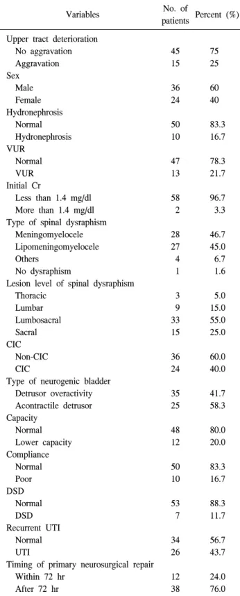
Table 1. Characteristics of patients with neurogenic bladder
Variables No. of
patients Percent (%) Upper tract deterioration
No aggravation Aggravation Sex
Male Female Hydronephrosis Normal Hydronephrosis VUR
Normal VUR Initial Cr
Less than 1.4 mg/dl More than 1.4 mg/dl Type of spinal dysraphism Meningomyelocele Lipomeningomyelocele Others
No dysraphism
Lesion level of spinal dysraphism Thoracic
Lumbar Lumbosacral Sacral CIC Non-CIC CIC
Type of neurogenic bladder Detrusor overactivity Acontractile detrusor Capacity
Normal Lower capacity Compliance Normal Poor DSD Normal DSD Recurrent UTI Normal UTI
Timing of primary neurosurgical repair Within 72 hr
After 72 hr
45 15 36 24 50 10 47 13 58 2 28 27 4 1 3 9 33 15 36 24 35 25 48 12 50 10 53 7 34 26 12 38
75 25 60 40 83.3 16.7 78.3 21.7 96.7 3.3 46.7 45.0 6.7 1.6 5.0 15.0 55.0 25.0 60.0 40.0 41.7 58.3 80.0 20.0 83.3 16.7 88.3 11.7 56.7 43.7 24.0 76.0 VUR: vesicoureteral reflux, CIC: clean intermittent catheterization, DSD: detrusor sphincter dyssynergia, UTI: urinary tract infection 대상 및 방법
1994년 1월부터 2007년 6월까지 신경인성 방광 환아 60명 (남아 36명, 여아 24명)을 대상으로 후향적으로 분석하였다.
방문 당시 평균 나이는 29.1개월 (신생아-14세)이었으며 평 균 추적관찰기간은 48개월 (1-120)이었다. 모든 환아에서 신경인성 방광으로 진단 시 신장 초음파와 배뇨요도방광 조영술을 시행하여 수신증 여부와 방광요관역류 여부를 확 인하였고, 필요에 따라 항콜린성제제와 예방적 항생제를 투여하였다. 3개월 간격으로 요 검사 및 요 배양검사를 시 행하였고 주기적으로 혈중 creatinine, 요역동학검사, 신장초 음파, 배뇨요도방광조영술, DMSA 신주사검사를 시행하였 다.
요역동학검사 시 방광내압 측정은 요도를 통하여 6 Fr 이 중관 카테터를 방광까지 삽입한 후 섭씨 37oC의 생리 식염 수를 사용하여 분당 환아의 방광 용적의 10% 주입하였으며 방광용적은 문헌 보고 <1세 이하: 방광용적 (ml)=38+
{2.5x나이 (개월)}, 1세 이후: 방광용적 (ml)={나이 (년)+
1}x30>에 따라 계산하였다 [9,10]. 이를 통해 방광 용적, 유 순도, 배뇨근 수축력, 표피전극을 사용한 요도 괄약근 활동 도 등을 관찰하였다. 먼저 배뇨 전 방광 충만 시 방광내압곡 선과 근전도의 모양을 관찰하며, 방광의 용적과 요의, 과반 사 방광 여부, 비억제 배뇨근 수축 시의 근전도의 감소 또는 상승 여부, 방광의 유순도, 비억제 배뇨근 수축 또는 기침 등으로 인한 복압 상승 시의 요의 누출, 요의 누출 시의 방 광 내압 등을 관찰하였다. 배뇨가 가능한 환아에서는 배뇨 시 방광 내압과 근전도 등을 관찰하였다. 요역동학검사 소 견으로 비억제 배뇨근 수축이 있는 경우 배뇨근과활동성 (detrusor overactivity), 배뇨근 수축이 관찰되지 않는 경우 무수축성배뇨근 (acontractile detrusor)으로 구분하였다. 배뇨 근 수축 시 괄약근의 이완 여부에 따라 배뇨근괄약근협조 장애를 구분하였다.
상부요로계 손상의 위험 인자로 진단 당시 수신증과 방 광요관역류 유무, 신경인성 방광의 원인이 되는 척수질환 의 종류, 척수 손상 위치, CIC 여부, 요역동학검사에서 신경 인성 방광의 형태, 방광용적, 방광유순도, 배뇨근괄약근협 조장애, 반복적인 요로감염, 척수 이분증에 대한 조기수술 여부에 대하여 알아보았다. 수신증은 진단 당시 시행한 초 음파에서 Society for Fetal Urology (SFU) 등급 2 이상으로 정의하였고 [11], 방광요관역류 유무는 배뇨요도방광조영 술을 시행하여 확인하였다. 요로감염은 요배양 검사에서 105 colony forming unit (CFU)/ml 이상인 경우로 정의하였 고, 2회 이상의 재발성 요로감염을 위험 인자로 고려하였
다. 상부요로계손상은 수신증이나 역류 정도가 악화되는 경우로 정의 하였다. 또한, DMSA 신주사 검사에서 5% 이
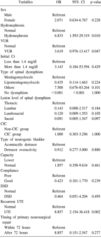
Table 3. Multivariate analyses for predicting upper tract de- terioration
Variables OR 95% CI p-value
Hydronephrosis Normal Hydronephrosis Recurrent UTI Normal UTI
Referent 2.181 Referent
5.810
1.191-11.941
1.200-28.192
0.036
0.029 logistic regression analysis (backward elimination), OR: odds ratio, CI: confidence interval, UTI: urinary tract infection
Table 2. Univariate analyses for predicting upper tract deterioration
Variables OR 95% CI p-value
Sex Male Female Hydronephrosis Normal Hydronephrosis VUR
Normal VUR Initial Cr
Less than 1.4 mg/dl More than 1.4 mg/dl Type of spinal dysraphism Meningomyelocele Lipomeningomyelocele Others
No dysraphism
Lesion level of spinal dysraphism Thoracic
Lumbar Lumbosacral Sacral CIC
Non-CIC group CIC group
Type of neurogenic bladder Acontractile detrusor Detrusor overactivity Capacity
Lower Normal Compliance Poor Good DSD Normal DSD Recurrent UTI Normal UTI
Timing of primary neurosurgical repair
Within 72 hours After 72 hours
Referent 2.071 Referent
6.833 Referent
3.619 Referent
3.143 Referent
0.435 7.500
<0.001 Referent
0.143 0.120 0.091 Referent
1.000 Referent
0.912 Referent
1.857 Referent
0.423 Referent
0.464 Referent
8.857
Referent 8.857
0.634-6.767
1.593-29.319
0.976-13.417
0.184-53.594
0.114-1.663 0.676-83.264
<0.001
0.008-2.517 0.009-1.553 0.005-1.547
0.303-3.296
0.277-3.000
0.358-9.634
0.101-1.770
0.051-4.204
2.154-36.418
0.151-2.547 0.228
0.010
0.047
0.429
0.224 0.101 1.000
0.184 0.105 0.097
1.000
0.880
0.461
0.239
0.495
0.002
0.277 logistic regression analysis, OR: odds ratio, CI: confidence interval, VUR: vesicoureteral reflux, CIC: clean intermittent catheterization, DSD: detrusor sphincter dyssynergia, UTI: urinary tract infection 상의 여과율이 감소하거나 신 실질의 반흔이나 신장 윤곽 의 결함이 확인될 경우도 상부요로계 손상으로 간주하였다 [12,13].
각각의 인자들은 로지스틱 회귀분석을 적용하여 단변량 및 다변량분석을 시행하였고 상부요로계 손상에 대한 상대 적 위험도를 구하였다. 통계분석은 PC-SPSS version 13.0 (SPSS, Inc, USA)을 이용하여 수행하였고, p값이 0.05 미만 인 경우에 통계학적 유의성이 있다고 판정하였다.
결 과 1. 신경인성 방광 환아의 특징
성별에 따라서는 상부요로계 손상의 유의한 차이가 없었 다. 신경인성 방광의 원인이 된 척수 손상의 원인은 46.7%
(28명)가 척수수막류였으며, 45% (27명)가 지방척수수막류 였다. 6.7% (4명)는 신경종 등의 척수 손상을 가진 환아였고 1.6% (1명)는 원인을 알 수 없었다. 척수 손상의 위치는 요 추-미추 손상이 55.0% (33명)로 가장 많았다. 40% (24명)의 환아가 지속적으로 CIC를 시행하였다. 신경인성 방광의 형 태는 배뇨근과활동성이 58.3% (35명), 무수축성배뇨근이 41.7% (25명)였다. 정상 방광용적보다 낮은 방광용적으로 가진 경우 20% (12명)였고, 방광 유순도가 낮은 환아는 16.7% (10명)였다. 11.7% (7명)에서 배뇨근괄약근협조장애 가 관찰되었다. 83.3% (50명)의 환아에서 척수 손상에 대한 수술이 시행되었고 이 중 12명이 72시간 내의 조기수술이 시행되었다 (Table 1).
2. 상부요로계 악화의 위험 인자 분석
진단 당시의 수신증 여부 (p=0.010)와 방광요관역류 (p=
0.047), 반복적인 요로감염 (p=0.002)만이 단변량 분석에서 유의한 인자였으며, 다변량 분석에서는 수신증 여부 [odds ratio (OR)=2.181, 95% confidence interval (CI) 1.191-11.941, p=0.036]와 반복적인 요로감염 [OR=5.810, 95% CI 1.200- 28.192, p=0.029]이 유의한 인자였다 (Table 2, 3).
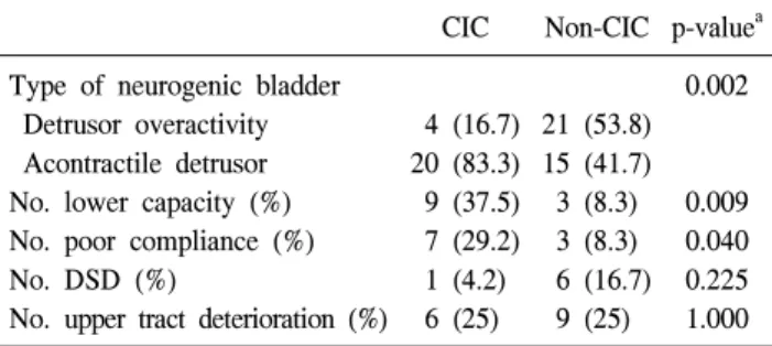
CIC는 주로 무반사 방광에서 많이 시행되었으며 (p=
0.002), 방광용적이 낮거나 (p=0.009), 유순도가 낮은 (p=
Table 4. Urodynamic and clinical characteristics between patients with CIC and those without CIC
CIC Non-CIC p-valuea Type of neurogenic bladder
Detrusor overactivity Acontractile detrusor No. lower capacity (%) No. poor compliance (%) No. DSD (%)
No. upper tract deterioration (%)
4 (16.7) 20 (83.3) 9 (37.5) 7 (29.2) 1 (4.2)
6 (25)
21 (53.8) 15 (41.7) 3 (8.3) 3 (8.3) 6 (16.7)
9 (25)
0.002
0.009 0.040 0.225 1.000 CIC: clean intermittent catheterization, DSD: detrusor sphincter dyssynergia, a: chi-square test
0.040) 환아에서 빈번하게 시행되었고 CIC시행 여부에 따 른 상부요로계 악화의 차이는 없었다 (Table 4).
고 찰
하부요로계 기능이상은 요저장 및 배설 장애로 나타나게 되고, 주로 척수수막류와 같이 신경학적인 원인에 의해서 나타나게 된다. 하부요로계 기능이상이 있는 환아에서 상 부요로계 기능 저하 및 신반흔의 형성은 5-50%까지 발생하 며, 이는 환아에게 매우 절박한 문제이다 [14-17]. 요역동학 검사에서 배뇨근괄약근협조장애, 높은 요누출압, 낮은 방광 유순도 등이 상부 요로계 기능 저하의 위험 인자라는 점은 보고되고 있다 [3,18]. Leonardo 등은 하부요로계 기능이상 이 있는 환아 120명을 대상으로 상부 요로계 기능이상의 위 험인자에 대한 연구에서 방광요관역류 및 요로감염, 잔뇨, 방광벽 비대 등이 단변량 분석에서 유의한 인자였으며, 다 변량 분석에서는 방광요관 역류만이 유의한 인자라고 보고 하였다 [19]. 저자들의 연구에서는 수신증과 방광요관 역류, 재발성 요로감염이 단변량에서 유의한 인자였고, 이중 수 신증과 재발성 요로감염이 다변량에서도 유의한 인자였다.
Holland 등은 재발성 요로감염이나 방광하부 폐색증상을 동반하지 않는 방광요관 역류는 신손상의 위험도가 떨어지 는 것으로 보고하였는데 [20], 이처럼 단순한 역류만으로 상부 요로계 이상을 가져오기 힘들다고 생각한다.
Almodhen 등은 보존적 치료를 시행한 척수수막류 환아 에서 사춘기 이후 환아들의 방광용적 및 배뇨근압이 증가 한다고 보고하였다 [21]. 또한 사춘기 이후 환아들이 유의 하게 요자제를 획득한다고 주장하였다. 하지만 이는 용적 증가와 관련 있으며 배뇨근압의 증가와는 관련성이 떨어지 는 것으로 생각한다. 그리고 흉추 손상이 있는 환아의 경우 요 자제 획득이 떨어지고 상부요로계 변화가 증가한다고 하였으나 본 연구에서는 위치에 따른 차이는 관찰되지 않
았다. 하지만 본 연구의 경우 흉추 손상 환아가 전체 환자의 3명으로 관련성을 찾기에는 제한이 있다고 생각한다.
Teichman 등은 척수수막류 환아에서 신기능 저하는 요로 감염과 방광요관역류와 관련이 있고 요역동학적검사 결과 와는 큰 관련이 없다고 주장하였다 [8]. 또한, CIC는 요 자 제가 없고 요로감염이 재발되거나 방광요관역류, 수신증이 있는 환아에서 시행되어야 하고, 그렇지 않은 경우 집중 추 적관찰만으로도 신기능 보존이 가능하며, 치료의 대안이 될 수 있다고 주장하였다. 저자들의 경우에도 요역동학적 검사 결과에 따른 상부요로계 이상의 차이는 없었다. 본 연 구에서 CIC는 주로 무반사 방광, 낮은 방광용적 및 유순도 가 낮은 환아에서 주로 시행되었다. 배뇨근과활동성의 경 우 CIC 권유에도 불구하고 외견상 배뇨하는 것처럼 보여 실천하지 않는 경우가 많았다. CIC 시행 여부에 따른 상부 요로계 악화의 차이가 없었지만 수신증과 반복적인 요로감 염이 있는 위험군 환아에서 CIC를 시행한 3명에서 모두 상 부요로계 악화가 관찰되지 않았다. 따라서 고위험군 환아 에서는 CIC 등의 적극적인 치료가 필요하다고 생각하며, 집중 추적관찰로 위험요소를 조기 발견하여 치료하는 것도 치료의 대안이 될 수 있다고 생각한다.
Tarcan 등은 생후 72시간 내의 조기 척수수술이 신경인성 방광 예후에 영향을 미친다고 주장하였다 [22]. 72시간이 지난 후 수술을 시행한 경우, 요누출압의 상승을 가져오므 로 조기 수술로 신경인성 방광 예후를 개선시킬 수 있다고 보고하였다. 저자들의 경우 수술을 시행한 50명 중 12명 (24%)이 72시간 내 조기 수술을 시행하였으나 72시간 이후 수술 환자와 상부요로계 이상의 차이는 관찰되지 않았다.
하지만 수술 전, 후 방광 기능의 변화가 발생할 수 있으므로 장기적인 추적 검사가 필요하다 [23].
Lee와 Greenfield는 항콜린성제제 등에 반응이 없는 척추 이분증 환아에서, 방광창냄술 등의 수술이 필요하다고 주 장하였다 [24]. 약물에 반응이 없는 경우 유아기에 높은 요 노출압과 방광벽의 비후로 상부 요로계 악화가 유발될 수 있으므로 방광창냄술을 통해 임상적 경과를 개선시키고, 적당한 나이가 되었을 때 요 자제를 가질 수 있는 재건술 시행하는 방법을 제시하였다. 본 연구의 경우에도 2명의 환 아에서 방광창냄술을 시행하였고 임상적 경과 호전을 보였 다.
신경인성 방광 환아에서 상부 요로계 악화 인자에 대한 보고는 여러 차례 있었지만 본 연구는 여러 악화 인자에 대한 위험도 평가 및 관련성을 확인하였다. 또한 신경인성 방광 환아에서 전통적으로 인정되고 있는 항콜린성제제와 청결 간헐적 도뇨 (CIC) 시행에 대하여 평가해보았다. 하지 만 제한된 환자에서 전향적 연구가 아닌 점은 본 연구의
제한점이라고 할 수 있다. 따라서 향후 보다 더 큰 환자군에 서 장기간의 추적 결과에 대한 연구가 필요하다고 생각한다.
결 론
신경인성 방광 환아에서 수신증과 반복적인 요로감염, 방광요관역류는 상부요로계 기능 악화의 중요한 위험 인자 로 여겨진다. 그러므로 이러한 위험 인자에 노출되어 있는 신경인성 방광 환아는 정기적인 요역동학검사와 방사선학 검사 등의 집중 추적관찰을 시행해야 하고 상부요로계 기 능악하를 예방하기 위해 적극적인 치료를 고려해야 한다고 생각한다.
REFERENCES
1. Feldman AS, Bauer SB. Diagnosis and management of dys- functional voiding. Curr Opin Pediatr 2006;18:139-47.
2. Ghoniem GM, Roach MB, Lewis VH, Harmon EP. The value of leak pressure and bladder compliance in the urodynamic evaluation of meningomyelocele patients. J Urol 1990;144:
1440-2.
3. McGuire EJ, Woodside JR, Borden TA, Weiss RM. Prognostic value of urodynamic testing in myelodysplastic patients. J Urol 1981;126:205-9.
4. Kaefer M, Pabby A, Kelly M, Darbey M, Bauer SB. Improved bladder function after prophylactic treatment of the high risk neurogenic bladder in newborns with myelomentingocele. J Urol 1999;162:1068-71.
5. Lee SL, Park WH, Shim HB. Long-term followup of clean intermittent catheterization in spinal cord injury patients.
Korean J Urol 1997;38:59-64.
6. Seo WK, Park CH, Kim CI, Kim KS. Long-term followup of clean intermittent catheterization in patients with neurogenic bladder. Korean J Urol 1995;36:645-50.
7. Wu HY, Baskin LS, Kogan BA. Neurogenic bladder dysfunc- tion due to myelomeningocele: neonatal versus childhood treatment. J Urol 1997;157:2295-7.
8. Teichman JM, Scherz HC, Kim KD, Cho DH, Packer MG, Kaplan GW. An alternative approach to myelodysplasia management: aggressive observation and prompt intervention.
J Urol 1994;152:807-11.
9. Hjalmas K. Micturition in infants and children with normal lower urinary tract. A urodynamic study. Scand J Urol Nephrol 1976;37(Suppl):1-106.
10. Holmdahl G, Hanson E, Hanson M, Hellstrom AL, Hjalmas K, Sillen U. Four-hour voiding observation in healthy infants.
J Urol 1996;156:1809-12.
11. Fernbach SK, Maizels M, Conway JJ. Ultrasound grading of hydronephrosis: introduction to the system used by the Society for Fetal Urology. Pediatr Radiol 1993;23:478-80.
12. Goldraich NP, Goldraich IH. Update on dimercaptosuccinic acid renal scanning in children with urinary tract infection.
Pediatr Nephrol 1995;9:221-6.
13. Kim BS, Kim HT, Chung SK. Clinical course of pediatric ureteropelvic junction obstruction according to the age at diagnosis. Korean J Urol 2007;48:1302-7.
14. Capitanucci ML, Iacobelli BD, Silveri M, Mosiello G, De Gennaro M. Long-term urological follow-up of occult spinal dysraphism in children. Eur J Pediatr Surg 1996;6(Suppl 1):
25-6.
15. Mostwin JL. Pathophysiology: the varieties of bladder over- activity. Urology 2002;60(Suppl 1):22-6.
16. Ottolini MC, Shaer CM, Rushton HG, Majd M, Gonzales EC, Patel KM. Relationship of asymptomatic bacteriuria and renal scarring in children with neuropathic bladders who are practic- ing clean intermittent catheterization. J Pediatr 1995;127:368- 72.
17. Silveri M, Capitanucci ML, Capozza N, Mosiello G, Silvano A, Gennaro MD. Occult spinal dysraphism: neurogenic void- ing dysfunction and long-term urologic follow-up. Pediatr Surg Int 1997;12:148-50.
18. Vega-P JM, Pascual LA. High-pressure bladder: an underlying factor mediating renal damage in the absence of reflux? BJU Int 2001;87:581-4.
19. Leonardo CR, Filgueiras MF, Vasconcelos MM, Vasconcelos R, Marino VP, Pires C, et al. Risk factors for renal scarring in children and adolescents with lower urinary tract dysfunction. Pediatr Nephrol 2007;22:1891-6.
20. Holland NH, Jackson EC, Kazee M, Conrad GR, Ryo UY.
Relation of urinary tract infection and vesicoureteral reflux to scars: follow-up of thirty-eight patients. J Pediatr 1990;116:
S65-71.
21. Almodhen F, Capolicchio JP, Jednak R, El Sherbiny M.
Postpubertal urodynamic and upper urinary tract changes in children with conservatively treated myelomeningocele. J Urol 2007;178:1479-82.
22. Tarcan T, Onol FF, Ilker Y, Alpay H, Simsek F, Ozek M. The timing of primary neurosurgical repair significantly affects neurogenic bladder prognosis in children with myelome- ningocele. J Urol 2006;176:1161-5.
23. Moon KH, Lee SK, Ra YS, Kim JB, Kim KS. The change of bladder function after neurosurgery in patients with lipom- yelomeningocele. Korean J Urol 2007;48:452-7.
24. Lee MW, Greenfield SP. Intractable high-pressure bladder in female infants with spina bifida: clinical characteristics and use of vesicostomy. Urology 2005;65:568-71.