98 http://e-aair.org © Copyright The Korean Academy of Asthma, Allergy and Clinical Immunology • The Korean Academy of Pediatric Allergy and Respiratory Disease
INTRODUCTION
Idiopathic hypereosinophilic syndrome (IHES) is a group of disorders characterized by the overproduction of eosinophils resulting in inflammatory damage to multiple organs, includ- ing skin, heart, lung, gastrointestinal tract, and nervous system.1 Signs and symptoms associated with lung involvement are rel- atively common, occurring in approximately 40% of IHES pa- tients.2,3 However, despite frequent lung involvement, acute re- spiratory distress syndrome (ARDS) is rarely seen in conjunction with IHES, with only a few cases described in the literature to date.4,5 This is the first case report of IHES presented with ARDS in Korea.
CASE REPORT
A 37-year-old male visited the emergency department at Sam- sung Medical Center, Seoul, Korea, presenting with a chief com- plaint of dyspnea. Initial symptoms had begun 1 week prior to his visit, characterized by fever and chills, followed by progres- sive respiratory difficulty. He appeared acutely ill. A crackling sound was heard upon chest examination. His heartbeat was regular without a murmur. A purplish discoloration of the right ankle was observed, but there were no local heat or tenderness.
Initial blood pressure was 163/91 mmHg, pulse rate was 114 beats/min, respiratory rate was 24 breaths/min, and body tem- perature was 38°C. Oxygen saturation was initially detected as
A Case of Idiopathic Hypereosinophilic Syndrome Presenting With Acute Respiratory Distress Syndrome
Kyung-Suk Lim, Jaehoon Ko, Seong Soo Lee, Beomsu Shin, Dong-Chull Choi, Byung-Jae Lee*
Department of Medicine, Samsung Medical Center, Sungkyunkwan University School of Medicine, Seoul, Korea
87%, and increased to 95% upon oxygen supplement via nasal cannula at 5 L/min. Following oxygen supplementation, arteri- al blood gas analysis (ABGA) showed a pH of 7.39, partial pres- sure of carbon dioxide (PaCO2) of 40.9 mmHg, partial pressure of oxygen (PaO2) of 113.7 mmHg, and oxygen saturation of 98%.
Laboratory tests revealed peripheral eosinophilia and throm- bocytopenia with a white blood cell count of 22,580/μL (50%
neutrophils, 31% eosinophils, 10% lymphocytes), Hb levels at 13.7 g/dL, and a platelet count of 26,000/μL. Prothrombin and activated partial thromboplastin times were 15 seconds, and 35.4 seconds respectively. Fibrinogen levels were within the normal range, but the concentration of D-dimer was markedly elevated at 26.69 μg/mL. Serum total IgE and ECP levels (939.4 IU/mL and 201.0 ng/mL, respectively) were both elevated, too.
Tests for both anti-nuclear antibodies and anti-neutrophil cyto- plasmic antibodies were negative. A test for parasites in the stool and serum were negative. Bone marrow biopsy showed normal cellularity with increased eosinophils. Fip1-like1 and platelet- derived growth factor receptor alpha (FIP1L1-PDGFRA) gene fusion was not detected. Patchy consolidation with ground-
Case Report
Allergy Asthma Immunol Res. 2014 January;6(1):98-101.
http://dx.doi.org/10.4168/aair.2014.6.1.98 pISSN 2092-7355 • eISSN 2092-7363
Although idiopathic hypereosinophilic syndrome(IHES) commonly involves the lung, it is rarely associated with acute respiratory distress syndrome (ARDS). Here we describe a case of IHES presented in conjunction with ARDS. A 37-year-old male visited the emergency department at Samsung Medical Center, Seoul, Korea, with a chief complaint of dyspnea. Blood tests showed profound peripheral eosinophilia and thrombocytopenia. Patchy areas of consolidation with ground-glass opacity were noticed in both lower lung zones on chest radiography. Rapid progression of dyspnea and hy- poxia despite supplement of oxygen necessitated the use of mechanical ventilation. Eosinophilic airway inflammation was subsequently confirmed by bronchoalveolar lavage, leading to a diagnosis of IHES. High-dose corticosteroids were administered, resulting in a dramatic clinical response.
Key Words: Idiopathic hypereosinophilic syndrome; eosinophilia; acute respiratory distress syndrome; pulmonary thromboembolism
This is an Open Access article distributed under the terms of the Creative Commons Attribution Non-Commercial License (http://creativecommons.org/licenses/by-nc/3.0/) which permits unrestricted non-commercial use, distribution, and reproduction in any medium, provided the original work is properly cited.
Correspondence to: Byung-Jae Lee, MD, PhD, Division of Allergy, Department of Medicine, Samsung Medical Center, Sungkyunkwan University School of Medicine, 81 Irwon-ro, Gangnam-gu, Seoul 135-710, Korea.
Tel: +82-2-3410-3427; Fax: +82-2-3410-3849; E-mail: [email protected] Received: February 7, 2013; Accepted: March 20, 2013
•There are no financial or other issues that might lead to conflict of interest.
Idiopathic Hypereosinophilic Syndrome With ARDS
Allergy Asthma Immunol Res. 2014 January;6(1):98-101. http://dx.doi.org/10.4168/aair.2014.6.1.98 AAIR
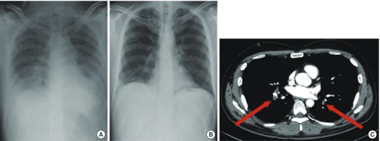
99 http://e-aair.org glass opacity in both lower lung zones was noticed in the initial
chest radiography (Fig. 1A).
During the initial workup, the patient became progressively tachypneic with increased oxygen demand to 15 L/min via fa- cial mask. The patient was transferred to the intensive care unit and oxygen supply was increased to 100% by high-flow nasal cannula. Despite supplementation, PaO2 levels remained low (49.9 mmHg) leading to a diagnosis of ARDS, and then the pa- tient underwent intubation and mechanical ventilation.
Bronchoalveolar lavage (BAL) revealed marked eosinophilia (187/200 evaluated cells, 93.5% eosinophils). Skin biopsy of the right ankle showed diffuse eosinophilic perivascular infiltration of the dermis (Fig. 2A) and eosinophilic abscess of subcutane- ous fat (Fig. 2B). Duplex sonography revealed deep vein throm- bosis in the left popliteal, soleal, and peroneal veins. Nerve con- duction test and electromyography showed neuropathy in the right posterior tibial nerve.
A diagnosis of IHES was reached based on the presence of pe- ripheral and tissue eosinophilia, along with the exclusion of
other causes of eosinophilia. High-dose corticosteroid (methyl- prednisone 1 mg/kg/day) therapy was initiated on the first day of admission. By day 3, eosinophil counts decreased to the nor- mal range (Fig. 3). Extubation was performed on day 4 as a re- sult of clinical and radiological improvements.
A chest CT scan was obtained on day 9 to evaluate symptoms of mild dyspnea despite the dramatic improvement seen by simple radiography (Fig. 1B). Pulmonary thromboembolism (PTE) was detected in the bilateral lobar and segmental branch- es of the pulmonary arteries (Fig. 1C), and we started anticoag- ulant therapy. On day 23, the patient was discharged from the hospital with a gradual tapering of corticosteroids.
DISCUSSION
In 1975, Chusid et al.6 established the basic guidelines for di- Fig. 1. (A) Patchy areas of consolidation with ground-glass opacity are observed in both lower lung zones on the initial chest radiograph. (B) Follow-up chest radiograph on hospital day 9 shows a dramatic decrease of lung infiltrate. (C) Chest CT scan shows bilateral acute pulmonary thromboembolism (arrows) involving both pulmonary arteries.
C
A B
Fig. 2. Skin biopsy of the purpuric lesions of the right ankle shows (A) diffuse eosinophilic perivascular infiltration in the dermis and (B) eosinophilic abscess
in the subcutaneous fat (H&E stain, ×40 magnification). Fig. 3. Platelet counts, eosinophil counts, and D-dimer levels during hospitaliza- tion. Following initiation of high-dose corticosteroid therapy, eosinophil counts decreased to normal levels by day 3, and platelet counts progressively recov- ered. D-dimer levels initially elevated, but began to decrease with anticoagulant therapy. HD, hospital day.
D-dimer (ug/mL) Eosinophil (×100/uL) Platelet (×1,000/uL)
80 70 60 50 40 30 20 10 0
250 200 150 100 50 HD1 HD4 HD7 HD10 HD13 HD17 HD20 HD23 0
D-dimer Eosinophil Platelet Corticosteroids
Anticoagulation
A B
Lim et al.
Allergy Asthma Immunol Res. 2014 January;6(1):98-101. http://dx.doi.org/10.4168/aair.2014.6.1.98 Volume 6, Number 1, January 2014
100 http://e-aair.org
agnosis of IHES, which is still in use today. Criteria include blood eosinophilia>1,500 cells/μL for longer than 6 months, lack of evidence for parasitic, allergic, or other known causes of eosin- ophilia, and the presence of organ damage or dysfunction relat- ed to hypereosinophilia. However, recently these criteria have been changed; when marked eosinophilia and obvious tissue damage such as cardiac involvement are observed, the initiation of treatment is recommended regardless of the duration of eo- sinophilia.7 Our case fulfills the diagnostic criteria for IHES ex- cept the duration of peripheral eosinophilia.
Lung involvement is relatively common in IHES. In a previous study, of 49 patients with IHES, 63% had more than one kind of respiratory symptoms, and 43% showed abnormal findings by chest radiography or CT scan.8 However, despite the fact that lung is frequently involved in IHES, there have been only a few cases that have presented ARDS. ARDS is an acute and diffuse inflammatory lung injury that leads to increased pulmonary vascular permeability, increased lung weight, and a loss of aer- ated tissue.9 Approximately 60 distinct etiologies including eo- sinophilic pneumonia have been recognized for ARDS, among which severe sepsis and bacterial pneumonia are the most common.10-14
Our patient presented fever and dyspnea initially, and chest radiography showed bilateral parenchymal infiltration. Intuba- tion was performed due to ARDS, and bronchoscopic examina- tion with BAL revealed profound alveolar eosinophilia. The main cause of ARDS was thought to be severe eosinophilic parenchy- mal inflammation of the lungs. This diagnosis was confirmed by rapid clinical and radiologic response to high-dose cortico- steroid therapy.
Thromboembolic complication is a common cause of mortal- ity and morbidity in patients with IHES. About 25% of IHES pa- tients experience thromboembolisms, with a mortality rate of 5%-10%.15 In this case, a chest CT scan on hospital day 9 revealed a thromboembolism in the bilateral pulmonary arteries. PTE was suspected considering the initial presentation, which in- cluded purplish discoloration of the right ankle, thrombocyto- penia, D-dimer elevation, and deep vein thrombosis in the left popliteal, soleal, and peroneal veins. However, the marked eo- sinophilia in BAL fluid and the dramatic response to corticoste- roid therapy prior to initiation of anticoagulant therapy suggest that primary cause of respiratory failure is parenchymal lung involvement of IHES, rather than PTE.
Corticosteroids are a first-line therapy for FIP1L1-PDGFRA- negative IHES.16,17 About 85% of patients on corticosteroid ther- apy reach partial or complete remission by 1 month.17 For pa- tients not responding to corticosteroids, second-line therapies such as hydroxyurea, interferon-α, anti-IL5 antibodies, or anti- CD52 antibodies can be considered.18 In the present case, pe- ripheral eosinophilia and involvement of the skin and lung were successfully treated by high-dose corticosteroid alone.
In summary, we describe a rare case of IHES with ARDS due
to eosinophilic lung involvement, which showed a dramatic re- sponse to high-dose corticosteroid therapy.
REFERENCES
1. Klion A. Hypereosinophilic syndrome: current approach to diagno- sis and treatment. Annu Rev Med 2009;60:293-306.
2. Fauci AS, Harley JB, Roberts WC, Ferrans VJ, Gralnick HR, Bjorn- son BH. NIH conference. The idiopathic hypereosinophilic syn- drome. Clinical, pathophysiologic, and therapeutic considerations.
Ann Intern Med 1982;97:78-92.
3. Spry CJ, Davies J, Tai PC, Olsen EG, Oakley CM, Goodwin JF. Clini- cal features of fifteen patients with the hypereosinophilic syndrome.
Q J Med 1983;52:1-22.
4. Roufosse F, Goldman M, Cogan E. Hypereosinophilic syndrome:
lymphoproliferative and myeloproliferative variants. Semin Respir Crit Care Med 2006;27:158-70.
5. Winn RE, Kollef MH, Meyer JI. Pulmonary involvement in the hy- pereosinophilic syndrome. Chest 1994;105:656-60.
6. Chusid MJ, Dale DC, West BC, Wolff SM. The hypereosinophilic syndrome: analysis of fourteen cases with review of the literature.
Medicine (Baltimore) 1975;54:1-27.
7. Simon HU, Rothenberg ME, Bochner BS, Weller PF, Wardlaw AJ, Wechsler ME, Rosenwasser LJ, Roufosse F, Gleich GJ, Klion AD. Re- fining the definition of hypereosinophilic syndrome. J Allergy Clin Immunol 2010;126:45-9.
8. Dulohery MM, Patel RR, Schneider F, Ryu JH. Lung involvement in hypereosinophilic syndromes. Respir Med 2011;105:114-21.
9. ARDS Definition Task Force, Ranieri VM, Rubenfeld GD, Thomp- son BT, Ferguson ND, Caldwell E, Fan E, Camporota L, Slutsky AS.
Acute respiratory distress syndrome: the Berlin definition. JAMA 2012;307:2526-33.
10. Pepe PE, Potkin RT, Reus DH, Hudson LD, Carrico CJ. Clinical pre- dictors of the adult respiratory distress syndrome. Am J Surg 1982;
144:124-30.
11. Hudson LD, Milberg JA, Anardi D, Maunder RJ. Clinical risks for development of the acute respiratory distress syndrome. Am J Respir Crit Care Med 1995;151:293-301.
12. Fowler AA, Hamman RF, Good JT, Benson KN, Baird M, Eberle DJ, Petty TL, Hyers TM. Adult respiratory distress syndrome: risk with common predispositions. Ann Intern Med 1983;98:593-7.
13. Villar J, Blanco J, Añón JM, Santos-Bouza A, Blanch L, Ambrós A, Gandía F, Carriedo D, Mosteiro F, Basaldúa S, Fernández RL, Kac- marek RM; ALIEN Network. The ALIEN study: incidence and out- come of acute respiratory distress syndrome in the era of lung pro- tective ventilation. Intensive Care Med 2011;37:1932-41.
14. Doyle RL, Szaflarski N, Modin GW, Wiener-Kronish JP, Matthay MA. Identification of patients with acute lung injury. Predictors of mortality. Am J Respir Crit Care Med 1995;152:1818-24.
15. Ogbogu PU, Rosing DR, Horne MK 3rd. Cardiovascular manifesta- tions of hypereosinophilic syndromes. Immunol Allergy Clin North Am 2007;27:457-75.
16. Park YM, Bochner BS. Eosinophil survival and apoptosis in health and disease. Allergy Asthma Immunol Res 2010;2:87-101.
17. Ogbogu PU, Bochner BS, Butterfield JH, Gleich GJ, Huss-Marp J, Kahn JE, Leiferman KM, Nutman TB, Pfab F, Ring J, Rothenberg ME, Roufosse F, Sajous MH, Sheikh J, Simon D, Simon HU, Stein ML, Wardlaw A, Weller PF, Klion AD. Hypereosinophilic syndrome: a
Idiopathic Hypereosinophilic Syndrome With ARDS
Allergy Asthma Immunol Res. 2014 January;6(1):98-101. http://dx.doi.org/10.4168/aair.2014.6.1.98 AAIR
101 http://e-aair.org multicenter, retrospective analysis of clinical characteristics and re-
sponse to therapy. J Allergy Clin Immunol 2009;124:1319-25.e3.
18. Park SM, Park JW, Kim SM, Koo EH, Lee JY, Lee CS, Choi DC, Lee
BJ. A case of hypereosinophilic syndrome presenting with multior- gan infarctions associated with disseminated intravascular coagu- lation. Allergy Asthma Immunol Res 2012;4:161-4.