56
C A S E REPO RT
pISSN: 2384-3799 eISSN: 2466-1899 Int J Thyroidol 2018 May 11(1): 56-59 https://doi.org/10.11106/ijt.2018.11.1.56
Received January 9, 2018 / Revised February 8, 2018 / Accepted February 14, 2018
Correspondence: Hwa Young Ahn, MD, PhD, Division of Endocrinology, Department of Internal Medicine, Chung-Ang University College of Medicine, 102 Heukseok-ro, Dongjak-gu, Seoul 06973, Korea
Tel: 82-2-6299-3152, Fax: 82-2-6299-2017, E-mail: [email protected]
Copyright ⓒ 2018, the Korean Thyroid Association. All rights reserved.
This is an open-access article distributed under the terms of the Creative Commons Attribution Non-Commercial License (http://creative- commons.org/licenses/by-nc/4.0/), which permits unrestricted non-commercial use, distribution, and reproduction in any medium, provided the original work is properly cited.
우측 갑상선결절로 오인된 젠커게실 1례
대한민국 공군 방공관제사령부1, 중앙대학교 의과대학 내과학교실, 내분비대사내과2
최창원
1, 안화영
2A Case of Zenker’s Diverticulum Mimicking a Right Side Thyroid Nodule
Chang Won Choi1 and Hwa Young Ahn2
Air Defense & Control Command, ROKAF1, Division of Endocrinology, Department of Internal Medicine, Chung-Ang University College of Medicine2, Seoul, Korea
Zenker’s diverticulum, a pulsion diverticulum of the hypopharynx, is a rare lesion that commonly occurs in left side of hypopharynx. The incidence of esophageal diverticula is much lower than that of focal lesions or nodules of thyroid. In an ultrasonography, the outpouching just like a focal thyroid lesion, may present as an oval or circular structure. The food remnants or gas bubbles present in the diverticulum may mimic microcalcifications presented in papillary thyroid carcinoma. We reported a case of right side Zenker’s diverticulum mimicking a thyroid cancer in thyroid ultrasonography.
Key Words: Thyroid nodule, Zenker diverticulum
서 론
Zenker 게실은 인두에서 상부 식도 이행부에 근육이 없는 부위에 생기는 압출성 게실로, 주로 식도 및 인두 좌측 부위에 생긴다. 고령의 환자에서 연하 장애, 음식 물의 역류, 만성기침, 흡인과 같은 증상으로 발견되는 경우가 대부분이지만, 특별한 증상 없이 젊은 연령에 서도 발생할 수 있다.1) 게실의 발생률이 갑상선결절의 발생률보다 낮으나, Zenker 게실은 해부학적 위치가 갑상선 위치와 가깝고, 초음파에서 원형 또는 타원형 으로 보이며, 내부의 음식 잔여물이나, 공기방울이 미 세석회화와 유사하게 보이는 경우가 많아 갑상선암으 로 오인되는 경우가 있다.2) Zenker 게실이 주로 좌측에 생기는데 반해 저자들은 우측에 우연히 발견된 갑상선 결절을 주소로 내원하여 초음파 및 식도조영술검사,
식도위내시경검사를 시행한 결과 Zenker 게실로 진단 되었던 증례를 경험하였기에 보고하는 바이다.
증 례
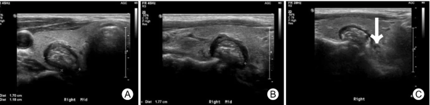
74세 남자 환자가 건강검진에서 우연히 발견된 갑상 선결절을 주소로 내원하였다. 갑상선질환의 과거력은 없었고, 이학적 검사 결과 특이 소견은 관찰되지 않았 다. 갑상선초음파 결과에서 우엽 중앙으로 17×12×18 mm 크기의 주변부 저에코성 가장자리를 지닌 동일 에 코 종괴가 관찰되었으며, 종괴 내부에 미세석회화와 거대석회화로 보이는 고에코의 침상 음영이 관찰되어 갑상선암이 의심되었다(Fig. 1A, B).
세포흡입 병리검사를 위한 추적 초음파검사에서 내 부에 다양한 음영선이 동반된 소견이 관찰되며, 환자 의 연하운동과 상관관계가 있어 보이고, 후방 음영 감
A Case of Zenker’s Diverticulum Mimicking a Right Side Thyroid Nodule
57 Int J Thyroidol
Fig. 1. The axial (A) and longitudinal (B) US scan of right thyroid lobe shows 17×12×18 mm sized isoechoic nodule with micro- and macrocalcification. The arrow shows communication of nodule and esophagus (C).
Fig. 2. (A, B) This esophago- gastroscopy shows an out- pouching lesion of upper eso- phagus. E: esophagus, Z:
Zenker’s diverticulum
소가 있는 것으로 보아 공기일 가능성이 고려되었으며, 식도와의 일부 연결성이 의심되어(Fig. 1C) 식도위내 시경검사(Fig. 2) 및 식도조영술검사(Fig. 3)를 시행하 였다. 식도위내시경검사 결과, 상부 식도 후방 쪽으로 Zenker 게실의 입구가 확인이 되었고, 식도조영술검사 결과, 상부식도 후방 부위로 약 16 mm 크기의 Zenker 게실에 바륨이 차있는 모습과 지연사진 영상에서도 바 륨이 배출은 되어있으나 아직 남아 있는 모습이 확인 되었다. 그 후, 환자는 별다른 증상이 없어 경과 관찰하 기로 하였다.
고 찰
식도게실은 위치에 따라 나뉘는데 가장 흔한 것으로 횡격막상부, 하인두, Zenker, 중부식도게실이 있다. 그 중 Zenker 게실은 1877년에 처음 Zenker와 von Ziemssen 이 23례의 하인두게실을 보고한 이후로 Zenker 게실로 불려왔다. 진성 게실은 장벽이 모두 포함되어 이탈되 는데 반해, Zenker 게실은 식도의 근육층을 관통해 점 막과 점막하층만 이탈하여 발생하는 가성 게실로, 식 도 원위부 폐쇄와 관련된 내강 압력의 증가로 발생하
고 윤상인두근(cricopharyngeus muscle) 주변의 경부 식 도 후방 벽의 해부학적으로 약해진 틈으로 돌출되어 형성되며, 킬리안위약부(Killian’s dehiscence)라고 불리 는 윤상인두근과 하인두수축근(inferior pharyngeal con- strictor) 사이에서 주로 발생한다.1) 그에 반해 Killian- Jamieson 게실은 Zenker 게실에 비해 유병률이 적고, 더 작으며, 윤상인두근 아래쪽으로 발생하여 경부 식 도 앞쪽 외측으로 돌출되게 된다. 본 증례는 식도조영 술 결과 상부 식도 후방 쪽으로 돌출된 게실이 확인되 었기에 Zenker 게실로 볼 수 있다. Zenker 게실의 경우 식도 상부에 발생하기 때문에 갑상선과 가깝게 위치하 여, 초음파검사에서 초음파 방향에 따라 갑상선 종양 으로 보일 수 있고 게실 내 존재하는 공기방울이나 음 식물 잔여물이 고에코의 침상음영으로 보여 갑상선 유 두 세포암에서 관찰되는 미세석회화로 오인될 수 있 다.2,3)
갑상선결절은 갑상선에 가장 흔한 질환으로 갑상선 초음파의 발달로 16-68%의 인구에서 관찰되며, 나이가 증가할수록 유병률이 늘어, 40세 이상에서는 50%에서 발견되고, 남성보다는 여성에서 흔히 발견된다. 갑상선 결절이 발견된 경우에, 결절의 크기 및 악성을 시사하
Chang Won Choi and Hwa Young Ahn
Vol. 11, No. 1, 2018 58 Fig. 3. The esophagography confirms a barium filled sac projecting from the upper esophagus, posterior aspects.
(A) Barium filling view, (B) mucosal relief view. Arrow:
Zenker's diverticulum.
Table 1. Literature review of ultrasonography of esophageal diverticulum
Reference Year Age/Sex Location Size (cm)
Sonographic findings
Echogenicity Internal echogenic foci
Boundary hypoechoic zone
Posterior shadowing
Kim et al.2) 2012 55/F Left 4.0 Hypoechoic Present Present Present
50/M Left 1.2 Hypoechoic Present Present Present
55/M Left 1.2 Hypoechoic Present Present Present
59/F Left 1.7 Hypoechoic Present Present Present
Cao et al.9) 2016 40/F Left 1.3 Hypoechoic Present Present Absent
Mimatsu et al.10) 2013 77/F Left 3.0 Isoechoic Present Present Present
Pang et al.11) 2009 54/M Left 1.1 Hypoechoic Present Present Absent
Kwak and Kim12) 2006 61/F Left 2.1 Isoechoic Present Present Present
56/F Left 2.0 Isoechoic Present Present Present
70/F Left 3.3 Hypoechoic Present Present Present
65/M Right 1.0 Hypoechoic Present Present Present
60/F Left 0.7 Round Absent Absent Absent
26/F Left 0.8 Round Absent Absent Absent
Present study 2017 74/M Right 1.7 Isoechoic Present Present Present
F: female, M: male
는 소견(저에코병변, 미세석회화, 불규칙하거나 침습 적 경계)을 보일 경우 진단을 위해 미세침흡인검사를 시행한다.4) 무증상 Zenker 게실을 인지하지 못한 상태 로 기관지삽관술이나 내시경을 할 경우 식도천공의 위
험성이 있을 수 있는데, 본 증례와 같은 경우 Zenker 게실을 갑상선결절로 오인하여 갑상선 미세침흡인검 사를 시행한다면 미세 식도천공 및 이로 인한 종격동 염과 같은 합병증이 발생할 수 있으므로, 갑상선결절
A Case of Zenker’s Diverticulum Mimicking a Right Side Thyroid Nodule
59 Int J Thyroidol
과 식도게실의 감별이 매우 중요하다.
Zenker 게실의 특징적인 초음파 소견은, 첫 번째로 병변의 내부에 고에코성 병변이 보이는데 이는 게실 내부의 공기 때문이고, 기존 보고된 증례들을 검토하 였을 때 거의 모든 경우에 있어 저에코성의 변연부를 지닌다는 점이다(Table 1). 두 번째는 병변의 벽이 다층 성의 패턴을 보이는 경우가 많은데, 이는 병변의 근원 이 소화관임을 시사하는 소견이라고 볼 수 있고, 특히 식도와 결절의심 병변 간의 연결이 의심되는 소견이 있다면 감별진단에 더욱 도움이 된다. 세 번째는 환자 에게 물을 삼키게 하거나 침을 삼키게 함으로써 갑상 선결절의 내부 에코변화 및 결절의 크기 변화가 있을 경우 Zenker 게실을 시사하는 소견으로 생각할 수 있 다.2,3,5-7)
Zenker 게실은 주로 좌측에 호발하는 양상을 보이는 데, 그 이유에 대하여 식도게실의 발생 위치가 주로 사 용하는 손의 방향에 따라 결정된다는 연구 결과가 있 다. 즉, 오른손잡이의 경우 식도게실은 주로 왼쪽에 생 기는데, 전체 인구 중 11% 정도가 왼손잡이이고 오른 손잡이가 훨씬 많기 때문에 식도게실이 주로 좌측에 호발하는 것으로 판단된다는 보고가 있으나8) 본 증례 의 환자는 오른손잡이인데 반해, 우측에 게실이 발생 한 것으로 보아 이러한 이론이 정확한 것은 아닌 것으 로 보인다.
Zenker 게실은 특별한 증상이 없을 경우에는 관찰만 으로 충분하나, 소화불량, 역류, 만성기침, 흡인 등의 증상이 발생하거나, 흡인성 폐렴 등의 합병증이 발생 한 경우에는 수술이나 내시경적 시술을 필요로 하는 경우가 있다.1) 본 증례의 환자는 아직 별다른 증상이 없어서 환자에게 발생할 수 있는 증상 및 합병증에 대 해 충분한 설명을 한 후 경과 관찰하기로 결정하였다.
Zenker 게실은 전체 식도조영술의 약 1% 정도에서 발견되는 식도의 가장 흔한 게실로 갑상선초음파검사 에서 갑상선결절로 오인될 수 있으나 환자의 연하운동 과 연관된 결절 내부 에코발생도의 변화 및 움직임의 관찰과 결절과 식도의 연결점을 찾는다면 불필요한 세
포검사를 피할 수 있고 이에 따른 합병증을 피할 수 있을 것으로 기대된다.
중심 단어: 갑상선결절, 젠커게실.
References
1) Law R, Katzka DA, Baron TH. Zenker's diverticulum. Clin Gastroenterol Hepatol 2014;12(11):1773-82; quiz e111-2.
2) Kim HK, Lee JI, Jang HW, Bae SY, Lee JH, Kim YS, et al. Characteristics of Killian-Jamieson diverticula mimicking a thyroid nodule. Head Neck 2012;34(4):599-603.
3) Kim SJ, Kim CH. The genetic studies of obsessive-compulsive disorder and its future directions. Yonsei Med J 2006;47(4):
443-54.
4) Haugen BR, Alexander EK, Bible KC, Doherty GM, Mandel SJ, Nikiforov YE, et al. 2015 American Thyroid Association Management Guidelines for adult patients with thyroid nodules and differentiated thyroid cancer: The American Thyroid Association Guidelines Task Force on thyroid nodules and differentiated thyroid cancer. Thyroid 2016;26(1):1-133.
5) Yoon HD, Shon HS. Killian-Jamieson diverticulum mimicking a thyroid nodule. Korean J Med 2005;68(4):467-8.
6) DeFriend DE, Dubbins PA. Sonographic demonstration of a pharyngoesophageal diverticulum. J Clin Ultrasound 2000;28(9):
485-7.
7) Komatsu M, Komatsu T, Inove K. Ultrasonography of Zenker's diverticulum: special reference to differential diagnosis from thyroid nodules. Eur J Ultrasound 2000;11(2):123-5.
8) Stafford ND, Moore-Gillon V, McKelvie P. Handedness and the side on which pharyngeal pouches occur. Br Med J (Clin Res Ed) 1984;288(6420):815-6.
9) Cao L, Ge J, Zhao D, Lei S. Killian-Jamieson diverticulum mimicking a calcified thyroid nodule on ultrasonography: a case report and literature review. Oncol Lett 2016;12(4):2742-5.
10) Mimatsu K, Oida T, Kano H, Kawasaki A, Fukino N, Kida K, et al. Killian-jamieson diverticula presenting synchronously with thyroid adenoma. Case Rep Gastroenterol 2013;7(1):
188-94.
11) Pang JC, Chong S, Na HI, Kim YS, Park SJ, Kwon GY.
Killian-Jamieson diverticulum mimicking a suspicious thyroid nodule: sonographic diagnosis. J Clin Ultrasound 2009;37(9):
528-30.
12) Kwak JY, Kim EK. Sonographic findings of Zenker diverticula.
J Ultrasound Med 2006;25(5):639-42.