ISSN 0378-6471 (Print)⋅ISSN 2092-9374 (Online)
http://dx.doi.org/10.3341/jkos.2016.57.6.999
Case Report
백내장 수술 후 발생한 스타필로코쿠스 루그두넨시스 안내염 1예
A Case of Staphylococcus lugdunensis Endophthalmitis after Cataract Surgery
한상윤⋅이태곤
Sang Youn Han, MD, Tae Gon Lee, MD, PhD
건양대학교 의과대학 김안과병원 안과학교실 명곡안연구소
Myunggok Eye Research Center, Department of Ophthalmology, Kim’s Eye Hospital, Konyang University College of Medicine, Seoul, Korea
Purpose: To report a case of Staphylococcus lugdunensis endophthalmitis following cataract extraction and intraocular lens implantation.
Case summary: A 59-year-old woman presented with unilateral vision impairment and eyeball pain in her left eye, thirteen days after phacoemulsification and posterior chamber intraocular lens implantation. Best-corrected visual acuity of her left eye was 20/200. Slit lamp examination of her left eye revealed a severe conjunctival injection, severe chamber reactions with exudative membranes, hypopyon (about 1 mm) in the anterior chamber, and the fundus was not visible. Before the patient was admitted to the hospital, we cultured samples of aqueous fluid and performed an intravitreal antibiotics injection (vancomycin 1.0 mg/0.1 mL, ceftazidime 2.0 mg/0.1 mL). However, on the next day, because the inflammatory reactions of the anterior chamber and vitreous cavity were not improved and Gram positive cocci was confirmed, we performed a pars plana vitrectomy and an additional intra- vitreal antibiotics injection (vancomycin 1.0 mg/0.1 mL, dexamethasone 0.5 mg/0.1 mL). Seven days after the surgery, Staphylococcus lugdunensis was identified in the aqueous fluids culture. 11 days after the surgery, her inflammation and symp- toms were improved and therefore, she could be discharged. Three months after the surgery, best-corrected visual acuity of her left eye was 20/20 and there was no evidence of recurrence of endophthalmitis and no abnormal findings in her fundus.
J Korean Ophthalmol Soc 2016;57(6):999-1003
Keywords: Cataract surgery, Endophthalmitis, Postoperative endophthalmitis, Staphylococcus lugdunensis
■Received: 2015. 7. 3. ■ Revised: 2015. 9. 9.
■Accepted: 2015. 10. 29.
■Address reprint requests to Tae Gon Lee, MD, PhD Kim's Eye Hospital, #136 Yeongsin-ro, Yeongdeungpo-gu, Seoul 07301, Korea
Tel: 82-2-2639-7811, Fax: 82-2-2639-9214 E-mail: [email protected]
ⓒ2016 The Korean Ophthalmological Society
This is an Open Access article distributed under the terms of the Creative Commons Attribution Non-Commercial License (http://creativecommons.org/licenses/by-nc/3.0/) which permits unrestricted non-commercial use, distribution, and reproduction in any medium, provided the original work is properly cited.
안내염은 백내장 수술 후에 생길 수 있는 매우 심각한 합 병증 중의 하나로, 적절한 치료에도 불구하고 영구적인 시 력손상을 초래할 수 있다. 백내장 수술 후 안내염의 발생률 은 0.05-0.68%로 보고되고 있으며,이를 성공적으로 치료하 기 위해서는 조기에 원인균을 동정하고 치료하는 것이 중요 하다.1-3 백내장 수술 후 발생하는 안내염 중에서 원인균이 동정된 경우는 44-75%로 보고되었고,4 이 중 Staphylococcus
epidermidis, Staphylococcus aureus, Streptococcus species
같은 그람양성균이 가장 흔하며, Psuedomonas, Haemophilus 와 같은 그람음성균도 약 6%를 차지한다.5Staphylococcus lugdunensis는 그람양성균으로, 눈꺼풀에
존재하는 정상세균총 중의 하나이며 공기 중의 오염물, 오 염된 세척액이나 수술도구, 인공수정체, 눈꺼풀과 눈물낭의 염증 등에 의해서 안내염의 원인균이 될 수 있다.6S.lugdun-
ensis에 의한 백내장 수술 후 안내염은 매우 드물고 예후가
좋지 않은 것으로 알려져 있으며, 국내에서는 아직 보고된 적이 없는데, 저자들은 백내장 수술 후 S.lugdunensis에 의 해 발생한 안내염 1예에서, 조기에 유리체절제술 및 유리체 강내 항생제주입술로 치료하여 좋은 결과를 경험하였기에, 이를 문헌고찰과 함께 국내 처음으로 보고하고자 한다.Figure 1. Pre- and intraoperative findings. (A) Just before vi-
trectomy, anterior segment shows severe conjunctival in- jection and hypopyon. (B) Thick, exudative membranes were found in anterior chamber.Figure 2. Intraoperative fundus finding. At the end of vi-
trectomy, there was no inflammation or retinal hemorrhages on her left fundus.증례보고
특별한 내과적 과거력과 외상력이 없는 59세 여자 환자 가 본원에서 좌안 초음파수정체유화술 및 인공수정체후낭 삽입술을 시행 받은 지 13일째 되는 날 아침부터 발생한 좌 안의 통증과 시력저하를 주소로 내원하였다. 백내장 수술 당시 후낭파열 등의 특이소견은 없었다. 내원 당시 측정한 최대교정시력은 우안 20/20, 좌안 20/200이었고, 비접촉식 안압계로 측정한 안압은 우안 10 mmHg, 좌안 11 mmHg였 다. 세극등현미경 검사에서 좌안의 결막충혈, 전방 내 다수 의 염증세포(+4), 1 mm 가량의 전방축농과 삼출성 막이 관 찰되었다. 매질의 혼탁으로 인해 시신경유두의 경계부 및 안저를 관찰하기 어려웠으나, 안저반사는 비교적 잘 관찰 되었다. 임상적인 소견에 따라, 백내장 수술 후 발생한 안 내염으로 진단하고 입원치료를 권유하였다. 입원 전 전방 천자를 시행하여 그람염색, 세균배양, 진균배양 및 세균동 정 검사를 의뢰하였고, 유리체강내항생제주입술(vancomy- cin 1.0 mg/0.1 mL, ceftazidime 2.0 mg/0.1 mL)을 함께 시 행하였다. 경구용 항생제로 ofloxacin 100 mg tid를 처방하 였고, 국소점안제로는 0.5% moxifloxacin 점안액과 1%
prednisolone acetate 점안액을 2시간 간격으로, 1% cyclo- pentolate HCl 점안액을 하루 2차례 쓰도록 처방하였다. 다 음 날, 그람양성구균으로 보이는 균주의 증식이 다수 관찰
되었고, 좌안의 전방 및 안저상태에도 호전이 없어, 국소마 취로 23게이지 유리체절제술을 시행하였다. 백내장 수술 당시 만들었던 각막절개창과는 다른 부위에 절개창을 내고 전방 내로 접근하여, 동공 및 후낭인공수정체를 덮고 있는 염증막을 hook과 intraocular forceps를 이용하여 제거하고, 유리체절제술을 시행하였다(Fig. 1). 수술 중 망막박리, 망 막괴사, 망막출혈 등 안저의 특별한 이상소견은 관찰되지 않 았고, 안저의 염증물질과 혼탁을 최대한 제거하였다(Fig. 2).
수술을 마치면서 vancomycin 1.0 mg/0.1 mL, dexame- thasone 0.5 mg/0.1 mL를 유리체강 내로 주사하였으며, 수 술 후 처방으로 전신적 항생제(vancomycin 500 mg IV bid) 투여를 시작하였다. 수술 후 2일째, 전방축농이 사라지고 전방 내 염증은 호전되었지만, 유리체강이 다시 혼탁해지 는 것으로 보여 유리체강내 항생제주입술(vancomycin 1.0 mg/0.1 mL)을 한 차례 더 시행하고 5% vancomycin 점안액 도 1일 6회 추가 처방하였다. 수술 후 7일째, 좌안의 최대 교정시력은 20/70으로 개선되었고 전방 및 유리체강 내 염 증은 호전되었으며, 안저에서도 특별한 이상 소견은 관찰 되지 않았고, 주관적인 증상도 많이 호전되었다. 같은 날, 균배양검사 결과 Staphylococcus lugdunensis가 동정되었고, 항균제 감수성 검사 결과 ciprofloxacin, gentamycin, vanco- mycin 등 대부분의 항균제에 감수성이 있었다(Table 1). 이 를 토대로 전신적 vancomycin의 투여를 중단하고, 경구용 항생제를 ofloxacin 100 mg tid에서 ciprofloxacin 250 mg bid로 변경하였으며, 0.5% moxifloxacin 점안액과 5% van- comycin 점안액을 1일 4회 점안하도록 하였다. 수술 후 11
A
B
Table 1. The result of antibiotic sensitivity test of Staphylococ- cus lugdunensis
Antibiotics Sensitivity
Ciprofloxacin Sensitive
Clindamycin Sensitive
Erythromycin Sensitive
Fusidic acid Sensitive
Gentamycin Sensitive
Habekacin Sensitive
Linezolid Sensitive
Nitrofurantoin Sensitive
Oxacillin Sensitive
Penicillin G Resistant
Quinupristin/dalfopristin Sensitive
Rifampin Sensitive
Teicoplanin Sensitive
Telithromycin Sensitive
Tetracycline Resistant
Trimethoprim/sulfamethoxazole Sensitive
Vancomycin Sensitive
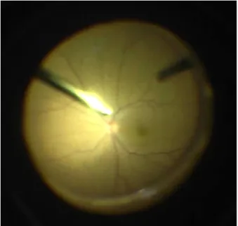
Figure 3. Postoperative fundus finding. At 3 months after vi-
trectomy, there was no inflammation or infection signs on fun- dus photo.Figure 4. Anterior segment photography. Anterior segment
photography on 3 months postoperative day shows clear cor- nea, normal anterior chamber, and posterior capsular intra- ocular lens was in place.일째, 상기와 같은 치료로 전방 및 유리체강내 염증과 주관 적 증상이 지속적으로 호전되어 퇴원하였으며, 수술 후 3개 월째 경과관찰 당시 좌안의 최대교정시력은 20/20으로 회 복되었고, 안내염의 재발 및 안저의 이상소견은 관찰되지 않았다(Fig. 3, 4).
고 찰
초음파 수정체 유화술 및 인공수정체 후낭삽입술은 안과 영역에서 가장 흔하게 행하는 수술 중 하나이며, 백내장 수 술 후 발생하는 안내염은 영구적인 시력손상 가능성은 물 론 적절한 치료 후에도 시력 예후가 좋지 않은 것으로 알려
진 심각한 합병증 중 하나이다.1,2 안내염의 치료 결과를 좋 게 하기 위해서는 빠른 진단과 적절한 치료를 통해서 균의 독소에 의한 안내 조직의 손상을 최소화해야 한다.4
안내염을 일으키는 흔한 원인균과 항균제 감수성 대해서 아는 것은 초기의 경험적 치료 방침을 결정하는 데 중요하 기 때문에, 여러 연구자들이 이에 대해 보고하였다. 1997년 발표된 Endophthalmitis Vitrectomy Study7에 따르면 백내장 수술 후 발생한 안내염 환자의 검체에서 Coagulase negative
staphylococci (CNS, 70.0%)가 가장 많이 자랐고, S.aureus
(9.9%), Viridans streptococci (3.7%), S.pneumonia (2.2%) 의 순서를 보였다. CNS 가운데는 S.epidermidis (81.9%)가 가장 많았고, S.lugdunensis (5.9%), S.warneri (2.7%) 순이 었다. CNS에 대한 항균제 감수성 검사에서는 Amikacin (86.1%), Vancomycin (100.0%)이 높은 감수성을 보였으며, 그람 음성균에 의한 감염에서 많이 사용되는 항생제인 Ceftazidime (62.1%)은 낮은 감수성을 보였다.6,8 Kunimoto et al9은 206명 206안의 수술 후 안내염 환자를 대상으로 연 구하여 총 112안에서 원인균주를 동정하였는데, 이 중S.epidermidis (33.3%)가 가장 많았으며, Pseudomonas spe- cies (19.8%), Aspergillus species (13.5%) 순이었다고 보고
했다. CNS에 대한 항균제 감수성 검사에서는 Cefazolin (93.2), Amikacin (89.5%), Ciprofloxacin (88.4%), Vanco- mycin (86.8%), Ceftazidime (80.8%) 모두 80% 이상의 감수 성을 보였다.S.lugdunensis는 CNS의 일종으로, 주로 피부와 연조직
염증의 원인균으로 알려져 있으며 뇌농양, 뇌수막염, 패혈증, 만성골수막염, 심내막염, 비뇨기계 감염, 창상감염 등도 일으킬 수 있다.10 S.lugdunensis는 눈꺼풀에도 존재하기 때 문에 안구의 침습적인 수술이나 시술 시 안구내로 이환되 어 안내염을 일으킬 가능성도 있다.6 S.lugdunensis는 배양 검사에서 황색의 색소침착을 보이며, 혈액배지에서 완전용혈되 는 특징이 있는데 이는 S.aureus의 형태학적인 특징과 비슷하 기 때문에 정확히 구분하기가 까다롭다. 따라서 S.lugdunensis 의 정확한 판별을 위해서는 일반적인 배양검사와 ornithine decarboxylase, pyrrolidonyl arylamidase phenotypic test 같 은 생화학적인 검사, eubacterial polymerase chain reaction amplification (PCR)을 함께 시행하는 것이 도움이 될 수 있 다.11
S.lugdunensis 감염증은 다른 종의 Staphylococcus 감염에
비해서 독성이 강한데, 이는 S.lugdunensis가 만들어 내는 세포 외 독소나 당질층이 중성구에 의한 탐식작용이나 효 소의 생산을 방해하기 때문이다.10 Chiquet et al11은 백내 장 수술 후에 발생한 S.lugdunensis 안내염 5예에 대해서 보 고하였다. 이들이 백내장 수술 후 안내염으로 진단 받기까 지의 기간은 평균 7.6일(범위 5-12일)이었으며, 이 중 3예에 서 일차 유리체절제술 후 망막박리가 발생하여 이차 유리 체절제술을 필요로 하였고 이 3예는 모두 최종시력이 안전 수동 이하였다. 이를 토대로, Chiquet et al11은 다른 CNS에 의한 안내염에 비해 S.lugdunensis에 의한 안내염이 최종적 인 시력예후가 더 나빴으며, 유리체절제술 후 망막박리가 생길 확률도 높다고 했고, 이는 S.lugdunensis의 독성이 후 극부뿐만 아니라 망막의 주변부까지 망막조직의 괴사와 같 은 손상을 유발하는 것과 연관이 있는 것 같다고 하였다.본 증례에서는 입원 전 전방천자와 함께 유리체강내항생제 주입술을 시행하였으나 염증이 곧바로 좋아지지는 않았고, 입원 다음 날 조기에 유리체절제술을 시행하여 후낭인공수 정체 주변 및 전방 내의 염증물질, 유리체강의 혼탁을 최대 한 제거하고 전신적 항생제 치료를 시작하면서 염증이 줄 어들고 시력이 회복됨을 확인하였다. 본 증례의 최종시력 은 20/20으로 좋았고, 수술 후에도 망막박리 등의 합병증이 발생하지 않았는데, 이는 진단 당시 초기 시력이 20/200으 로 비교적 좋았던 점과 입원 다음 날 조기에 유리체절제술 을 시행하여 감염균과 독소를 없애고, S.lugdunensis의 독성 으로 인한 안내조직의 손상을 최소화하였던 점 때문일 것 으로 생각한다. Han et al8과 Kunimoto et al9의 연구에서 CNS에 의한 안내염은 그람양성구균 감염에 널리 쓰이는 경험적 항생제에 비교적 높은 감수성을 갖는 것으로 보고 하였다. 본 증례에서도 초기 배양검사에서 그람양성구균이
보고되어 경험적으로 vancomycin 점안액과 vancomycin 정 맥주사를 추가 처방하였으며, 이 또한 염증을 호전시키는 데 중요한 역할을 했을 것으로 생각된다.
요약하면, S.lugdunensis는 눈꺼풀의 정상세균총을 이루 는 균주 중 하나로 안내염을 유발할 수 있고, 독성이 강하므 로 신속하게 배양검사 및 균동정 검사를 시행해야 하며, 그람 양성구균이 자랄 경우 드물지만 S.lugdunensis 안내염의 가능 성도 고려해야 한다. 백내장 수술 후 발생한 S.lugdunensis 안 내염을 적절한 국소적, 전신적 그리고 유리체강내 항생제 주입술과 조기 유리체절제술로 치료한 1예를 경험하였기에 이를 보고하는 바이다.
REFERENCES
1) Lalwani GA, Flynn HW JR, Scott IU, et al. Acute-onset endoph- thalmitis after clear corneal cataract surgery (1996-2005). Clinical features, causative organisms, and visual acuity outcomes.
Ophthalmology 2008;115:473-6.
2) Doft BH, Kelsey SF, Wisniewski S, et al. Treatment of endoph- thalmitis after cataract extraction. Retina 1994;14:297-304.
3) Laatikainen L, Tarkanen A. Early vitrectomy in the treatment of post-operative purulent endophthalmitis. Acta Ophthalmol (Copenh) 1987;65:455-60.
4) Jung JY, Ko BY, Kim BY. Factors associated with a poor visual result in acute endophthalmitis after cataract surgery. J Korean Ophthalmol Soc 2008;49:1242-7.
5) Han YS, Chung IY, Park JM. A case of Alcaligenes xylosoxidans endophthalmitis after cataract extraction. J Korean Ophthalmol Soc 2005;46:186-9.
6) Bannerman TL, Rhoden DL, McAllister SK, et al. The source of coagulase-negative staphylococci in the Endophthalmitis Vitrectomy Study. A comprarison of eyelid and intraocular isolates using pulsed- field gel electrophoresis. Arch Ophthalmol 1997;115:357-61.
7) Results of the Endophthalmitis Vitrectomy Study. A randomized trial of immediate vitrectomy and of intravenous antibiotics for the treatment of postoperative bacterial endophthalmitis. Endophthalmitis Vitrectomy Study Group. Arch Ophthalmol 1995;113:1479-96.
8) Han DP, Wisniewski SR, Wilson LA, et al. Spectrum and suscepti- bilities of microbiologic isolates in the Endophthalmitis Vitrectomy Study. Am J Ophthalmol 1996;122:1-17.
9) Kunimoto DY, Das T, Sharma S, et al. Microbiologic spectrum and susceptibility of isolates: part I. postoperative endophthalmitis.
Endophthalmitis Research Group. Am J Ophthalmol 1999;128:
240-2.
10) von Eiff C, Peters G, Heilmann C. Pathogenesis of infections due to coagulase-negative staphylococci. Lancet Infect Dis 2002;2:677-85.
11) Chiquet C, Pechinot A, Creuzot-Garcher C, et al. Acute post- operative endophthalmitis caused by Staphylococcus lugdunensis.
J Clin Microbiol 2007;45:1673-8.
= 국문초록 =
백내장 수술 후 발생한 스타필로코쿠스 루그두넨시스 안내염 1예
목적: 백내장 수술 후 발생한 Staphylococcus lugdunensis 안내염 1예를 경험하였기에 이를 보고하고자 한다.
증례요약: 59세 여자 환자가 본원에서 좌안 백내장 수술을 받은 후 13일째 발생한 좌안의 통증과 시력저하를 주소로 내원하였다. 좌안 의 최대교정시력은 20/200이었고, 세극등현미경 검사에서 좌안의 결막충혈, 전방 내 다수의 염증세포, 1 mm 가량의 전방축농과 삼출 성 막이 관찰되었으며 안저는 흐려 잘 보이지 않았다. 전방천자 및 유리체강내항생제주입술(vancomycin 1.0 mg/0.1 mL, ceftazidime 2.0 mg/0.1 mL)을 시행하고 입원하였는데, 다음 날 증상의 호전이 없고 그람양성구균이 자라는 것이 확인되어, 유리체절제술과 함께 유리체강내항생제주입술(vancomycin 1.0 mg/0.1 mL, dexamethasone 0.5 mg/0.1 mL)을 추가로 시행하였다. 수술 7일째, 균 배양검 사 결과 Staphylococcus lugdunensis가 동정되었고, 수술 11일째, 염증이 조절되고 증상이 호전되어 퇴원하였다. 수술 3개월 후, 좌 안의 최대교정시력은 20/20이며, 안내염의 재발 및 안저의 이상소견은 관찰되지 않았다.
<대한안과학회지 2016;57(6):999-1003>