INTRODUCTION
Recently, the clinical use of [18F]FDG PET or PET/CT has rapidly increased as a management tool for sta ging or localization of
metastatic disease in patients with various malignancies. Uptake of FDG in the normal thyroid gland is very low and is usually not visualized on whole-body PET or PET/CT [1]. Occasionally, thyroid incidentalomas are shown on PET or PET/CT as a focal or diffuse increase in FDG uptake. The clinical issue is whether or not thyroid in ci dentalomas are benign or malignant. Consi- dering the con venience and in creased use of PET or PET/CT in pre-treatment assess ment of patients with cancer, the clinical sig ni fi cance of thyroid inci den talomas is an important issue.
Until now, many investigators have shown the results which have focused on the incidence of thyroid incidentalomas and the rate of malignancies associated with thyroid incidentalo- mas on PET or PET/CT in patients with malignancy. Based on our experience, the incidence of thyroid incidentalomas in pa-
Characteristics of thyroid incidentalomas detected by pre-treatment [ 18 F]FDG PET or PET/CT in patients with cervical cancer
Won-Moo Lee1,*, Beob-Jong Kim1, Moon-Hong Kim1, Seok-Cheol Choi1, Sang-Young Ryu1, Ilhan Lim2, Kidong Kim1,†
Departments of 1Obstetrics and Gynecology and 2Nuclear Medicine, Korea Cancer Center Hospital, Korea Institute of Radiological
& Medical Sciences, Seoul, Korea
Received Aug 25, 2011, Revised Oct 11, 2011, Accepted Oct 17, 2011
*The current affiliation of Won-Moo Lee is Department of Obstetrics and Gynecology, Hanyang University Hospital, Seoul, Korea.
†The current affiliation of Kidong Kim is Department of Obstetrics and Gy- ne cology, Seoul National University Bundang Hospital, Seongnam, Korea.
Correspondence to Kidong Kim
Department of Obstetrics and Gynecology, Seoul National University Bundang Hospital, 300 Gumi-dong, Bundang-gu, Seongnam 463-802, Korea. Tel: 82- 31-787-7262, Fax: 82-31-787-4054, E–mail: [email protected]
Objective: Considering the increased use of [18F]FDG PET or PET/CT, the clinical significance of thyroid incidentalomas is the subject of controversy. The aim of this study was to determine the incidence of malignancies associated with thyroid incidentalomas detected by pre-treatment PET or PET/CT in patients with cervical cancer.
Methods: We retrospectively reviewed the medical records of patients with cervical cancer who had thyroid incidentalomas de- tected by pre-treatment PET or PET/CT and were treated at our institute between January 2001 and December 2009.
Results: Of 327 patients who underwent pre-treatment PET or PET/CT, 33 patients had thyroid incidentalomas (10.1%) and 4 patients were diagnosed with thyroid malignancies by percutaneous needle aspiration (PCNA) or surgery. To put it concretely, of 33 patients with thyroid incidentaloma, 16 patients had a diffuse uptake and 17 patients had a focal uptake. Four of 17 patients with focal uptake were diagnosed with thyroid malignancies (23.5%). One patient with a focal uptake had an atypical cell based on PCNA, but did not undergo additional studies. The mean SUVmax of thyroid malignancies did not differ from that of benign thyroid diseases.
Conclusion: Thyroid incidentalomas are frequently detected by pre-treatment PET or PET/CT in patients with cervical cancer.
Focal uptake on PET or PET/CT has a high risk of thyroid cancer.
Keywords: Cervical cancer, Fluorodeoxyglucose F18, Positron-emission tomography, Positron-emission tomography and computed tomography, Histological confirmation, Thyroid incidentalomas
tients with cervical cancer is higher than expected. However, there are no studies which have focused on the patients with cervical cancer.
In the current study we determined the incidence of thyroid incidentalomas and the rate of malignancies associated with thyroid incidentalomas detected by pre-treatment PET or PET/
CT in patients with cervical cancer.
MATERIALS AND METHODS 1. Patients
The medical records of patients with cervical cancer who had thyroid incidentalomas by pre-treatment PET or PET/CT and were treated at our institute between January 2001 and December 2009 were reviewed retrospectively. Age, cervical cancer stage, type of thyroid incidentalomas (focal and dif- fuse), the maximal standardized uptake value (SUVmax) of the thyroid incidentalomas, thyroid ultrasonography (USG) find- ings, serum thyroid-stimulating hormone (TSH) level, percuta- neous needle aspiration (PCNA) of the thyroid incidentalomas results, and final biopsy results from lobectomy or thyroidec- tomy specimens were obtained. Approval from the Institu- tional Review Board was obtained for this retrospective study (K-1009-002-071).
2. PET or PET/CT evaluation
Until September 2005, PET was performed on an advance HR+ Scanner (General Electric, Waukesha, WI, USA). After Sep- tember 2005, we used the following PET/CT scanners: Bio graph 6 (Siemens Medical Solutions, Malvern, PA, USA); or Discovery LS (General Electric Medical Systems, Milwaukee, WI, USA).
Each scan was obtained following the protocol. Patients were fasted at least 6-hour before the PET acquisition. Intravenous injection of 440±60 MBq (range, 165 to 758 MBq) of FDG was followed by a tracer up take phase of about 60 minutes, dur- ing which the patients sat in a quiet room without talking.
The CT scan was performed before emission PET scans. The current of the CT tube was adjusted according to patient weight. The CT data were resized from a 512×512 matrix to a 128×128 matrix to match the PET data in order to generate a CT transmission map and to fuse images. PET emission data were acquired for five to seven bed positions, typically from the base of the skull through the upper thigh. PET images were reconstructed using CT for attenuation correction with the ordered-subsets expectation maximization algorithm (two iterations, eight subsets) and a 5-mm Gaussian filter using a 128×128 matrix.
Thyroid incidentalomas was defined as a newly identified
thyroid lesion on PET or PET/CT in a patient without a previ- ous, known history of thyroid disease. FDG uptake in less than 1 lobe was defined as a focal lesion, whereas uptake in the entire thyroid gland was considered a diffuse pattern [2].
3. Ultrasonographic evaluation
Thyroid nodules satisfying any one of the following condi- tions were classified as malignant: 1) hypoechogenicity, 2) taller shape, 3) ill-defined margin, 4) central vascularity, 5) incomplete halo, and 6) micro- or macro-calcification [3,4].
Cases that did not satisfy any of those six criteria were consid- ered as benign nodule on USG examinations.
4. Criteria for thyroid PCNA cytology
PCNA was performed using a 21-guage needle on a 10-mL syringe under US guidance. Specimens were smeared on glass slides and stained using the Papanicolaou method. A cytolog- ic diagnosis was made by experienced cytopathologists in our institution. Cytologic diagnosis was as followings; 1) nondiag- nostic applies to specimens that are unsatisfactory owing to blood, overly thick or air dried smears, or an inadequate num- ber of follicular cells; 2) benign was made by cytologic find- ings of an adequately cellular specimen composed of varying proportions of colloid and benign follicular cells arranged as macrofollicles and macrofollicle fragments; 3) atypia of unde- termined significance (AUS) was diagnosed when the results of PCNA cytology are not easily classified into the benign, suspicious, or malignant categories; 4) follicular neoplasm was made by cytologic findings of high cellularity and colloid is scant or absent; 5) suspicious for malignancy was classified when only 1 or 2 characteristic features of papillary thyroid carcinoma are presented or a malignant diagnosis cannot be made with certainty; 6) malignant was used whenever the cy- tomorphologic features are conclusive for malignancy [5].
5. Statistical analysis
Data were expressed as the percentage of focal and diffuse incidental thyroid FDG uptake among the population. The difference in SUVmax between benign and malignant groups was analyzed by a Student’s t-test, and significance was set at a p<0.05. Data were analyzed using SPSS ver. 13.0 (SPSS Inc., Chicago, IL, USA).
RESULTS
Total 1,271 patients were newly diagnosed as cervical cancer, and 327 patients underwent pre-treatment PET or PET/CT dur- ing the study period. We recommended PET or PET/CT to all
patients before starting treatment; however, 75% of all patients did not undergo pre-treatment PET or PET/CT due to long wait- ing time for evaluation or their own economic pro blems. Of 327 patients, 33 patients had thyroid incidentalo mas (10.1%).
Twenty nine of 33 patients (89.7%) had cervical cancer with early stage (Table 1). The mean SUVmax of patients with thyroid
incidentaloma was 4.24 for diffuse uptake and 8.40 for focal uptake, without a statistical difference between these groups (p=0.08).
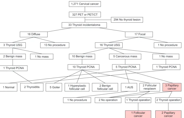
Sixteen of 17 patients with focal uptake underwent thyroid USGs and PCNAs. Only one patient with diffuse uptake under- went a thyroid PCNA and was diagnosed with thyroiditis. One patient with focal thyroid incidentalomas had an AUS based on the PCNA result, but did not undergo additional studies (Fig. 1).
The results of PCNA in patients with focal uptake were as follows: nine patients had benign lesions; three patients had malignancies; two patients had follicular neoplasms; one patient had AUS; and one patient had normal thyroid tissue.
Three patients with malignancies and one patient with follicu- lar neoplasm based on PCNA underwent total thyroidectomy or lobectomy, all of whom were confirmed to have thyroid cancer. Therefore, 4 of 17 patients with focal uptake were diagnosed with thyroid cancer by PCNA or surgery (23.5%).
One patient with follicular neoplasm refused additional treat- ment. Three of four patients with thyroid malignancies had a
Fig. 1. Flow chart of patient selection process. PET, [18F]FDG PET; USG, ultrasoundgraphy; PCNA, percutaneous needle aspiration; AUS, atypia of undetermined sig ni ficance.
Table 1. Clinicopathologic characteristics of patients with thyroid incidentalomas on PET or PET/CT
Diffuse
(n=16) Focal
(n=17) p-value Age (yr), mean±SD 53.81±10.80 56.29±10.69 NS Stage (%)
Stage I 5 (31.3) 6 (35.3)
Stage II 11 (68.7) 7 (41.2)
Stage III+IV 0 (0) 4 (23.5)
SUVmax, median (range) 3.6 (2.2-7.6) 4.3 (2.5-36.9)
PET, [18F]FDG PET; SD, standard deviation; NS, not signi ficant; SUVmax, maximal standard unit value.
relatively high SUVmax on pre-treatment PET or PET/CT (Table 2). The mean SUVmax of patients with thyroid malignancies was higher than that of patients with benign masses, but the dif- ference of SUVmax was statistically insignificant (17.8 vs. 6.16, respectively, p=0.056).
DISCUSSION
Thyroid incidentalomas are defined as newly identified focal thyroid lesions encountered during imaging studies, including CT, MRI, and USG. Although some studies using high-resolution USG have reported the risk of cancer of thyroid incidentalo- mas 1.5-10% [6], these modalities (CT, MRI, and USG) are not specific for thyroid malignancy [7].
The current widespread use of PET or PET/CT has resulted in an increase in the detection of thyroid incidentalomas. The in- cidence of thyroid incidentalomas and the rate of malignancy associated with thyroid incidentalomas on PET or PET/CT var- ies with the study population. Generally, the incidence of thy- roid incidentalomas is 2-9% [3,8,9], and the rate of malignancy associated with focal thyroid incidentalomas is as high as 28- 64% [2,10]. Especially, some authors reported that the preva- lence of thyroid incidentaloma in cancer screening group was about 3% and this value was not differ from metastasis work-
up group [9]. In another study, the authors showed the results that the incidence of thyroid incidentalomas in patients with gynecologic cancer is 10.4% and the rate of malignancy of dif- fuse and focal thyroid incidentalomas is 9.7% [11].
In our study, thyroid incidentalomas were identified in 33 of 327 patients with cervical cancer, corresponding to a preva- lence of 10.1%. This finding is similar or higher than previously reported in the literature [3,8,9,11]. However, it was remark- able that the incidence of malignancy in focal FDG uptake in patients with cervical cancer is 23.5% and which is higher than previous reports included all gynecologic malignancies [11].
Some studies have reported that the average SUVmax of malig- nant lesions is significantly higher than benign lesions [9,11,12].
A recent study even showed the result that the size and visual grade on the PET/CT were the potent predictors for differenti- ation of malignancy in focal thyroid incidentaloma rather than the mean SUVmax [13]. In this study, the mean SUVmax was 6.16 for benign lesions and 17.8 for malignant lesions, with a mar- ginal difference between the two groups (p=0.056). Although some authors suggest that the mean SUVmax of thyroid malig- nancies were significantly higher than those of benign tumors (p<0.001) [9], the role of SUVmax in differentiating benign from malignant lesions is controversial, because the SUVmax overlaps between benign and malignancy and the study sample num- Table 2. Characteristics of patients with focal thyroid incidentalomas on PET or PET/CT
No. SUVmax Thyroid USG Thyroid PCNA TSH Surgery Histology
1 2.5 Benign Benign (goiter) Normal Not done -
2 2.6 Benign Follicular neoplasm Normal Not done -
3 2.7 Cancer Malignancy (PC) Normal Not done -
4 3.2 Benign Benign (goiter) Normal Not done -
5 3.2 Benign Benign (HFC) Not done Not done -
6 3.6 Benign Benign (goiter) Not done Not done -
7 3.9 Not done Not done Not done Not done -
8 3.9 No mass Benign (thyroiditis) Elevation Not done -
9 4.3 Benign Benign (BFC) Not done Not done -
10 5.0 Benign Benign (goiter) Normal Not done -
11 5.4 Cancer AUS Normal Not done -
12 5.5 Cancer Normal Not done Not done -
13 9.2 Cancer Malignancy (PC) Normal TT+LND PC
14 9.6 Benign Benign (BFC) Not done Not done -
15 18.9 Benign Benign (goiter) Normal Not done -
16 22.4 Cancer Malignancy (PC) Normal TT+LND PC
17 36.9 Benign Follicular neoplasm Normal Lobectomy FC
PET, [18F]FDG PET; SUVmax, maximal standard unit value; USG, ultrasonography; PCNA, percutaneous needle aspiration; TSH, thyroid stimulating hormone; PC, papillary carcinoma; HFC, hyperplastic follicular cell; BFC, benign follicular cell; AUS, atypia of undetermined significance; TT, total thyroidectomy; LND, lymph node dissection; FC, follicular car cinoma.
ber has been small in most previous studies.
We admit that our study has some limitations. First, PET or PET/CT produce functional images that reflect increase rates of glucose metabolism in tumor, and it has many pit-falls in clinical use. Second, the number of patients enrolled in this study was too small to induce a strong conclusion. Therefore, more data and large-scale studies are required to determine the clinical significance of thyroid incidentalomas in patients with cervical cancer.
In conclusion, the incidence of thyroid incidentalomas de- tected by PET or PET/CT in patients with cervical cancer was 10.1%; approximately one-half of thyroid incidentalomas have focal uptake and one-half of thyroid incidentalomas have dif- fuse uptake. The rate of malignancy of focal thyroid inciden- talomas in patients with cervical cancer was 23.5% and this result was much higher than that in other gynecologic ma- lignancies reported in the previous literature. Because of the high rate of malignancy, histological confirmation including PCNA in patients with focal thyroid incidentalomas is neces- sary to distinguish between benign and malignant thyroid incidentalomas. SUVmax could be helpful to distinguish malig- nant thyroid incidentaloma from benign ones.
CONFLICT OF INTEREST
No potential conflict of interest relevant to this article was reported.
ACKNOWLEDGMENTS
We thank our staffs and research nurses (Hee Sook Lee and Younha Kim) for their contribution in data collection for this study.
REFERENCES
1. Nakamoto Y, Tatsumi M, Hammoud D, Cohade C, Osman MM, Wahl RL. Normal FDG distribution patterns in the head and neck: PET/CT evaluation. Radiology 2005;234:879-85.
2. Eloy JA, Brett EM, Fatterpekar GM, Kostakoglu L, Som PM,
Desai SC, et al. The significance and management of inci- dental [18F]fluorodeoxyglucose-positron-emission tomo- graphy uptake in the thyroid gland in patients with cancer.
AJNR Am J Neuroradiol 2009;30:1431-4.
3. Kang BJ, O JH, Baik JH, Jung SL, Park YH, Chung SK. Inci den tal thyroid uptake on F-18 FDG PET/CT: correlation with ultra- sonography and pathology. Ann Nucl Med 2009;23:729-37.
4. Rago T, Vitti P. Role of thyroid ultrasound in the diagnostic evaluation of thyroid nodules. Best Pract Res Clin Endocrinol Metab 2008;22:913-28.
5. Cibas ES, Ali SZ; NCI Thyroid FNA State of the Science Con- fe rence. The Bethesda system for reporting thyroid cyto- pathology. Am J Clin Pathol 2009;132:658-65.
6. Burguera B, Gharib H. Thyroid incidentalomas: prevalence, diagnosis, significance, and management. Endocrinol Metab Clin North Am 2000;29:187-203.
7. Mazzaferri EL. Thyroid cancer in thyroid nodules: finding a needle in the haystack. Am J Med 1992;93:359-62.
8. Cohen MS, Arslan N, Dehdashti F, Doherty GM, Lairmore TC, Brunt LM, et al. Risk of malignancy in thyroid incidentalomas identified by fluorodeoxyglucose-positron emission tomo- graphy. Surgery 2001;130:941-6.
9. Kang KW, Kim SK, Kang HS, Lee ES, Sim JS, Lee IG, et al. Pre- valence and risk of cancer of focal thyroid incidenta lo ma identified by 18F-fluorodeoxyglucose positron emission tomography for metastasis evaluation and cancer scree- ning in healthy subjects. J Clin Endocrinol Metab 2003;88:
4100-4.
10. Chen W, Parsons M, Torigian DA, Zhuang H, Alavi A. Evalu a- tion of thyroid FDG uptake incidentally identified on PET/
CT imaging. Nucl Med Commun 2009;30:240-4.
11. Bae JS, Chae BJ, Park WC, Kim JS, Kim SH, Jung SS, et al. Inci- dental thyroid lesions detected by PET/CT: pre valence and risk of thyroid cancer. World J Surg Oncol 2009;7:63.
12. Choi JY, Lee KS, Kim HJ, Shim YM, Kwon OJ, Park K, et al.
Focal thyroid lesions incidentally identified by integrated 18F-FDG PET/CT: clinical significance and improved cha- rac terization. J Nucl Med 2006;47:609-15.
13. Kim BH, Na MA, Kim IJ, Kim SJ, Kim YK. Risk stratification and prediction of cancer of focal thyroid fluoro deoxy- glu cose uptake during cancer evaluation. Ann Nucl Med 2010;24:721-8.