422
접수일:2009년 2월 6일, 게재확정일:2009년 5월 27일 교신저자:이 준 영
광주시 동구 서석동 588 조선대병원 정형외과
TEL: 062-220-3147ㆍFAX: 062-226-3379 E-mail: [email protected]
Correspondence to Jun-Young Lee, M.D.
Department of Orthopaedic Surgery, Chosun University Hospital, 588, Seosuk-dong, Dong-gu, Gwangju 501-717, Korea
Tel: +82-62-220-3147, Fax: +82-62-226-3379 E-mail: [email protected]
후외방 도달법에 의한 족관절 삼과골절에서 후과 골절편의 치료
- 예비 보고-
이준영ㆍ하상호ㆍ노경환*ㆍ이상준 조선대학교 의과대학 정형외과학교실, *광주보훈병원 정형외과
Treatment of the Posterior Malleolar Fragment of Trimalleolar Fracture Using Posterolateral Approach
- Preliminary Report -
Jun-Young Lee, M.D., Sang-Ho Ha, M.D., Kyung-Hwan Noh, M.D.*, and Sang-Jun Lee, M.D.
Department of Orthopaedic Surgery, College of Medicine, Chosun University,
*Department of Orthopaedic Surgery, GwangJu Vaterans Hospital, Gwangju, Korea
Purpose: We wanted to evaluate the effectiveness of posterolateral approach for open reduction and internal fixation of posterior malleolar fragment with trimalleolar fracture of ankle joint.
Materials and Methods: There were 27 cases of trimalleolar fracture in our hospital from Jan. 2005 to Dec. 2007. We investigated 10 patients who underwent operation with the posterolateral approach. The mean follow up period was 20 (6-36) months. Preoperative posterior malleolar fragment involved above 25% of articular surface in 10 cases and displaced more than 2 mm in 4 cases. We analyzed the radiologic type of posterior malleolar fragments and complications, and evaluated the AOFAS score.
Results: All cases showed primary union mean 11.8 (8-14) weeks. The complication focal skin necrosis in one case and all patients showed excellent AOFAS score.
Conclusion: The posterolateral approach may be a useful for open reduction and internal fixation of posterior malleolar fragment with trimalleolar fracture, especially simultaneous management of lateral malleolar fracture.
Key Words: Ankle joint, Trimalleolar fracture, Posterior malleolar fragment, Posterolateral approach
서 론
족관절 삼과골절에서 후과 골절편은 회전 및 수직 부 하에 의해 발생하고, 불안정한 골절로 관혈적 정복술이 필요한 경우가 많다.1) 또한 후과 골절을 포함하는 족관절 골절은 기능상 예후가 불량한 경우가 많으며, 그 치료법 에 대해서도 논란이 많다.2) 이러한 불안정성을 결정하는 요소 중 가장 중요한 것은 골편의 크기로 골절편의 크기 가 25-30% 이상인 경우 내고정을 시행하는 지침이 일반 적으로 받아들여지고 있다.3,4) 골절의 정복시 간접적 정
복법을 사용한 경우는 기술적으로 어렵고, 관절 내 감입 된 골절편을 정복하지 못한다면 골절편의 이차적 탈구를 유도하여 더 나쁜 예후를 갖게 되며 즉각적인 수술적 치 료가 이루어지지 않는 경우는 혈종이나 가골이 형성되어 정확한 정복이 어려워진다.5) 삼과 골절의 치료에서 정확 한 정복을 위하여 골절편에 대한 노출이 필요한 경우에는 일반적으로 후내측 도달법이 많이 사용되어지고 있으 나6,7) 이 도달법을 통해서는 후과 골절편에 대한 노출이 제한적이라는 단점이 있어 여러 저자들에 의해 변형된 도
Fig. 1. Posterolateral approach. (A) The longitudinal incision is placed just medial to the posterior border of the fibula. (B) Retracting the peroneal tendons medially gives access to the posterior aspect of the lateral malleolus. (C) View of the posterior fragment in the interval between the peroneal tendons and the flexor hallucis longus. (D) Posterior fragment was reduced and fixed with 1 or 2 of 4.0 mm cannulated screw.
Fig. 2. Transverse computed tomographic scan at the level of the tibial plafond showing posterolateral fragment of the posterior malleolus. We measured the area of the posterior malleolar frag- ment and the remaining cross-sectional area of the tibia.
달법이 제안되어 왔다.5) 이 중 한 방법인 후외방 도달법 에 의한 삼과골절의 치료는 우수한 결과를 보이고 있으
나,5,8,9) 아직 국내에는 단편적인 내용 이외에 족관절 삼
과 골절을 후외방 접근법을 이용하여 치료한 경우에 대한 보고가 없는 상태로, 저자들은 삼과 골절에서 후외방 도 달법을 이용하여 후과 골절을 치료한 결과를 분석하고 그 유용성에 대하여 알아보고자 하였다.
대상 및 방법
1. 연구대상
2005년 1월부터 2007년 12월까지 본 교실에서 경험 한 족관절 삼과 골절 27예 중 후과의 수술적 치료가 요하 다고 생각되는 10예에 대하여 모두 후외방 도달법을 이 용한 내고정을 시행하였고 최소 6개월 이상 추시하였다.
나머지 17예는 모두 보존적 치료를 하였거나 경피적 유 관나사 고정술을 시행하였기에 본 연구에서는 제외하였 다. 먼저 외측과에 대한 정복술을 시행한 후 후과의 전위 가 2 mm 이상인지를 확인하였으며 수술 전 CT 상 25%
이상의 관절면을 포함한 골절을 수술의 대상으로 하였다.
2. 수술방법 및 술 후 처치
모든 경우에 측와위에서 수술을 시행하였고, 후과 골절 과 내과 골절은 4.0 mm 유관나사를 사용하여 고정하였 으며, 외과 골절에 대해서는 1/3 반원형 금속판을 이용한 antiglide plate 고정을 시행하였다. 수술 방법은 먼저 비골의 후연을 따라서 종으로 피부 절개를 시행한 후 비 복신경 손상에 주의하며 비골건을 내측으로 당겨 비골의 후면을 노출시킨 후 외과 골절에 대해 정복 및 금속판 고
정을 시행하였다. 다음으로 다시 비복건을 외측으로 당 겨 비복건과 장무지 굴근 사이로 후과 골절편을 노출시켜 이 골절편에 대하여 정복을 시행한 후 4.0 mm 유관나사 를 이용하여 고정을 시행하였다(Fig. 1). 내과 골절에 대 해서는 다리를 외회전 시킨 상태에서 내과 후면을 따라 3 cm의 피부 절개를 시행하고 정복을 시행한 후 4.0 mm 유관나사를 이용하여 고정하였다. 수술 후 가능한 빨리 목발을 이용한 비체중 부하 보행을 시작하였고 적극적인 관절 운동을 시작하였다. 수술 후 4주째 부분 체중 부하 보행을 시작하였고, 수술 후 8주째 전 체중부하 보행을 목표로 재활을 시행하였다.
3. 평가방법
수술 전 방사선학적 평가는 족관절 전후면, 측면, mortise면 사진을 이용하여 Lauge-Hansen과 Dennis-
Table 1. The Degree of Posterior Malleolar Displacement
Degree of displacement Cases
Minimal displacement (<2 mm) Displacement (>2 mm)
6 4
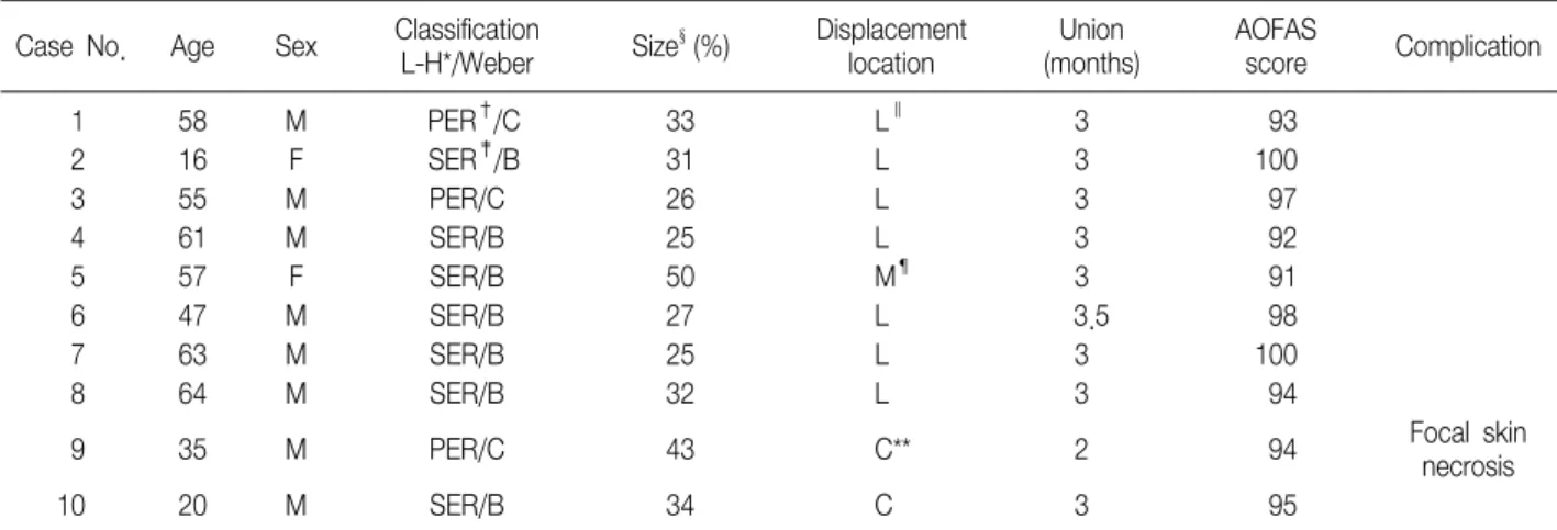
Table 2. Data about the Patients
Case No. Age Sex Classification
L-H*/Weber Size§(%) Displacement location
Union (months)
AOFAS
score Complication
1 58 M PER†/C 33 L∥ 3 93
2 16 F SER‡/B 31 L 3 100
3 55 M PER/C 26 L 3 97
4 61 M SER/B 25 L 3 92
5 57 F SER/B 50 M¶ 3 91
6 47 M SER/B 27 L 3.5 98
7 63 M SER/B 25 L 3 100
8 64 M SER/B 32 L 3 94
9 35 M PER/C 43 C** 2 94 Focal skin
necrosis
10 20 M SER/B 34 C 3 95
*, Lauge-Hansen; †, pronation-external rotation; ‡, supination-external rotation; §, joint involvement ratio of the posterior malleolar fragment;
∥, posterolateral; ¶, posteromedial; **, central.
Weber분류를 하였으며, 각 예에서 CT촬영을 시행하 였고 분석 프로그램(Display Workstation Version 2.0.73.315, copyright ⓒ RAYPAX, Co., Ltd. 1997- 2008)을 이용하여 후과 골절편의 크기, 전위정도 및 후 과 골절편이 관절면을 침범한 정도에 대해 분석하였다 (Fig. 2). 후과 골절편의 위치는 Haraguchi 등2)의 분류 를 참고하여 후외측, 후내측, 중앙부로 분류하였다.
수술 후 기능의 평가는 AOFAS score를 이용하였으 며,10) 수술 후 방사선상 평가는 수술 후 전후면 및 측면 사진과 Mortise면 촬영을 시행하였으며, 추시 방사선 사 진상에서 수술 후 골유합 시기, 정복의 유지 여부, 불유 합 및 지연유합 등의 합병증을 조사하였다.
결 과
Lauge-Hansen 분류상 회외-외회전 손상은 7예였 고, 회내-외회전 손상은 3예였다. Dennis-Weber 분류 상 B형이 7예였고, C형이 3예였다. 10예 모두에서 후과 골절편이 관절면의 25% 이상을 차지하였고 후과 골절편 의 전위 정도는 2 mm 이하가 6예였으며, 2 mm 이상이 4예였다(Table 1). 후과 골절편의 위치는 후외측이 7예
였고, 중앙부가 2예, 후내측이 1예였다.
10예 모두에서 술 후 단순 방사선 검사상 골절편의 정 확한 정복이 이루어져 있었으며 추시 결과 일차적인 골 유합을 얻을 수 있었다. 방사선 추시는 수술 후 2주 간격 으로 시행하였고 골유합까지의 기간은 8주에서 14주 사 이로 평균 11.8주를 보였으며 추시 도중 고정된 골절편 의 전위는 관찰되지 않았다. 합병증으로는 수상 시 심한 타박이 있던 환자에서 피부 조직의 부분적인 괴사가 발생 하였으나 피판술이나 피부 이식없이 변연 절제술 후 일차 봉합으로 치료하였다. 기능적 평가는 AOFAS score상 총 100점에서 통증 평균 37점, 기능 평균 48.4점, 정렬 평균 10점으로 총 평균 95.4점의 매우 우수한 결과를 보 였다(Table 2).
고 찰
족관절의 후과 골절은 주로 삼과골절의 형태로 나타나 며, 회전력에 의한 골절에서 비골 원위부의 골절과 이에 따른 후 원위 경비 인대의 견인력에 의해 후과 골절편이 생기게 된다. Cooper11)는 이러한 경골 후과를 침범한 족 관절 후과 골절을 처음으로 언급 하였고, Cotton12)은 족 관절 골절의 새로운 형이라 보고하였으며, Henderson13) 은 삼과 골절이라는 용어를 처음으로 사용하였다. Bur- well과 Chanley 등14)은 회외 외회전 손상이 가장 많았다 고 보고하고 있고, 본 연구에서도 회외 외회전 손상이 10 예 중 7예로 많았다. 족관절은 기립 및 보행에 중요한 역 할을 담당하며, 후 경골 면의 체중부하 및 족관절 안정성
Fig. 3. (A, B) Preoperative anteroposterior and lateral radiograph of the left ankle of a sixty-four-year-old man who vehicle accident, revealing an supination-external rotation injury and Weber B trimalleolar fracture with posterior malleolar fragment of 32% joint surface involved. He has a osteoporosis of T-score −3.8. (C, D) There is postoperative radiograph. (E, F) Radiograph made three months postoperatively, showing complete bony union.
에 있어서 생역학적 기능의 중요성으로 해부학적 정복이 실패한다면 종종 나쁜 기능상 결과를 가져올 수 있다. 골 절편의 치료에 대해서는 수술 여부 및 수술 방법 등에 있 어서 많은 논란이 있다. Dickson15)은 종골에 골 견인술 을 3주간 시행 후 석고 붕대로 고정하였고, Dieterle16)는 K-강선을 이용한 경피적 고정술을 시행하였다. 많은 저 자들이 관절면의 5-10%를 차지하는 작은 골편은 고정이 필요하지 않다고 주장하였다.3,17) 그러나 다른 저자들에 서 10% 이하의 골절편의 경우에도 해부학적 정복을 시행 한 경우 그 결과가 더 좋았다는 주장도 있었다.18) McLaughlin19)은 관절면의 25% 이상 침범시 관혈적 정 복 후 내고정이 필요하다고 하였고, McDaniel과 Wilson 은 관절면의 25% 이상을 침범하는 후과 골절을 동반하는 삼과 골절의 경우 비관혈적 정복후 거골의 아탈구가 남게 되어 외상성 관절염의 빈도가 증가하므로 이런 경우 관혈 적 정복 및 내고정이 필요하다고 기술하였다.4,20) Jas- kulka 등3)은 관절면의 25% 이상의 골절편인 경우 해부 학적 정복이 이루어졌다고 하더라도 고정이 되지 않는 경 우보다 고정을 시행한 경우가 기능적 결과가 더 좋았다고 주장하였다. 저자들의 경우에는 보존적 치료를 시행한 예가 없어 그 결과를 비교해 볼 수는 없었지만, 수술 전 계측상 후과 골절편이 관절면의 25% 이상을 침범하였으 나 2 mm 이하의 전이를 보였던 6예에서 관혈적 정복 및 내고정을 통해서 AOFAS score상 매우 우수한 결과를 얻 을 수 있었다(Fig. 3). 일반적으로 대부분의 저자들은 후 과 골절편이 경골 원위 관절면의 25-33% 이상을 차지하 는 비교적 큰 골절편에 있어서 관혈적 정복을 통한 해부
학적 정복 및 내고정술을 시행하는 것을 추천하고 있 다.3,4) 저자들의 경우에도 25% 이상의 관절면을 포함하 는 후과 골절편에 대해서 관혈적 정복 및 유관 나사를 이 용한 내고정을 통해 AOFAS score 상 매우 우수한 결과 를 얻을 수 있었다(Fig. 4).
족관절 삼과골절에서 관혈적 정복을 위한 접근 방법으 로 Jergeson21)은 후내방 도달법을 소개하며 내과와 외과 의 골절면을 직접 볼 수 있고, 내과 및 후과 골절편을 동 시에 정복하고 고정할 수 있는 장점이 있다고 하였다. 그 러나 외과 골절이 심한 분쇄를 보이거나 내과 및 후과 골 절의 고정 후에도 외과 골절이 정복이 되지 않은 경우에 는 추가적인 접근법이 필요하게 되고, 이때 추가적인 절 개를 시행함으로 창상 문제가 발생할 가능성이 높아지는 단점이 있으며, 많은 경우에 후과 골절은 후외측에 골절 편이 위치함으로 이에 대한 정복 및 고정에 어려움이 있 을 수 있다.21) 이에 반해 후외방 도달법은 족관절 및 후과 골절의 직접적인 관찰이 가능하고 동시에 해부학적 정복 및 고정을 시행할 수 있으며, 외과의 골절에 대해서 동시 에 정복 및 고정을 시행할 수 있는 장점이 있다. 또한 내 과 골절은 작은 피부 절개를 통한 고정이 가능하여 피부 문제가 발생할 가능성이 적다.5,22)
삼과 골절에서 후외방 도달법을 이용한 치료에 대한 보고는 많지 않으나, Miller8)는 5예의 삼과 골절에서 후 과 골절에 대하여 후외방 도달법을 이용한 치료를 시행하 여 좋은 결과를 보고하였고, Heim9)은 16예에 대하여 후 외방 도달법을 이용한 수술적 치료를 시행하여 우수한 결 과를 보고하였다. 또한 Talbot5)은 후외방 도달법에 대한
Fig. 4. (A, B) Preoperative anteroposterior and lateral radiograph of the left ankle of a sixty-one-year-old man who slip down, revealing an supination-external rotation injury and Weber B trimalleolar fracture with posterior malleolar fragment of 25% joint surface involved.
(C, D) Preoperative CT of the sagittal and axial view. It has posterolateral fragment. (E) There is postoperative radiograph. (F, G) Radiograph made six months postoperatively showing complete bone union. (H, I) Normal ankle joint motion of twelve months postoperatively.
기술적인 언급과 함께 그 우수성에 대하여 보고하였다.
후과 골절편은 인대 신연술(ligamentotaxis)에 의한 기 전으로 내과 및 외과의 정복 및 고정이 이루어지면 저절 로 정복이 이루어지는 경우가 많아 수술 시행 시 외과나 내과 골절부터 고정을 시행하는 경우가 많았다. 그러나, 관절면의 25% 이상을 차지하는 후과 골절편의 경우에 인 대 신연술에 의한 정복이 안되는 경우가 많고 후외방 도 달법을 시행하는 경우 후과 골절편과 관절면을 직접 보면 서 정복을 시행하기 위해서 후과 골편을 먼저 고정한 후 외과 골절편을 고정하고 내과를 고정하고 있다.2-4,23,24) 저자들의 경우에는 외과 골절을 먼저 정복 및 고정을 시 행하고 후과 골절편의 정복 및 고정을 시행하였는데 심한 전이를 보이는 후과 골절편의 경우에는 완전한 정복이 되 지는 않지만 외과를 먼저 정복시 후과 골절편이 해부학적 위치로 접근하여 정복이 용이하였으며, 경골 후면과 후 과 골절편의 피질골의 모양을 맞춰서 정복한다면 굳이 관 절면을 확인 하지 않아도 비간접적으로 정확한 관절면의 정복을 얻을 수 있었다. 후외방 도달법을 사용한 경우 후 외측 골편에 접근하는데 매우 유용하였으며 후내측이나
후중심부에 골절편이 있는 경우도 적절한 견인을 통하여 골절편을 확인할 수 있어 골절편의 위치에 상관없이 유용 한 도달법으로 생각되었다. 그러나 후외방 도달법이 아 직까지 골다공증이 심한 환자에 적용하는데 논란이 있겠 으며, 본 논문에서 10예에 대하여 후외방 도달법을 이용 한 치료를 시행하여 양호한 결과를 얻었지만, 그 증례수 가 많지 않아 결과를 일반화 시킬 수 없는 한계점이 있었 다. 또한 추시기간이 길지 않아 향후 발생할 수 있는 장기 합병증에 대해 지속적인 추시가 필요할 것으로 사료된다.
결 론
족관절 삼과골절의 수술적 치료에 있어 후외방 도달법 은 후과 골절편의 관혈적 정복 및 고정을 용이하게 하며, 또한 외과 골절의 정복 및 고정을 동시에 시행할 수 있는 유용한 도달법 중의 하나로 사료된다.
감사의 글
이 논문은 2007년도 조선대학교 연구비의 지원을 받 아 연구되었습니다.
참고문헌
1. Nugent JF, Gale BD. Isolated posterior malleolar ankle fractures. J Foot Surg. 1990;29:80-3.
2. Haraguchi N, Haruyama H, Toga H, Kato F. Pathoana- tomy of posterior malleolar fractures of the ankle. J Bone Joint Surg Am. 2006;88:1085-92.
3. Jaskulka RA, Ittner G, Schedl R. Fractures of the posterior tibial margin: their role in the prognosis of malleolar fractures.
J Trauma. 1989;29:1565-70.
4. McDaniel WJ, Wilson FC. Trimalleolar fractures of the ankle. An end result stydy. Clin Orthop Relat Res. 1977;
(122):37-45.
5. Talbot M, Steenblock TR, Cole PA. Posterolateral appro- ach for open reduction and internal fixation of trimalleolar ankle fractures. Can J Surg. 2005;48:487-90.
6. Carr J. Skeletal trauma: basoc science, management and reconstruction. In: Browner B, Jupiter J, Levine A, Trafton P ed. Philadelphia: Elsevier Science; 2003. 371-401.
7. Marsh JL, Saltzman C. Rockwood and Green's fractures in adults. In: Bucholz R, Heckman J ed. Philadelphia: Lippincott, Williams & Wilkins; 2001. 2001-90.
8. Miller AJ. Posterior malleolar fractures. J Bone Joint Surg Br.
1974;56:508-12.
9. Heim UF. Trimalleolar fractures: late results after fixation of the posterior fragment. Orthopedics. 1989;12:1053-9.
10. Kitaoka HB, Alexander IJ, Adelaar RS, Nunley JA, Myerson MS, Sanders M. Clinical rating systems for the ankle-hindfoot, midfoot, hallux, and lesser toes. Foot Ankle Int.
1994;15:349-53.
11. Cooper A. A treatise on dislocations and on fractures of the joints: fractures of the neck of the thigh-bone. 1823. Clin Orthop Relat Res. 2007;458:6-7.
12. Cotton FJ. A new type of ankle fracture. JAMA. 1915;64:318-
21.
13. Henderson MS. Trimalleolar fracture of the ankle. Surg Clin North AM. 1932;12:867-72.
14. Burwell HN, Chanley AD. The treatment of displaced fractures at the ankle by rigid internal fixation and early joint movement. J Bone Joint Surg Br. 1965;47:634-60.
15. Dickson FJ. Posterior marginal fracture of the tibia. Surg Gynecol Obstet. 1933;56:525-8.
16. Dieterle J. The use of kirschner wires in maintaining reduc- tion of fracture dislocation of the ankle joint: a report of two cases. J Bone Joint Surg Am. 1935;17:990-5.
17. Broos PLO, Bisschop APG. Operative treatment of ankle fractures in adults: correlation between types of fracture and final results. Injury. 1991;22:403-6.
18. van Laarhoven CJHM. Fractures of the ankle joint:
Retrospective and prospective studies on the (long-term) results of protocolled treatment. Utrecht, The Netherlands: Acade- misch Ziekenhuis; 1994. 112-8.
19. McLaughlin HI. Trauma. Philadelphia: WS Saunders Co;
1960. 357.
20. Sachs W, Kanat IO, McLaughlin E, Burns DE. A surgical approach to a displaced ankle fracture. J Foot Surg. 1984;
23:302-7.
21. Jergeson F. Open reduction of fractures and dislocation of the ankle. Am J Surg. 1959;98:136.
22. Henry AK. Extensile Exposure. Endinburgh: Churchill Livingstone; 1973.
23. Jeong HJ, Kim KC, Chung SW. Treatment of the posterior malleolar fracture. J Korean Fracture Soc. 1998;11:924-31.
24. Kim SJ, Choi IY, Ahn TK. A clinical study of the trimalleolar fractures of the ankle. J Korean Fracture Soc.
1989;2:145-54.
= 국문초록=
목 적: 족관절 삼과골절에서 후과 골절편의 크기가 관절면의 25% 이상을 차지하는 경우에 대하여 후외방 도달법 에 의한 정복 및 내고정술로 치료한 결과를 분석하고 그 유용성에 대하여 알아보고자 하였다.
대상 및 방법: 2005년 1월부터 2007년 12월까지 족관절 삼과골절로서 본원에서 수술한 27예 중 후외방 도달법 을 이용한 10예를 대상으로 하였으며, 평균 추시 기간은 20 (6-36)개월이었다. 술전 후과 골절편이 관절면의 25% 이상을 침범한 경우가 10예에서 관찰되었고, 4예에서 2 mm 이상 전이를 보였다. 후과 골절편의 방사선학 적 양상에 대하여 분석하였고, 술 후 기능적 평가는 AOFAS 점수를 사용하였으며, 그 외 임상적 평가 및 합병증 등에 대하여 조사하였다.
결 과: 모든 환자에서 평균 11.8 (8-14)주에 일차적인 골유합을 얻었다. 합병증으로 피부 조직의 부분적인 괴사가 1예에서 관찰되었으며, AOFAS 점수상 우수한 결과를 보였다.
결 론: 족관절 삼과골절의 수술적 치료에 있어 후외방 도달법을 이용한 후과 골절편의 관혈적 정복 및 고정방법 은 외과부 골절의 정복 및 고정을 동시에 시행할 수 있는 유용한 도달법 중의 하나로 사료된다.
색인 단어: 족관절, 삼과 골절, 후과 골절편, 후외방 도달법