Since an infra-optic course of a proximal precommuni- cating artery was first described by Robinson and demonstrated on angiography by Isherwood and Dutton, 44 cases have been reported (1, 2). Most cases demonstrate unilateral involvement and only 10 cases of bilateral involvement have been reported out of the 45 cases, including the case described in this report.
Although cerebral aneurysms are frequently associat- ed with this congenital anomaly, an infra-optic course of
the anterior cerebral artery (ACA) and association with aortic coarctation is rare (3). To the best of our knowl- edge, this is the third case documented for an associa- tion with aortic coarctation in a patient an infra-optic course of the ACA (2, 4). We report a case of bilateral in- fra-optic ACAs where a middle cerebral artery aneurysm and aortic coarctation are associated. The sig- nificance of recognition of this anomaly for patient man- agement is discussed.
Case Report
A 28-year-old woman was admitted to our hospital af- ter a sudden onset of severe headache and vomiting dur- ing sleeping. A physical examination of the patient re- vealed moderate neck stiffness. A brain computed to- mography (CT) examination performed on admission
Infra-optic Course of Both Anterior Cerebral Arteries Associated with a Middle Cerebral Artery
Aneurysm and an Aortic Coarctation
1Cheol Ji, M.D., Jae-Geun Ahn, M.D., Song-Mee Cho, M.D.2
1Department of Neurosurgery, St. Paul’s Hospital, The Catholic University of Korea, College of Medicine, Seoul, Korea
2Department of Radiology, St. Paul’s Hospital, The Catholic University of Korea, College of Medicine, Seoul, Korea
Received February 23, 2009 ; Accepted March 19, 2009
Address reprint requests to : Song-Mee Cho, M.D., Department of Radiology, St. Paul’s Hospital, The Catholic University of Korea, 620-56 Jeonnong-dong, Dongdaemun-gu, Seoul 130-709, Korea.
Tel. 82-2-958-2084 Fax. 82-2-960-4568 E-mail: [email protected]
A ruptured aneurysm at the bifurcation of the left middle cerebral artery with an in- fra-optic course of the bilateral anterior cerebral arteries was found in a 28-year-old woman. Both abnormal anterior cerebral arteries arose from the ipsilateral internal carotid arteries, at the level of the origin of ophthalmic arteries, passed underneath the ipsilateral optic nerves and turned upward at the ventral portion of the optic chiasm.
In addition, an aortic coarctation was found with the use of thoracic aortography. An infra-optic course of the bilateral anterior cerebral arteries is an extremely rare anom- aly. An infra-optic course of the bilateral anterior cerebral arteries is frequently associ- ated with cerebral aneurysms and possibly with a coarctation aorta. The clinical fea- tures, radiological findings and possible genesis of this anomaly are presented.
Index words :Anterior cerebral artery Intracranial aneurysm Aortic carctation Optic nerve
Tomography, X-Ray computed
aneurysm at the bifurcation of the left middle cerebral artery (MCA) and both ACAs that originated from the ipsilateral internal carotid artery (ICA) at the level of the origin of the ophthalmic arteries. Both ACAs ran under the medial side of each optic foramen and then proceed- ed in a cephalic course (Fig. 2). Conventional angiogra- phy demonstrated a saccular aneurysm at the bifurca- tion of the left middle cerebral artery and the low bifur- cation of both ICAs. Both ACAs arose from each side of the ICAs at the level of the ophthalmic artery, coursing medially and superiorly and coursing at the midline of the brain (Fig. 3). Thoracic aortography, which was per- formed just prior to cerebral angiography, showed the aortic coarctation located at the descending aorta, distal to the origin of the left subclavian artery (Fig. 4).
Thoracic aortography also demonstrated multiple collat- erals seen in the upper chest.
When the carotid and chiasmatic cisterns were opened for surgery of the left MCA aneurysm, the left A1 segment was found to originate from the left ICA at the level of the ophthalmic artery. Furthermore, the left A1 segment coursing under the left optic nerve and both distal A1 segments anterior and medial to the optic chi- asm were observed (Fig. 5). Aortic surgery was per- formed at another institution.
An anomalous infra-optic course of the ACA is associ- ated with a low bifurcation of the ipsilateral internal carotid artery and the absence of a contralateral precom- municating tract. For an infra-optic course, the ACA usually arises from the intradural ICA at or near the ori- gin of the ophthalmic artery, although an extradural ori- gin proximal to the ophthalmic artery has also been re- ported (3). The A1 segment then passes medially below either the ipsilateral optic nerve or the optic chiasm. The ascending course is variable as the artery may turn su- periorly anterior or posterior to the optic chiasm, and may continue and ascend to the lateral side of the con- tralateral optic nerve or the contralateral side of the op- tic chiasm, or may even perforate the ipsilateral optic nerve. An anomalous vessel joins either the distal por- tion of the normal A1 or the anterior communicating artery (ACoA) to supply the circulation to the territory of the ACA.
An infra-optic course of ACA has a characteristic ap- pearance as seen on conventional angiography: appar- ent low bifurcation of the ICA and a horizontal-medial
Fig. 1. A CT scan obtained at the level of the basal cistern in a 28-year-old woman shows diffuse subarachnoid hemorrhage
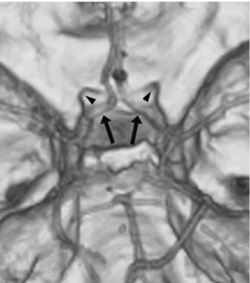
Fig. 2. A cranial view of cerebral CT angiography with volume rendering demonstrates both anterior cerebral arteries (ar- rows) originating from the internal carotid artery at the level of the ophthalmic artery and then courses under the medial side
A B
C
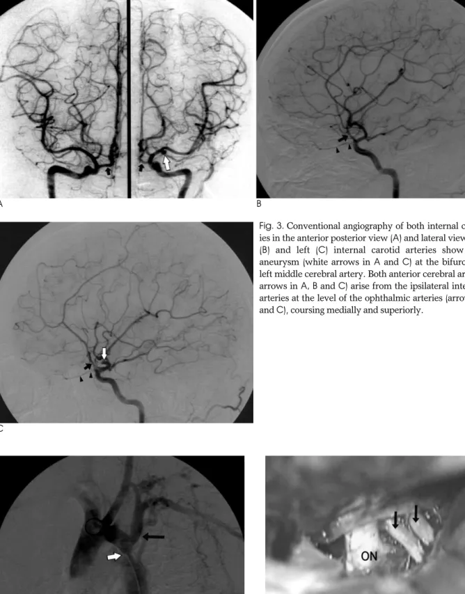
Fig. 3. Conventional angiography of both internal carotid arter- ies in the anterior posterior view (A) and lateral view of the right (B) and left (C) internal carotid arteries show a saccular aneurysm (white arrows in A and C) at the bifurcation of the left middle cerebral artery. Both anterior cerebral arteries (black arrows in A, B and C) arise from the ipsilateral internal carotid arteries at the level of the ophthalmic arteries (arrowheads in B and C), coursing medially and superiorly.
Fig. 4. Thoracic aortography shows the coarctation aorta (white arrow) at the descending aorta, distal to the origin of the left subclavian artery (black arrow).
Fig. 5. An intraoperative finding demonstrates both anterior cerebral arteries (arrows) at the medial side of the left optic nerve (ON).
mirrors the findings seen with conventional angiogra- phy, but a review of source images or obtaining high- spatial-resolution images of the suprasellar region pro- vides additional information not available by the use of conventional angiography, such as the relationship of the ACA to the optic chiasm (3). Routine MR imaging of the brain enables identification of this variation without concurrent MR angiographic sequences, showing the A1 segment between both optic nerves before the optic chiasm.
The exact embryogenesis of this vascular anomaly is unclear, but the most logical explanation seems to be re- lated to persistence of an anatomic loop around the optic nerve of the primitive dorsal ophthalmic artery that nor- mally disappears as the ophthalmic artery is formed (1, 5). According to Bosma (6), the anomaly might represent persistence of the primitive maxillary artery that sup- plies the optic stalk and cup and terminates at the pros- encephalon during the early embryonic stage.
An infra-optic course of an ACA is frequently accom- panied by other vascular abnormalities such as carotid agenesis, fused pericallosal arteries, a variant of carotid artery-basilar artery anastomosis, Moyamoya disease and coarctation of the aorta. This case showed an associ- ation of aortic coarctation with the infra-optic course of the ACA in addition to a cerebral aneurysm. Most vas- cular diseases have a tendency to affect both the heart and brain. Intracranial aneurysms are found more often in patients with aortic coarctation as compared to the general population (7). Aortic coarctation diagnosed in adulthood is rare, but poses special problems.
Treatment with angioplasty is possible, but is complicat- ed by aneurysms of the isthmus in 20% of cases, and currently surgery is considered as the treatment of choice.
Although association of a cerebral aneurysm and aor- tic coarctation is well known and association of the in- fra-optic course of the ACA and cerebral aneurysm is high (60%, 27/45), association of both a cerebral aneurysm and aortic coarctation in this anomaly has
management of the patient.
Vascular anomalies associated with intracranial aneurysms, even if rare, must be recognized in order to plan for appropriate treatment of a patient with intracra- nial aneurysms (1). In addition, diagnosis of aortic coarc- tation should be considered in adolescent and young adult patients that present with intracranial aneurysms.
Awareness of the possibility of an infra-optic course of the ACA and aortic coarctation is important in the set- ting of aneurysm surgery.
In conclusion, an infra-optic course of the anterior cerebral artery is an extremely rare anomaly and is fre- quently associated with intra-cranial aneurysms.
Considering that a cerebral aneurysm can be accompa- nied with a coarctation aorta, it would be advantageous to predict the association of these anomalies from preop- erative imaging studies.
References
1. Robinson LR. An unusual human anterior cerebral artery. J Anat 1959;93:131-133
2. Isherwood I, Dutton J. Unusual anomaly of anterior cerebral artery. Acta Radiol Diagn 1969;9:345-351
3. Given CA 2nd, Morris PP. Recognition and importance of an in- fraoptic anterior cerebral artery: case report. AJNR Am J Neuroradiol 2002;23:452-454
4. Lehmann G, Vincentelli F, Ebagosti A. Rare abnormalities of the circle of Willis: infra-optic pathway of the anterior cerebral arter- ies. Neurochirurgie 1980;26:243-246
5. Spinnato S, Pasqualin A, Chioffi F, Da Pian R. Infraoptic course of the anterior cerebral artery associated with an anterior communi- cating artery aneurysm: anatomic case report and embryological considerations. Neurosurgery 1999;44:1315-1319
6. Bosma NJ. Infra-optic course of anterior cerebral artery and low bifurcation of the internal carotid artery. Acta Neurochir 1977;38:
305-312
7. Connolly HM, Huston J 3rd, Brown RD Jr, Warnes CA, Ammash NM, Tajik AJ. Intracranial aneurysms in patients with coarctation of the aorta: a prospective magnetic resonance angiographic study of 100 patients. Mayo Clin Proc 2003;78:1491-1499
8. Wong ST, Yuen SC, Fok KF, Yam KY, Fong D. Infraoptic anterior cerebral artery: review, report of two cases and an anatomical clas- sification. Acta Neurochir 2008;150:1087-1096
대한영상의학회지 2009;60:377-381
중뇌동맥 뇌동맥류 파열과 대동맥 협착에 동반된 양측 전뇌동맥의 시신경하 주행1
1가톨릭대학교 의과대학 성바오로병원 신경외과
2가톨릭대학교 의과대학 성바오로병원 영상의학과
지 철∙안재근∙조송미2
스물 여덟 살 여자 환자에서 중뇌동맥 뇌동맥류 파열과 동반된 양측 전뇌동맥의 시신경하 주행이 관찰되었다. 양 측 전뇌동맥이 시상동맥 기시부 직 상방의 동측의 내경동맥에서 기시하여 시신경의 하부와 시신경교차 내측으로 진 행하였다. 전뇌동맥의 시신경하 주행은 매우 드문 기형으로 종종 뇌동맥류와 동반되고 대동맥 협착과는 드물게 동반 된다. 이 증례에서 방사선학적 소견과 발생학적 고찰을 하고자 한다.