Received: February 28, 2013; Accepted: March 25, 2013 Corresponding author : Yun Kyung Shin
Tel: +82 (51) 740-2421 e-mail: [email protected] 1225-3480/24464
This is an Open Access article distributed under the terms of the Creative Commons Attribution Non-Commercial License with permits unrestricted non-commercial use, distribution, and reproducibility in any medium, provided the original work is properly cited.
납(Pb)에 노출된 바지락,
Ruditapes philippinarum
의 생물지표 변화신윤경, 박정준, 임현식1, 이정식2 국립수산과학원 전략양식연구소 양식관리과
1목포대학교 해양수산자원학과, 2전남대학교 수산생명의학과
Changes of Biomarker in Manila clam, Ruditapes philippinarum Exposed to Lead
Yun Kyung Shin, Jung Jun Park, Hyun Sig Lim1 and Jung Sick Lee2
Aquaculture Management Division, Aquaculture Research Institute, NFRDI, Busan 619-902, Korea
1Department of Marine and Fisheries Resources, Mokpo National University, Muan 534-729, Korea
2Department of Aqualife Medicine, Chonnam National University, Yeosu 550-749, Korea
ABSTRACT
This study was conducted to find out biological response of Manila clam, Ruditapes philippinarum exposed to lead (Pb). Experimental period was four weeks. Experimental groups were composed of one control condition and three lead exposure conditions (0.25, 0.50 and 1.00 mg/l). The results of the study confirmed that lead induces reduction of survival rate and oxygen consumption rate and histopathology of organ structure of the bivalve. Oxygen consumption rate was observed exposure groups lower than control decline by 25%-72%. Histological analysis of organ system illustrated expansion of hemolymph sinus, disappearance of epidermal layer and degeneration of connective tissue layer of the mantle. Also, histological degenerations as epithelial necrosis and hyperplasia of mucous cells are recognized in the gill and it was observed expansion of hemolymph sinus, disruption of epithelial layer, decrease of mucous cell and degeneration of connective tissue layer in the foot. In the digestive diverticulum, it was showed atrophy of basophilic cell and degeneration of epithelial cell in the digestive tubules, and as the concentration of lead increased the accumulation of lipofuscin increased.
Key words: Ruditapes philippinarum, Lead (Pb), Survival, Oxygen consumption, Histopathology, Lipofuscin
서 론
수서생태계에서 생물에게 미치는 스트레스는 그 특성에 따 라 물리, 화학 및 생물학적 요인으로 구분된다. 화학적 요인 가운데 중금속은 비중 4.0 이상의 금속 원소로서 필수원소와 비필수원소로 구분한다. 필수원소들은 생물의 대사조절 등 다 양한 생화학적 과정에서 중요한 역할을 하며, 구리 (Cu), 철
(Fe), 아연 (Zn), 망간 (Mn) 등이 속한다. 비필수원소들은 생 물 체내에서 뚜렷한 생리기능이 알려져 있지 않은 카드뮴 (Cd), 납 (Pb), 수은 (Hg), 비소 (As), 크롬 (Cr) 등으로 이들 은 수서생태계의 주요 환경 오염원으로 작용한다 (Viarengo, 1985; Fichet et al., 1998).
중금속 가운데 납에 의한 수서 무척추동물의 생물독성은 Artemia salina, Crassostrea gigas 및 Paracentrotus lividus 유생 발생과정의 이상 (Fichet et al., 1998), Ceriodaphnia dubia와 Daphnia carinata의 생식저해 (Cooper et al., 2009), Perna viridis의 아가미와 소화선의 구조적 이상에 따른 기능의 저하 (Vasanthi et al., 2012) 등 이 보고되고 있다.
환경요인들의 영향으로 인해 생물이 받는 위해도를 평가하 는 방법은 위험성 확인 (hazard identification), 노출평가 (exposure assessment), 용량-반응 평가 (dose-response
assessment) 및 위해도 결정 (risk characterization) 의 주 요 4단계이다 (NRC, 1983). 위와 같은 방법에 따라 위해도를 평가하기 위해서는 지표생물 (indicator organism) 과 생물 지표 (biomarker) 의 선정이 중요하다.
지표생물 가운데 이매패류들은 주로 여과섭식에 의해 먹이 를 섭이하기 때문에 환경오염원에 쉽게 노출된다. 또한 이동성 이 낮아 일생동안 서식범위가 국한되어 있고, 체내로 유입된 오염원들의 축적은 용이하지만 체내에서 해독과 체외방출이 낮아 특정 지역의 오염상태를 알아보기 위한 지표종으로 많이 사용되고 있다 (Marin et al., 2006; Schintu et al., 2008;
Husmann et al., 2012).
생물지표는 생물체에 미치는 외인성 요인들의 영향을 측정 할 수 있는 세포 또는 개체수준의 생리, 생화학 및 구조 등의 지표를 지칭하는 용어이다. 생물지표에는 생리학적, 생화학적, 생식생물학적, 조직학적, 유전학적 지표 등이 있다 (Huggett et al., 1992).
우리나라에서 바지락, Ruditapes philippinarum은 생산 량이 높은 중요한 생물자원 가운데 하나로서 해양생태계의 관 리, 생물자원의 보존 및 수산물 안전성 측면에서 관리가 필요 한 종이다. 본 연구에서는 바지락에 미치는 납의 독성을 사망 률, 호흡 및 조직학적 지표 등을 이용하여 평가하고자 하였다.
재료 및 방법 1. 재료
본 연구에 사용된 바지락, Ruditapes philippinarum은 각장 3.0-4.0 cm의 성체였으며, 외형 및 운동성에 이상이 없 는 개체를 실험에 사용하였다.
시험물질인 납은 PbCl2 (Lead Chloride, Shinyo Pure Chem. Co., Japan) 를 증류수로 1000 mg/l의 표준용액을 만든 다음, 실험 농도별로 희석하여 조제하였다.
2. 방법 1) 농도구
급성독성 실험의 농도구는 Watling and Watling (1982) 과 Viarengo (1985) 의 보고를 이용하여 13개 농도구를 설정 하였다. 각 농도구에서 2회 반복실험을 실시하여 LC50-96 hr 을 구한 다음, 이를 기준으로 1개의 대조구와 3개의 노출구 (0.25 mg/l, 0.50 mg/l, 1.00 mg/l) 를 설정하였다.
2) 사육조건
본 실험에서 사용된 수조는 유리로 제작된 30l의 지수형수 조였다. 실험생물들을 채집한 후, 1주일 동안 순치시킨 다음 노출실험을 실시하였다. 실험기간 동안 광주기는 자연조건으 로 하였으며, 수온은 17 ± 0.5℃였다.
만성독성 실험의 노출실험 기간은 4주였다. 각 농도구당 노 출되어진 개체들은 밀도를 고려하여 각각 20-30개체였다. 실 험기간 동안 광주기는 자연조건으로 하였으며, 수온은 17 ± 0.5℃였다. 먹이는 식물플랑크톤인 Isochrysis galbana를 개 체 당 1-2 × 10⁶ cell/ml씩 3일에 1회 공급하였고, 실험 중 실험 개체들의 분비물로 인한 사육수의 변화와 중금속 농도변 화를 최소화하기 위해 3일에 한 번씩 사육수를 교환하였다.
3) 사망률 분석
실험생물의 사망여부는 패각을 벌리고 있는 개체들 가운데 외부에서 인위적인 자극을 가하여 반응이 없는 개체를 사망한 개체로 판단하였다. 사망개체는 매일 오전 오후 각 1회씩 확인 하여 전체 실험 개체 수에 대한 누적사망 개체수를 사망률로 환산하였다.
4) 산소 소비율 분석
산소 소비율은 Shin et al. (2006) 의 방법에 따라 실험생 물의 호흡에 의한 산소 소비량을 측정하여 표시하였다. 산소 소비량은 실험생물의 크기에 따라 0.5-1.6 l의 respirometer chamber와 산소검량기 (Isteck, 915PDC) 를 이용하여 측정 하였다.
5) 조직학적 분석
바지락의 각장 등 측정형질을 계측한 후, 해부하여 외투막, 아가미, 발 그리고 소화맹낭이 포함된 내장낭 일부를 적출하였 다. 광학현미경 조직표본제작은 적출한 시료들을 Bouin 용액 에 24시간 동안 고정하고, 48시간 동안 흐르는 물에 수세하였 다. 파라핀 절편법에 의해 4-6 μm 두께로 연속 절편하여 슬 라이드 글라스에 부착하였다. 그 후, Mayer's hematoxyline- eosin (H-E) 염색 및 alcian blue-periodic acid and Schiff's solution (AB-PAS, pH 2.5) 반응을 실시하였다. 그 리고 lipofuscin을 확인하기 위해 Long Ziehl-Neelsen 염색 을 시행하였으며, lipofuscin의 분포비율은 Lee and Park (2007) 의 방법에 따라 현미경 화상분석장치 (IMT, Visus, USA) 를 이용하여 분석하였다.
6) 통계분석
대조구에 대한 노출구들의 유의적인 차이 (P < 0.05) 를 알아보기 위해 SPSS 통계 프로그램을 이용하여 paired sample t-test를 하였다.
결 과 1. 사망률
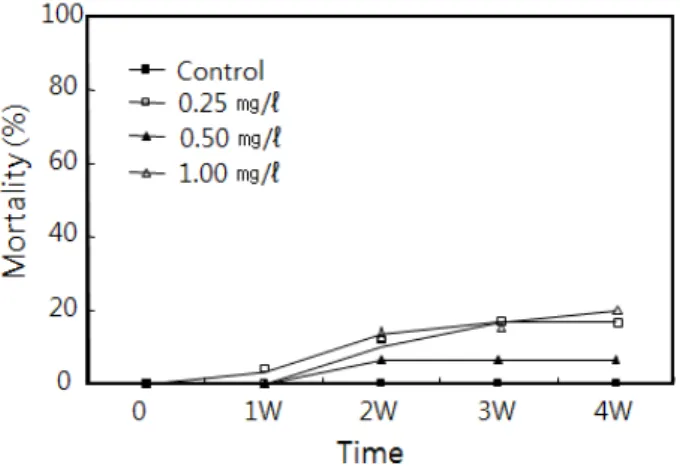
납 노출에 의한 바지락의 4주 동안의 사망률은 Fig. 1에 나
Fig. 1. Mortality of Ruditapes philippinarum exposed to lead for 4 weeks.
Fig. 2. Oxygen consumption rate of Ruditapes philippinarum exposed to lead for 4 weeks.
Fig. 3. Histology of organs of Ruditapes philippinarum. A: Mantle. B: Gill. C: Foot. D: Digestive diverticulum. C, cilia; Dt, digestive tubule; Ep, epithelium; Fc, frontal cilia; Hc, hemocytes; Lc, lateral cilia; Sc, secretory cell.
타낸 것처럼 대조구에서 0%였다. 납 노출구인 0.25, 0.50 및 1.00 mg/l의 농도구에서는 7.0-20.0%의 누적사망률을 보였 으나 유의차는 없었다.
2. 산소 소비율
납의 노출에 의한 바지락의 산소 소비율 변화는 Fig. 2에 나타내었다. 대조구에 비해 모든 납 노출구에서 산소 소비율은
노출 3주에 가장 낮았으며, 농도구 별로 대조구에 비해 25%-72%의 감소를 나타내었다.
3. 기관계의 조직학적 변화
바지락 외투막은 조직학적으로 바깥상피층, 결합조직층 및 안쪽상피층으로 구성된다. 상피층은 단층으로 섬모원주형세포 와 분비세포들로 이루어져 있다. 결합조직층은 교원섬유와 일 부 근섬유들로 구성되며, 혈림프동을 가진다 (Fig. 3A). 납에 노출된 바지락의 외투막은 가장 낮은 노출구인 0.25 mg/l의 농도구에서 상피층의 변형과 섬모의 부분적인 탈락과 혈구의 증가가 관찰되었다. 1.0 mg/l의 농도구에서는 혈림프동의 확 장과 결합조직층의 파괴 및 부분적인 상피층의 변형과 섬모의 탈락이 관찰되었다 (Fig. 4A, B, C).
아가미는 판형으로 다수의 새엽들로 구성되는데, 각각의 새 엽 상피층은 단층으로 섬모원주형 상피세포와 분비세포들로 이루어져 있다 (Fig. 3B). 납에 노출된 바지락의 아가미에서 는 0.25 mg/l와 0.5 mg/l 농도구에서 50% 새엽 상피층의 자 유면에 존재하는 섬모의 탈락과, 1.0 mg/l의 농도구에서는 부 분적인 새엽 상피층의 탈락과 새엽의 붕괴가 나타났다 (Fig.
4D, E).
발의 상피층 역시 다른 기관계와 마찬가지로 단층으로 섬모 원주형 상피세포와 분비세포들로 이루어져 있다 (Fig. 3C). 납 에 노출된 바지락의 발에서는 0.25 mg/l의 농도에서 대부분의 개체들은 상피층의 선조연과 섬모의 탈락, 상피층의 다층화 그 리고 근육층 혈림프동의 확장과 근섬유다발의 변성이 관찰되 었으며, 0.5 mg/l의 농도구에서는 상피층의 파괴 및 점액세포 의 위축 그리고 결합조직층의 변성이 관찰되었다 (Fig. 4F, G).
소화맹낭은 다수의 소화선세관들로 구성되는데, 소화선세관 은 단층으로 섬모원주형 상피세포와 호염기성세포들로 이루어 져 있다 (Fig. 3D). 납에 노출된 바지락의 소화맹낭에서는 0.25 mg/l의 농도구의 경우 소화선세관 상피층의 변형과 내강 의 폐쇄가 나타났다. 0.5 mg/l와 1.0 mg/l의 농도구에서는 호 염기성세포의 위축과 lipofuscin의 분포가 뚜렷하였으며, 1.0 mg/l의 농도구에서는 소화선세관 상피층의 다층화가 관찰되었 다 (Fig. 4H).
Fig. 4. Histopathology of organs of Ruditapes philippinarum exposed to lead for 4 weeks. A: Mantle. 0.25 mg/l. B: Mantle. 1.0 mg/l. C: Mantle. 1.0 mg/l. D: Gill. 0.25 mg/l. E: Gill. 1.0 mg/l. F: Foot. 0.5 mg/l. G: Foot. 0.25 mg/l. H: Digestive diverticulum. 1.0 mg/l. C, cilia; Ct, connective tissue; El, epithelial layer; Gf, gill filament; Hc, hemocytes; Hs, hemolymph sinus.
Fig. 5. Lipofuscin (Lf) distributions in the digestive diverticulum of Ruditapes philippinarum exposed to lead for 4 weeks. Dt, digestive tubule.
4. Lipofuscin 분포비율
납 노출에 의한 바지락의 소화맹낭에서 Long Ziehl- Neelsen 염색 결과 자색을 나타내는 lipofuscin의 분포비율은 대조구와 가장 낮은 농도구인 0.25 mg/l에서는 0%였다. 하지 만, 0.5 mg/l와 1.0 mg/l의 농도구에서 lipofuscin분포는 각각 3.33%와 7.04%로 농도 의존적 분포비율을 보였다 (Fig. 5).
고 찰
생물은 화학물질과 접촉하였을 때 화학적 인지로 인한 생물 학적 반응이 시작된다. 따라서 화학물질을 평가하는데 있어 생 물지표는 매우 유용한 수단 가운데 하나이다 (Hebel et al., 1997).
생물지표는 종 특이성과 오염원 특이성을 가지는데 (Huggett et al., 1992), 본 연구에서 사용한 생물지표 가운
데 하나인 산소 소비율 역시 이러한 경향을 보인다. 구리에 노 출된 Pseudocalanus sp.와 Calanus sp.의 산소 소비율은 변화 없이 일정하게 유지된다 (Reeve et al., 1977). 그리고 TBTCl, 수은, 카드뮴에 노출된 Littorina brevicula의 산소 소비율은 감소하다가 증가하는 경향을 보인다 (Chin et al., 1999). 하지만, 구리에 노출된 진주담치, Mytilus edulis (Brown and Newell, 1972), 카드뮴과 아연에 노출된 Perna viridis (Cheung and Cheung, 1995), 카드뮴에 20 일간 노출된 Neomysis awatschensis (Kang et al., 1997) 그리고 은과 크롬에 노출된 P. viridis (Vijayavel et al., 2007) 의 산소 소비율은 감소한다. 본 연구에서 납에 노출된 바지락의 산소 소비율은 감소하다가 다시 증가하는 변화 경향 을 보여 TBTCl, 수은, 카드뮴에 노출된 L. brevicula와 유사 하게 나타났다.
이매패류에서 화학물질 및 중금속 등의 오염원들은 외투막,
아가미, 신장, 발 그리고 소화맹낭에 주로 축적되며 (Morrison, 1993). 직간접적으로 이들은 수서생물의 기관계 구조의 변화를 일으켜 생리학적 불균형을 초래한다 (Watling and Watling, 1982; Gregory et al., 1999; Otludil et al., 2004).
이매패류에서 외투막은 감각, 패각형성 및 외투강 정화 기능 을 수행하는 기관계이다 (Eble, 2001). 수중오염원에 의한 이 매패류의 외투막 이상은 상피층의 정단부에 존재하는 선조연 과 섬모들이 탈락, 점액세포들의 활성 감소, 결합조직층에서 교원섬유와 근섬유 다발의 변성을 들 수 있다. TBTCl에 노출 된 대복, Gomphina veneriformis의 외투막 상피세포의 변 형 및 상피층과 결합조직층에 존재하는 점액물질의 변화 등이 관찰되었다 (Park and Lee, 2008). 또한, 노닐페놀에 노출된 바지락의 외투막에서 혈림프동의 확장과 혈구의 증가, 상피층 자유면의 선조연 탈락, 결합조직층의 근섬유 다발의 분절과 산 성 점액세포의 증가가 확인되었다 (Park and Lee, 2011). 본 연구에서도 납에 노출된 바지락의 외투막에서 다양한 조직학 적 이상을 보였는데, 이러한 외투막의 이상은 정상적인 외투강 정화기능의 장애 및 패각 형태의 기형 등을 유발할 것으로 판 단된다.
이매패류의 아가미는 호흡뿐 만 아니라 여과섭식 기능을 수 행하는 중요한 기관계 가운데 하나이다 (Benninger et al., 1988; Eble, 2001). 수중의 구리, 염소, 철에 노출된 굴, 진주 담치, 바지락 아가미에서는 새엽 상피층의 분리, 혈림프동의 확장, 새엽 섬모대의 소실 그리고 새엽 상피층의 분리와 파괴 등 아가미로써의 기능을 상실한 조직상들이 관찰되었다 (Kim and Lee, 1988). DDT와 PCB에 오염된 핀란드의 해안에서 채집된 진주담치의 아가미에서는 정단상피와 측면상피의 붕 괴와 측면섬모의 탈락이 관찰되었다 (Sunila, 1988). 수은에 노출된 P. viridis (Gregory et al., 1999) 와 중금속 오염지 역에서 채집된 P. viridis의 아가미에서도 이와 유사한 구조적 이상이 보고되고 있다 (Vasanthi et al., 2012). 본 연구 결과, 납에 노출된 바지락의 아가미 이상현상은 호흡률의 변화 및 여과섭이 기능의 장애를 유발할 것으로 생각된다.
이매패류의 발은 단층의 상피세포로 구성되어진 상피층이 조밀하게 발달한 근육층을 둘러싸고 있는 구조를 하고 있다. 이들 근육층은 저질에 잠입 시 이완, 수축하면서 저질에 파고 들면서 이동한다 (Eble, 2001). 0.4와 0.8 mg/l의 endosulfan에 30일 동안 노출된 Planorbarius corneus의 발은 노출 10일에는 점액세포와 단백질세포가 증가한 후 노출 20일 이후에는 다시 감소하며, 상피세포가 탈락된다. 사망 직 전의 개체들에서는 상피세포의 탈락과 더불어 근섬유들이 위 축되는 조직상이 관찰된다 (Otludil et al., 2004). 또한, 아연 에 노출된 대복의 발은 노출 초기에는 상피층 선조연의 일부가
소실되고 혈림프동이 확장된다. 그리고 결합조직층에 존재하 던 점액세포들이 근육층까지 확장된다. 그리고 농도 의존적으 로 상피층의 박리과 상피세포의 괴사, 근육층을 구성하는 환상 근층과 종주근층의 근섬유 다발의 분절화가 나타난다 (Ju et al., 2006). 노닐페놀에 노출된 바지락의 발은 0.05 mg/l 농 도구에서 상피층이 비후되며, 0.01과 0.02 mg/l 농도구에서는 상피층 선조연의 일부가 소실된다 (Park and Lee, 2011). 납 에 노출된 바지락의 발에서도 다양한 구조적 이상현상들이 관 찰되었는데, 이는 바지락의 이동과 저질잠입 능력의 저하로 이 어질 수 있는 결과들이다.
이매패류에서 소화맹낭의 소화선세관은 소화, 흡수 및 오염 원의 해독기능을 수행하는데, 소화맹낭은 다른 기관에 비해 오 염원의 축적성이 높다 (Moore and Allen, 2002). 특히, 아 연, 구리, 카드뮴, 망간의 농도는 아가미나 외투막 보다 소화맹 낭에서 높다 (Husmann et al., 2012). 소화맹낭을 구성하는 세포들의 손상은 독성영향을 평가하는데 중요한 지표가 된다 (Depledge and Hopkin, 1995). 0.08 mg/l의 구리, 아연 및 카드뮴에 41일 동안 노출된 지중해담치의 소화선세관을 구성 하는 상피세포들은 모두 섬모가 소실되었다 (Soto et al., 1996). 30일 동안 0.227 mg/l의 니켈에 노출된 Lammellidaens marginalis의 소화선세관은 상피층의 파괴 와 기저막 분리현상을 보인다 (Andhale et al., 2011). 노르 웨이의 심각한 구리 오염지역에서 채집한 진주담치의 소화선 세관에서 내강의 팽창이 관찰되며, 오염의 정도가 심한 지역에 서 채집한 개체들은 호염기성 세포의 비율이 높고, 소화세포의 비율은 낮다 (Zorita et al., 2006). 본 연구 결과, 소화선에서 소화, 흡수, 분비 및 해독작용 등의 대사활동에 관여하는 소화 선세관의 상피세포와 호염기성세포의 변화와 파괴가 관찰되었 는데, 이러한 결과로 인해 납에 노출된 바지락의 소화선에서는 정상적인 대사활동이 불가능할 것으로 판단된다.
이매패류에서 lipofuscin은 구리, 아연, 철, 망간, 마그네슘 과 같은 중금속 이온들을 포획가능한 제3의 용해소체로 여겨 지기 때문에 중금속이나 저산소증에 인한 스트레스의 영향의 지표로 이용되어 왔다 (Krishnakumar et al., 1990;
Viarengo et al., 1990). 진주담치를 15주 동안 0.8, 5.0, 20.0 mg/l의 구리에 노출 시킨 결과, 고농도에서 용해소체의 확장과 lipofuscin의 축적이 증가하였고, PAHs, PCBs와 중 금속에 오염된 지역에서 채집된 개체들도 지방과립이 증가하 고 lipofuscin의 비율도 증가하였다 (Moore, 1988; Regoli, 1992; Da Ros et al., 2000). Tapes semidecussatus의 경 우에도 고농도의 중금속에 오염된 저질에 노출시킨 후 대조구 와 비교한 결과, 대조구의 소화맹낭에서는 lipofuscin이 관찰 되지 않았지만 중금속 노출구에서는 lipofuscin이 다량으로 축적되었다 (Byrne and O'Halloran, 1999). 그리스
Thermaikos만의 지중해담치의 소화선세관 상피세포에 존재 하는 자가용해소체와 잔여소체들은 오염지역으로 갈수록 크기 와 분포정도가 높아졌으며, 중금속을 함유한 용해소체 또한 더 많이 관찰되었다 (Domouhtsidou and Dimitriadis, 2001).
본 연구의 결과를 종합해보면, 납에 노출된 바지락에서도 대복 을 아연 (Ju et al, 2006), 노닐페놀 (Lee and Park, 2007) 에 노출시킨 결과, 그리고 바지락을 노닐페놀에 노출시킨 결과 (Park and Lee, 2011) 등과 마찬가지로 소화선세관 상피세 포들의 구조적 변성과 함께 농도 의존적으로 lipofuscin의 분 포가 높게 나타났다.
요 약
본 연구는 납에 노출된 바지락, Ruditapes philippinarum 의 생물학적 반응을 알아보고자 하였다. 실험기간은 4주였으 며, 실험구는 대조구 1개와 납 노출구 3개 (0.25, 0.50 and 1.00 mg/l) 였다. 실험 결과 납은 바지락의 생존율과 산소 소 비율의 저하 및 기관계의 조직학적 변성을 유도하는 것이 확인 되었다. 납 노출구에서 산소소비율은 25-72% 감소하였다. 기 관계의 조직학적 분석 결과, 외투막 상피층과 결합조직층의 변 성, 아가미 점액세포의 증가와 상피세포의 괴사, 발에서는 상 피층의 붕괴, 점액세포의 감소 및 혈림프동의 확장과 결합조직 층의 변성을 나타냈다. 소화맹낭의 소화선세관에서는 호염기 성세포와 상피세포의 위축 및 농도 의존적으로 lipofuscin의 축적이 확인되었다.
사 사
본 연구는 국립수산과학원 수산시험연구사업 (RP-2013- AQ-012) 으로 수행되었습니다.
참고문헌
Andhale, A.V., Bhosale, P.A. and Zambare, S.P. (2011) Histopathological study of nickel induced alterations in the fresh water bivalve, Lammellidens marginalis. Journal of Experimental Sciences, 2: 1-3.
Beninger, P.G., Le Pennec, M. and Salaun, M. (1988) New observations of the gills of Placopecten magellanicus (Mollusca: Bivalvia), and implications for nutrition. Marine Biology, 98: 61-70.
Brown, B.E. and Newell, R.C. (1972) The effect of copper and zinc on the metabolism of the mussel Mytilus edulis. Marine Biology, 16: 108-118.
Byrne, P.A. and O'Halloran, J. (1999) Aspects of assaying sediment toxicity in Irish estuarine ecosystems. Marine Pollution Bulletin, 39: 97-105.
Cheung, S.G. and Cheung, R.Y. (1995) Effects of heavy metals on oxygen consumption and ammonia excretion in green-lipped mussels (Perna viridis).
Marine Pollution Bulletin, 31: 381-386.
Chin, P., Lee, J.A., Shin, Y.K. and Lee, J.S. (1999) Environmental tolerance for pollutants in Littorina brevicula (Philippi). 2. The growth, metabolism and histological changes exposed to TBTCl and heavy metals in Littorina brevicula. Journal of Korean Fisheries Society, 32: 593-600. [in Korean]
Cooper, N.L., Bidwell, J.R. and Kumar, A. (2009) Toxicity of copper, lead, and zinc mixtures to Ceriodaphnia dubia and Daphnia carinata.
Ecotoxicology and Environmental Safety, 72: 1523–
1528.
Da Ros, L., Nasci, C., Marigómez, I. and Soto, M.
(2000) Biomarkers and trace metals in the digestive gland of indigenous and transplanted mussels, Mytilus galloprovincialis, in Venice Lagoon, Italy.
Marine Environmental Research, 50: 417–423.
Depledge, M.H. and Hopkin, S.P. (1995) Methods to assess effects on brackish, estuarine and near-coastal water organisms. In: Methods to assess the effects of chemicals on ecosystems. (ed. by Linthurst, R.A., Bourdeau, P. and Tardiff, R.G.). pp.
125-149. Wiley, Chichester, UK.
Domouhtsidou, G.P. and Dimitriadis, V.K. (2001) Lysosomal and lipid alterations in the digestive gland of mussels, Mytilus galloprovincialis (L.) as biomarkers of environmental stress. Environmental Pollution, 115: 123-137.
Eble, A.F. (2001) Anatomy and histology of Mercenaria mercenaria. In: Biology of the hard clam. (ed. by Kraeuter, J.N., Castagna, M.). pp. 117-220. Elsevier, New York.
Fichet, D., Radenac, G. and Miramand, P. (1998) Experimental studies of impacts of harbour sediments resuspension to marine invertebrates larvae: Bioavailability of Cd, Cu, Pb and Zn and toxicity. Marine Pollution Bulletin, 36: 509-518.
Gregory, M.A., George, R.C., Marshall, D.J., Anandraj, A. and Mcclurg, T.P. (1999) The effects of mercury exposure on the surface morphology of gill filament in Perna perna (Mollusca: Bivalvia). Marine Pollution Bulletin, 39: 116-121.
Hebel, D.K., Jones, M.B. and Depledge, M.H. (1997) Responses of crustaceans to contaminant exposure:
a holistic approach. Estuarine, Coastal and Shelf Science, 44: 177-184.
Huggett, R.J., Kimerle, R.A., Mehrle, P.M. Jr and Bergman, H.L. (1992) Biomarkers: biochemical, physiological, and histological markers of anthropogenic stress. pp. 1-347. Lewis Publishers, Boca Raton, FL.
Husmann, G., Abele, D., Monien, D., Monien, P., Kriews, M. and Philipp, E.E.R. (2012) The influence of sedimentation on metal accumulation and cellular oxidative stress markers in the Antarctic bivalve Laternula elliptica. Estuarine Coastal and Shelf Science, 111: 48-59.
Ju, S.M., Lee, J.W., Jin, Y.G., Yu, J. and Lee, J.S. (2006) Effect of zinc bioaccumulation on survival rate,
activity, growth and organ structure of the equilateral venus, Gomphina veneriformis (Bivalvia:
Veneridae). Journal of Environmental Toxicology, 21:
115-126. [in Korean]
Kang, J.C., Kim, H.Y. and Chin, P. (1997) Toxicity of cooper, cadmium and chromium on survival, growth and oxygen consumption of the mysid, Neomysis awatschensis. Journal of Korean Fisheries Society, 30: 874-881. [in Korean]
Kim, S.Y. and Lee, T.Y. (1988) The effects of pollutants effluent from a steam-power plant on coastal bivalves. Ocean Research, 10: 47-56. [in Korean]
Krishnakumar, P.K., Asokan, P.K. and Pillai, V.K. (1990) Physiological and cellular responses to copper and mercury in the green mussel Perna viridis (Linnaeus). Aquatic Toxicology, 18: 163-173.
Lee, J.S. and Park, J.J. (2007) Risk assessment of nonylphenol using the sex ratio, sexual maturation, intersex and lipofuscin accumulation of the equilateral venus, Gomphina veneriformis (Bivalvia:
Veneridae). Journal of Korean Fisheries Society, 40:
16-23. [in Korean]
Marin, M.G., Boscolo, R., Cella, A., Degetto, S. and Ros, L.D. (2006) Field validation of autometallographical black silver deposit (BSD) extent in three bivalve species from the Lagoon of Venice, Italy (Mytilus galloprovincialis, Tapes philippinarum, Scapharca inaequivalvis) for metal bioavailability assessment.
Science of Total Environment, 371: 156-167.
Moore, M.N. (1988) Cytochemical responses of the lysosomal system and NADPH-ferrihemoprotein reductase in molluscan digestive cells to environmental and experimental exposure to xenobiotics. Marine Ecology Progress Series, 46: 81-89.
Moore, M.N. and Allen, J.I. (2002) A computational model of the digestive gland epithelial cell of the marine mussel and its simulated responses to oil-derived aromatic hydrocarbons. Marine Environmental Research, 54: 579-584.
Morrison, C.M. (1993) Histology and cell ultrastructure of the mantle and mantle lobes of the eastern oyster, Crassostrea virginica, Gmelin: A summary atlas. American Malacological Bulletin, 10: 1-24.
NRC (National Research Council). (1983) Risk assessment in the federal government: managing the process. pp. 1-192. National Academy Press, Washington, D.C.
Otludil, B., Cengiz, E.I., Yildirim, M.Z., Unver, O. and Unlu, E. (2004) The effects of endosulfan on the great ramshorn snail, Planorbariua corneus (Gastropoda, Pulmonata): a histopathological study.
Chemosphere, 56: 707-716.
Park, J.J. and Lee, J.S. (2008) Tributyltin chloride (TBTCI) toxicity on the growth and mantle structure of the equilateral venus, Gomphina veneriformis (Bivalvia: Veneridae). The Korean Journal of Malacology, 24: 229-241. [in Korean]
Park, J.S. and Lee, J.S. (2011) Change of reproductive
and histological biomarkers of Ruditapes philippinarum (Bivalvia: Veneridae) exposed to nonylphenol. The Korean Journal of Malacology, 27:
181-190. [in Korean]
Reeve, M.R., Gamble, J.C. and Walter, M.C. (1977) Experimental observation on the effects of cooper on copepod and other zooplankton: controlled ecosystem pollution experiment. Bulletin of Marine Science, 27: 92-104.
Regoli, F. (1992) Lysosomal responses as a sensitive stress index in biomonitoring heavy metal pollution.
Marine Ecology Progress Series, 84: 63-69.
Schintu, M., Durante, L., Maccioni, A., Meloni, P., Degetto, S., and Contu, A. (2008) Measurement of environmental trace-metal levels in mediterranean coastal areas with transplanted mussels and DGT techniques. Marine Pollution Bulletin, 57: 832-837.
Shin, Y.K., Kim, B.H. Oh, B.S., Jung, C.G., Sohn, S.G.
and Lee, J.S. (2006) Physiological responses of the ark shell, Scapharca broughtonii (Bivalvia: Arcidae) to decrease in salinity. Journal of Fisheries Science and Technology, 9: 153-159. [in Korean]
Soto, M., Cajaraville, M.P. and Marigómez, I. (1996) Tissue and cell distribution of copper, zinc and cadmium in the mussel, Mytilus galloprovincialis, determined by autometallography. Tissue and Cell, 28: 557-568.
Sunila, I. (1988) Acute histological responses of the gill of the mussel, Mytilus edulis, to exposure by environmental pollutants. Journal of Invertebrate Pathology, 52: 137-141.
Vasanthi, L.A., Revathi, P., Arulvasu, C. and Munuswamy, N. (2012) Biomarkers of metal toxicity and histology of Perna viridis from Ennore estuary, Chennai, south east coast of India. Ecotoxicology and Environmental Safety, 84: 92–98.
Viarengo, A. (1985) Biochemical effects of trace metals.
Marine Pollution Bulletin, 16: 153-158.
Viarengo, A., Canesi, L., Pertica, M., Poli, G., Moore, M.N. and Orunesu, M. (1990) Heavy metal effects on lipid peroxidation in the tissues of Mytilus galloprovincialis Lam. Comparative Biochemistry and Physiology - part C, 97: 37-42.
Vijayavel, K., Gopalakrishnan, S. and Balasubramanian, M.P. (2007) Sublethal effect of silver and chromium in the green mussel Perna viridis with reference to alterations in oxygen uptake, filtration rate and membrane bound ATPase system as biomarkers.
Chemosphere, 69: 979–986.
Watling, H.R. and Watling, R.J. (1982) Comparative effects of metals on the filtering rate of the brown mussel, Perna perna. Bulletin of Environmental Contamination and Toxicology, 29: 651-657.
Zorita, I., Ortiz-Zarragoitia, M., Soto, M., Cajaraville, M.P. (2006). Biomarkers in mussels from a copper site gradient (Visnes, Norway): an integrated biochemical, histochemical and histological study.
Aquatic Toxicology, 78: S109-S116.