Received August 9, 2011 ; Accepted September 28, 2011 Corresponding author: Jung Sick Lee
Tel: +82 (61) 659-7172 e-mail: [email protected] 1225-3480/24393
노닐페놀에 노출된 바지락,
Ruditapes philippinarum
의 생식 및 조직학적 지표 변화박지선, 이정식
전남대학교 수산생명의학과Change of Reproductive and Histological Biomarkers of Ruditapes philippinarum (Bivalvia: Veneridae) Exposed to
Nonylphenol
Ji Seon Park and Jung Sick Lee
Department of Aqualife Medicine, Chonnam National University, Yeosu 550-749, Korea
ABSTRACT
This study was investigated to find out the change of reproductive and histological biomarkers of Ruditapes
philippinarum exposed to nonylphenol (NP). Experimental period was 16 weeks. Experimental groups werecomposed of one control condition and three NP exposure conditions (0.05, 0.1, 0.2 mg NP L
-1). Gonad index was higher in exposure group than control in female and higher in control than exposure group in male. Intersexuality in the NP exposure group was 14.56% with male exhibiting higher ratio than female. Histological analysis of organ system illustrated expansion of hemolymph sinus, loss of striated border of inner epidermis and degeneration of mucous cell in the mantle. In the gill partially disappearance of frontal and lateral cilia, degeneration of mucous cell were observed. Also, hyperplasia of epithelium and disappearance of striated border were observed in the foot. As the concentration of NP increased, the accumulation of lipofuscin increased in the digestive gland. The results of this study suggest that NP induces reduction of survival rate, histopathology and intersex of R. philippinarum.
Furthermore, it is indentified that NP functions as an estrogenic effector on the reproduction of R. philippinarum.
Key words: nonylphenol, reproductive and histological biomarkers, Ruditapes philippinarum
서 론
해양생태계에서 생물요소에 영향을 주게 되는 스트레스의 종류에는 광, 수온, pH, 염분과 같은 물리적인 요인과 미생물, 바이러스, 기생충과 같은 생물학적 요인 그리고 중금속과 내분 비계장애물질 (endocrine dirupting chemicals: EDCs), 난 분해성화학물질 (persistant organic pollutants: POPs) 등 과 같이 환경오염원으로 작용하는 화학적인 요인이 있다 (Rand and Petrocelli, 1985).
이 가운데 EDCs는 인간의 산업활동 결과로 만들어진 화학 물질로서 다이옥신, 페놀류, 폴리염화페닐 (PCB), 유기주석
(TBT), 과불화화합물 (PFCs) 등이 이에 포함되는데, 이들 화 합물질은 동물체내로 유입되어 마치 호르몬처럼 작용하여 본 래의 조절기작을 방해한다 (Fenner-Crisp, 1997).
그 중 노닐페놀 (nonylphenol: NP)은 nonylphenol ethoxylates (NPEs)의 생산, NP phosphites, aminocarb 살충제, 스프레이에 사용된다 (Granmo et al., 1989;
Maguire, 1999). NP phosphites는 고무나 플라스틱 산업에 서 방부제로 사용되고 있으며, NPEs는 비이온화 계면활성제 로서 플라스틱, 라텍스페인트, 윤활유, 유화제에 사용되고 있 다. 이들의 광범위한 사용 결과, 간접적으로는 하수처리장에 방출되고 있으며 (Ekelund et al., 1990; Maguire, 1999), 최종적으로 해양에 유입된다. 또한 NP는 소수성 화합물질로 수중에서 쉽게 분해되지 않고 부유입자에 흡착되어 저층에 퇴 적물로 쌓여 수질과 저질의 오염원으로 작용한다 (Ekelund et al., 1990).
이매패류들은 주로 저질 내에 서식하는 생물로서 환경수 및
물질의 유입이 쉬운 연안에 서식하기 때문에 각종 화학물질에 의한 영향을 받을 가능성이 매우 높아 해양저질과 수질의 오염 상태를 알아보기 위한 지표종으로 많이 사용되고 있다 (Pipe and Coles, 1995; Siah et al., 2003).
해양생물을 이용하여 EDCs의 독성을 평가하는데 사용되는 생물 지표로는 사망률, 성장, 행동, 분자생물학적 지표, 생화학 적 지표, 생리학적 지표, 생식지수, intersex 및 imposex등의 생식 지표 및 조직학적 지표 등이 있다 (Alzieu and Heral, 1984; Timmermans et al., 1996; Gauthier-Clerc et al., 2002; Lee and Park, 2007).
이 연구는 바지락, Ruditapes philippinarum의 사망률, 생식 및 조직학적 지표를 이용하여 노닐페놀의 위해성을 검토 하고자 하였다.
재료 및 방법
1. 재 료이 연구에 사용된 바지락, Ruditapes philippinarum은 2009년 1월에 전남 남해안 해창만에서 채집하였다. 크기는 각 장 3.0-4.0 cm의 성체였으며, 약 500개체를 채집하여 외형 및 운동성에 이상이 없는 320개체를 실험에 사용하였다.
2. 방 법 1). 실험조건
채집한 시료는 실험실내 지수형 반순환여과식 유리수조에 약 2주 동안 순응시켰다. 실험기간은 생식소의 발달단계가 비 활성기인 2009년 1월 20일에 시작하여 2009년 5월 12일까 지 16주였다. 이 실험에 사용한 수조는 30 L의 사각 유리수조 로써 수조 내부는 바지락의 서식환경과 비슷한 저질환경을 유 지하기 위해서 채집지역의 저질을 두께 10 cm 정도 깔아 주 었다. 실험기간동안 광주기와 온도는 자연조건으로 설정하였 으며, 먹이는 Pavlova lutheri를 바지락 개체당 1-2 × 10⁶ cell mL-1 을 3일에 1회씩 공급하였고, 사육수 교환은 먹이공 급 시 사육수의 2/3를 교환하였다. 각 실험구별 개체수는 40 개체였으며, 동시 2회 반복하였다.
실험용액은 노닐페놀 (nonylphenol: C15H24O, Aldrich Chem. Co. Inc., USA)을 아세톤으로 1000 mg L-1의 표준 용액을 만든 다음, 실험농도별로 희석하여 조제하였다. 노닐페 놀이 첨가되지 않은 여과해수를 대조구로 하였고, 노출구는 급 성독성실험 결과와 기존의 연구결과 (Matozzo and Marin, 2005) 를 참고하여 각각 0.05, 0.1, 0.2 mg NP L-1로 설정 하였다.
2) 생존율
생존은 실험기간 동안 매일 오전 10시와 오후 6시에 관찰하
패각을 닫지 않는 개체, 입·출수관의 반응이 없는 개체를 사망 개체로 판단한 후, 누적사망률을 구하여 생존율로 환산하였다.
3) 생식지표
생식소지수 (gonad index: GI)는 조직표본 관찰 결과, 생 식소 발달단계를 5단계로 나누고 발달단계별 상수 (In=0, Ea
Ⅰ=1, Ea Ⅱ=2, Ea Ⅲ=3, La=4)를 곱한 전체의 합을 개체 수로 나누었다.
GI =
(N of In × 0) + (N of Ea Ⅰ × 1) + (N of Ea
Ⅱ × 2) + (N of Ea Ⅲ × 3) + (N of La × 4)
Total number (N)
이성생식세포 발현율은 생식소 조직표본에서 반대 성의 생 식세포가 관찰되는 것만을 기준으로 조사하였다.
4) 조직학적 분석
바지락의 각장, 각고, 전중량, 육중량을 측정한 후, 해부하여 외투막, 아가미, 발 그리고 생식소와 중장선이 포함된 내장낭 일부를 적출하였다. 광학현미경 조직표본제작은 적출한 시료 들을 Bouin 용액에 24시간 동안 고정하고, 48시간 동안 흐르 는 물에 수세하였다. 파라핀 절편법에 의해 4-6 μm 두께로 연속 절편하여 슬라이드 글라스에 부착하였다. 그 후, Mayer's hematoxyline-eosin (H-E) 염색, Masson 삼중염 색, alcian blue-periodic acid and Schiff's solution (AB-PAS, pH 2.5) 반응 및 lipofuscin을 관찰하기 위해 Long Ziehl-Neelsen 염색을 시행하였다.
5) 유의성 검증
대조구에 대한 노출구들의 유의적인 차이 (P < 0.05) 를 알아보기 위해 통계처리는 SPSS 통계 프로그램을 이용하여 paired sample t-test를 하였다.
결 과
1. 생 존실험기간 동안 생존율은 Fig. 1에 나타냈다. 실험종료시점 에 대조구의 생존율은 약 78%였으며, 노닐페놀 노출구의 평 균 생존율은 48.75%였다. 각 노출구 농도구인 0.05, 0.1, 0.2 mg NP L-1의 생존율은 각각 53.75, 43.75, 48.75% 로 감소 하였다.
2. 생식지표의 변화
1) 생식소지수 (gonad index: GI)
생식소 발달단계를 근거로 GI의 변화를 Fig. 2에 나타냈다.
모든 실험구에서 GI는 수컷보다 암컷에서 낮게 나타났다. 대
0 20 40 60 80 100
0 2 4 6 8 10 12 14 16
Weeks
Survival rate(%)
Control 0.05 ㎎ NP L-1 0.1 ㎎ NP L-1 0.2 ㎎ NP L-1
Fig. 1. Survival rate of Ruditapes philippinarum exposed to nonylphenol for 16 weeks.
Fig. 2. Gonad index (GI) of Ruditapes philippinarum exposed to nonylphenol for 16 weeks. Vertical bar: SD.
Fig. 3. Photomicrographs of intersex gonad of Ruditapes philippinarum exposed to nonylphenol for 16 weeks. A:
ovary. H-E stain. B: testis. H-E stain. Oc: oocyte, Sc:
spermatocyte.
Fig. 4. Intersexuality of Ruditapes philippinarum exposed to nonylphenol for 16 weeks.
조구에서 암컷과 수컷의 GI는 각각 1.46과 3.0이었다. 노출구 의 암컷과 수컷의 평균 GI는 각각 1.57과 2.71로 대조구에 비 해 암컷은 증가하였지만 수컷은 감소하였다. 노출구의 GI는 암·수 모두 0.05 mg NP L-1와 0.1 mg NP L-1 농도구에서 대조구와 유의적인 차이를 보였는데, 이들 농도구에서 암컷의 GI는 대조구보다 높았으나 수컷의 GI는 대조구보다 낮았다 (P < 0.05).
2) 이성생식세포 발현
생식소 조직표본을 광학현미경으로 관찰한 결과, 암컷의 생식소에서 수컷의 생식세포가 관찰되고 수컷의 생식세포 에서 암컷의 생식세포가 관찰된 이성생식세포 발현 현상이 확인되었다 (Fig. 3).
대조구의 이성생식세포 발현율은 7.7%였는데, 암컷에서 는 5.41%, 수컷에서는 13.33%였다. 노출구의 평균 이성생 식세포 발현율은 14.56%로 대조구에 비해 증가하였으며, 암컷은 10.3%, 수컷은 25.77%였다. 농도구별 성에 따른 이 성생식세포 발현율은 암컷은 3.13%, 5.56%, 22.2%로 농도
의존적으로 증가하였으며, 수컷은 33.33%, 16.7%, 27.3%
로 가장 낮은 농도구인 0.05 mg NP L-1에서 가장 높게 나 타났다 (Fig. 4).
3. 조직학적 지표의 변화 1) 외투막
바지락의 외투막은 외부상피층과 내부상피층 사이에 결합조 직층이 존재하였다. 외부상피층은 단층구조이며, 섬모의 발달 이 미약한 원주형 세포들로 구성되어 있었다. 결합조직층은 교 원섬유와 근섬유 다발로 이루어진 소성결합조직으로 다수의 혈림프동을 가지고 있었다. 내부상피층은 단층의 섬모원주형 세포들로 구성되어 있었다 (Fig. 5A). 점액세포들은 AB-PAS (pH 2.5) 반응결과, alcian blue에 보라색과 푸른색으로 반응 하였다.
노닐페놀에 노출된 바지락의 외투막은 0.05 mg NP L-1 농도구에서는 혈림프동의 확장이 뚜렷했다 (Fig. 5B). 0.1 mg NP L-1 농도구에서는 AB-PAS (pH 2.5) 반응에서 푸 른색으로 반응한 점액세포의 감소가 관찰되었다 (Fig. 5C and D). 0.2 mg NP L-1 농도구에서는 내부상피층의 선조
Fig. 5. Histopathological changes of mantle of Ruditapes philippinarum exposed to nonylphenol for 16 weeks. A:
Control. Cross section showing the outer epidermal layer (Oel) and inner epidermal layer (Iel). H-E stain. B:
0.05 mg NP L-1. Showing the expanded hemolymph sinus (Hs). H-E stain. C and D: 0.1 mg NP L-1. Showing the mucous cell (Mc) of alcian blue positive. AB-PAS (pH 2.5). E: 0.2 mg NP L-1. Cross section showing the partially disappearance of striated border (Sb) in inner epidermal layer. F: 0.2 mg NP-1. Showing the destruction of the muscle fiber bundle. AB-PAS (pH 2.5).
Fig. 6. Histopathological changes of gill of Ruditapes philippinarum exposed to nonylphenol for 16 weeks. A:
Control. Longitudinal section showing the plica. H-E stain. B: 0.05 mg NP L-1. Showing the extended hemolymph sinus (Hs), decreased frontal cilia (Fc) and partially disappearance of lateral cilia (Lc). H-E stain. C and D: 0.1 mg NP L-1. Showing the mucous cell (Mc) of alcian blue positive. AB-PAS (pH 2.5) reaction. E: 0.2 mg NP L-1. Showing the destruction of interlamella junction. H-E stain. F: 0.2 mg NP-1. Showing the increased hemocytes. E, epithelium; F, filament.
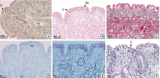
Fig. 7. Histopathological changes of foot of Ruditapes philippinarum exposed to nonylphenol for 16 weeks. A and B: Control. Showing the epithelial layer (El), connective tissue layer (Ctl), circular muscle layer (Cml) and longitudinal muscle layer (Lml). Masson`s trichrome stain. C: Control. Longitudinal section showing the reticular muscle layer of longitudinal and longitudinal muscle fiber bundles. Masson's trichrome stain. D: 0.1 mg NP L-1. Showing the mucous cell (Mc) of alcian blue positive. AB-PAS (pH 2.5). E: 0.2. mg NP L-1 Showing the fragmentation of muscle fiber bundle. Masson's trichrome stain. F: 0.2 mg NP L-1. Showing the partially disappearance of cilia (C). Masson's trichrome stain.
연의 탈락과 박리 그리고 결합조직층의 근섬유 다발의 분절 이 관찰되었다 (Fig. 5E and F).
2) 아가미
아가미는 다수의 새엽이 연결된 판새형이었다. 각각의 새판 은 여러 개의 새엽다발로 구성되어 있었으며, 새엽다발은 근섬 유에 의해 연결되어 있는 다수의 새엽으로 구성되어 있었다.
각각의 새엽다발 안에는 혈림프동이 존재하고 있었다. 새엽의 상피층은 단층으로 기저부와 중간부에서는 편평형 상피세포 또는 입방형 상피세포가 관찰되었으며, 정단면에서는 섬모원 주형 상피세포가 관찰되었다 (Fig. 6A). 점액세포들은 AB-PAS (pH 2.5) 반응시 푸른색으로 반응하는 점액물질을 가지고 있었다.
노닐페놀에 노출된 바지락의 아가미는 0.05 mg NP L-1 농 도구에서는 혈림프동의 확장과 정단섬모 (frontal cilia) 와 측 면섬모 (laterlar cilia) 의 부분적 소실이 관찰되었고 (Fig.
6B), 0.1 mg NP L-1 농도구의 경우, AB-PAS (pH 2.5) 에 반응시킨 결과, alcian blue에 푸른색으로 반응한 점액물질이 interlamella junction 주위와 기저부에서 관찰되었다 (Fig.
6C and D). 0.2 mg NP L-1 농도구의 경우, interlamella junction의 붕괴와 섬유다발의 절단이 관찰되었으며 (Fig.
6E), 혈구의 증가도 관찰되었다 (Fig. 6F).
3) 발
발은 외부상피층, 결합조직층, 근육층으로 구성되어 있었다.
외부상피층은 다수의 주름을 가지는 단층으로 섬모의 발달이 뚜렷한 원주형 상피세포로 구성되어 있었다. 상피층의 자유면 에는 선조연의 발달이 뚜렷하였다. 결합조직층은 근섬유 다발 로 구성된 소성결합조직이었으며, 근육층은 환상근층과 종주 근층으로 구성되어 있었고, 혈림프동이 존재하고 있었다 (Fig.
7A, B and C). 상피층에 존재하고 있는 원형의 점액세포들은 AB-PAS (pH 2.5) 반응 시 alcian blue에 푸른색으로 반응 하였다.
노닐페놀에 노출된 바지락의 발은 0.1 mg NP L-1 농도구 에서는 AB-PAS (pH 2.5) 반응 시 alcian blue에 푸른색으 로 반응한 점액세포들이 상피층 뿐만 아니라 결합조직층, 근육 층까지 관찰되었다 (Fig. 7D). 0.2 mg NP L-1 농도구의 경 우, 환상근층과 종주근층이 분절되었으며, 섬모들이 부분적으 로 소실되었다 (Fig. 7E and F).
4) 소화선
소화선세관은 상피세포와 호염기성세포의 두 종류 세포로 구성되어 있었다. 상피세포들은 키가 큰 원주형 세포로 단층의 원을 이루고 있으며, 핵은 세포 기저부에 존재하고 있었다. 호 염기성세포 또한 키가 큰 원주형 세포로서 핵은 세포의 중앙에 존재하고 있었다 (Fig. 8A). 이들 소화선세관을 둘러싸고 있
Fig. 8. Histopathological changes of digestive gland of Ruditapes philippinarum exposed to nonylphenol for 16 weeks. A: Control. Digestive tubule (Dt), showing the basophilic cell (Bpc) and epithelium (E) in the epithelial layer. H-E stain. B: 0.05. mg NP L-1. Showing partially hypertrophy of epithelial layer. H-E stain. C: 0.1 mg NP L-1. Showing partially destruction of epithelial layer. D: 0.2 mg NP L-1. Showing the lipofuscin granules (Lf) in the digestive tubule.
Long-Ziehl Neelsen stain.
Fig. 9. Lipofuscin distribution on digestive gland of Ruditapes philippinarum exposed to nonylphenol for 16 weeks.
는 결합조직층은 AB-PAS (pH 2.5) 반응 시 옅은 보라색으로 반응하였다.
노닐페놀에 노출된 소화선은 0.05 mg NP L-1 농도구의 경 우, 소화선세관의 상피층이 부분적으로 파괴되었으며 (Fig.
8B), 0.1 mg NP L-1 농도구의 경우, 소화선세관의 세포질에 다량의 소화물질들이 관찰되었다 (Fig. 8C). 0.2 mg NP L-1 농도구의 경우, 상피층의 파괴와 상피세포들의 변성이 관찰되
었다 (Fig. 8D).
소화선에서 지방갈색소의 분포비율을 알아보기 위해 Long-Ziehl Neelsen 염색을 시행한 결과, 소화선의 상피세 포에서 세포질 내에 자주색의 지방갈색소가 관찰되었다 (Fig.
7D). 소화선에서 지방갈색소의 분포비율은 대조구의 경우 0.67%였지만, 0.05, 0.1, 0.2 mg NP L-1 농도구에서는 각각 0.78, 1.03, 3.68%로 농도의존적으로 증가하였으며, 0.1, 0.2
mg NP L-1 농도구에서는 대조구와 유의적인 차이를 보였다 (P < 0.05, Fig. 9).
고 찰
노닐페놀은 암컷화 특성이 강한 물질로서 다양한 수서동물 에 생존, 생식 및 구조적 이상 등 여러 가지 생물학적 이상현 상을 유발하며, 그로 인해 섭이, 호흡, 생식에 영향을 주기 때 문에 최종적으로는 종의 감소를 초래한다 (Nimrod and Benson, 1998; Gimeno et al., 1998; Servos, 1999;
Ackermann et al., 2002).
노닐페놀에 50일 동안 노출된 zebra mussel, Dreissena polymarpha의 경우, 노출 종료시점에서 0.1 mg L-1의 농도 구에서 생존율은 100%였다. 1.0 mg L-1의 농도구에서는 실 험 종료시점에서 사망률이 다소 증가하였지만 대조구와 비교 하였을 때 유의적인 차이를 보이지 않았다. 하지만 5.0 mg L-1의 농도구에서는 노출 25일째, 10 mg L-1 의 농도에서는 노출 15일째 모든 개체가 사망하였다. 50일 동안의 LC10은 0.68 mg L-1이었다 (Quinn et al., 2006). 노닐페놀에 24주 동안 노출된 대복, Gomphina veneriformis의 경우에는 노 출 종료시점에서 대조구와 노출구의 생존율은 각각 대조구에 서 94.3% 였으며, 노출구 1.0, 2.5, 5.0 μg L-1에서 생존율은 각각 92.5, 90.6, 94.3%로 나타났다 (Lee and Park, 2007).
노닐페놀에 50일 동안 노출된 zebra mussel, Dreissena polymarpha와 이 실험에서 8주 동안 노출된 바지락을 0.1 mg L-1의 농도구에서 서로 비교하였을 때, 생존율이 각각 100%와 63.75%로 차이나는 것을 보아 바지락이 노닐페놀에 더 민감하다고 생각되며, 대복, Gomphina veneriformis과 는 0.05 mg L-1 농도구에서 비교하였을 때, 대복 생존율이 94.3%인 반면, 바지락은 53.75%인 것으로 보아 바지락이 대 복보다 노닐페놀에 더 민감하다고 생각된다.
무척추동물의 생식소발달은 여러 가지 환경요인 가운데 수 온이 가장 중요한 요인이며, 여러 가지 물리, 화학, 생물학적 요인들이 작용한다. 이 외에도 화학물질을 비롯한 다양한 수중 오염원은 수서동물의 생식에 중요한 영향을 미친다 (Gibbs et al., 1988; Jobling et al., 1996).
노닐페놀에 장기간 (115일) 노출된 D. polymorpha의 수 컷 생식소는 정자형성소낭의 크기가 작아지고, 정원세포와 정 모세포의 수가 감소하였으며, 소낭과 소낭 사이가 많이 벌어지 는 현상을 나타냈다 (Quine et al., 2004). Horiguchi et al.
(2000) 은 비오염지역인 Tsushima에 서식하는 전복류인 Haliotis madaka을 유기주석 오염지역인 Jogashima로 옮 겨와 7개월 동안 노출시킨 결과, 비오염지역과 오염지역 개체 들의 생식소지수는 매우 큰 차이를 보였다고 보고하였다.
노닐페놀에 24주 동안 노출된 대복의 생식소지수는 암컷의
경우 1.0 μg L-1 농도구에서 가장 높게 (1.32) 나타났으며, 대조구에 비해 노출구에서 높거나 유사하였다. 반면, 수컷의 경우에는 대조구 (1.41) 에 비해 1.0, 2.5, 5.0 μg L-1에서는 각각 1.14, 1.0, 1.0으로 낮은 값을 보였다 (Lee and Park, 2007).
이 실험에서 노닐페놀에 노출된 바지락의 생식소지수는 암 컷의 경우 대조구보다 높거나 유사한 반면, 수컷의 경우에는 모두 노출구에서 대조구보다 낮게 나타났다. 따라서 노닐페놀 에 24주 동안 노출된 대복 역시 수컷보다 암컷에서 대조구보 다 노출구에서 높거나 유사했으므로, 노닐페놀은 암컷 특성 유 발물질로 생각된다.
EDCs는 다양한 수서동물의 intersex를 유도하는 기능을 가진다. 경골어류에서는 Gambusia affinis에서의 intersex (Drysdale and Bortone, 1989)와 Oryzias latipes에서 testis-ova (Gray and Metcalfe, 1997) 현상 등이 보고되었 다. 노닐페놀에 24주 동안 노출된 대복에서는 23.6%의 intersex가 확인되었다 (Lee and Park, 2007).
이 연구에서도 바지락의 intersex 분석 결과, 대조구에서는 7.7%였으며, 노닐페놀 노출구 전체에서 14.56%의 intersex 가 확인되었다. 이러한 연구결과, 바지락에서 intersex 현상은 수온, 염분도 변화 등의 서식환경의 변화에 따라 유도될 수 있 는 것으로 생각되지만, 적어도 노닐페놀이 이러한 현상의 유발 을 촉진하는 것으로 판단된다.
노닐페놀은 생물체 내에서 암컷의 특징을 유발하는 물질로 알려져 있다 (Tolls et al., 1994; Ackermann et al., 2002).
본 연구에서도 바지락 암컷의 생식소지수는 대조구에 비해 노 닐페놀 노출구에서 높았으나 수컷에서는 반대현상을 보였으며, intersex 발현율도 수컷에서 높게 나타나 노닐페놀의 암컷화 특징의 유도기능을 확인할 수 있었다.
이매패류에서 화학물질 및 중금속 등의 오염원들은 외투막, 아가미, 신장, 발 그리고 중장선에 주로 축적되며 (Morrison, 1993), 직간접적으로 이들은 수서생물의 기관계 구조의 변화 를 일으켜 생리학적 불균형을 초래한다 (Watling and Watling, 1982; Gregory et al., 1999; Otludil et al., 2004).
수중오염원에 의한 이매패류의 구조적 이상으로는 외투막의 경우 상피층의 정단부에 존재하는 선조연과 섬모들이 탈락, 점 액세포들의 활성 감소, 결합조직층에서 교원섬유와 근섬유 다 발의 변성을 들 수 있다. 아가미에서는 새엽 상피층의 분리, 혈림프동의 확장, 새엽 섬모대의 소실 그리고 새엽 상피층의 분리와 파괴 등 아가미로써의 기능을 상실한 조직상이 관찰되 었다 (Kim and Lee, 1988; Gregory et al., 1999).
TBTCl에 노출된 대복 외투막은 가운데 주름부위에 바깥분 지에서 점액세포가 증가하고, 외투막 상피층과 결합조직층의
주름과 바깥쪽 주름의 상피세포의 변형 및 상피층과 결합조직 층에 존재하는 점액물질의 변화 등이 관찰되었다 (Park and Lee, 2008).
본 실험에서도 노닐페놀에 노출된 바지락의 외투막에서 혈 림프동의 확장과 혈구의 증가, 상피층 자유면의 선조연 소실 및 탈락, 결합조직층의 근섬유 다발의 분절과 산성 점액세포의 증가가 확인되었다.
이매패류의 아가미는 호흡뿐 만 아니라 순린과 함께 여과섭 식의 기능을 담당한다 (Jorgensen, 1981; Benninger et al., 1988). 또한 이매패류 아가미의 구조, 점액 분비세포와 섬모 운동의 특성은 환경변화 및 섭이생태와 연관하여 많은 연구가 수행되어지기도 했다 (Axiak and George, 1987; Gregory et al., 1999).
수중의 중금속 (Cu, Cl, Fe) 오염원에 의한 굴, Crassostrea gigas, 진주담치, Mytilus edulis, 바지락, Ruditapes philippinarum의 아가미의 구조적 이상으로는 새엽 상피층의 분리, 혈림프동의 확장, 새엽 섬모대의 소실 그 리고 새엽 상피층의 분리와 파괴 등 아가미로써의 기능을 상실 한 조직상들이 관찰되었다 (Kim and Lee, 1988). DDT와 PCB에 오염된 핀란드의 해안에서 채집된 진주담치의 아가미 에서는 정단상피와 측면상피의 붕괴와 측면섬모의 탈락이 관 찰되었다 (Sunila, 1988).
본 연구에서도 노닐페놀에 노출된 바지락의 경우, 새엽 혈림 프동의 확장과 정단섬모와 측면섬모의 부분적 소실이 관찰되 었으며, 점액세포의 변성과 섬유다발의 절단이 관찰되어 기존 의 보고들과 유사한 결과를 나타냈다.
바지락의 발은 단층의 상피세포로 구성되어진 상피층이 조 밀하게 발달한 근육층을 둘러싸고 있는 구조를 하고 있다. 이 들 근육층은 저질에 잠입 시 이완, 수축하면서 저질에 파고들 면서 이동한다.
0.4와 0.8 mg L-1의 endosulfan에 30일 동안 노출된 Planorbarius corneus의 발은 노출 10일에는 점액세포와 단 백질세포가 증가한 후 노출 20일 이후에는 다시 감소하였으 며, 상피세포가 탈락되었다. 사망 직전의 개체들에서는 상피세 포의 탈락과 더불어 근섬유들이 위축되는 조직상이 관찰되었 다 (Otludil et al., 2004). 또한 아연에 노출된 대복의 발은 노출 초기에는 상피층에 존재하는 선조연의 일부가 소실되었 고, 섬모들이 탈락되었으며 혈림프동이 확장되었다. 그리고 결 합조직층에 존재하던 점액세포들이 근육층까지 확장되었다. 그리고 농도의존적으로 상피층의 박리과 상피세포의 괴사, 근 육층을 구성하고 있는 환상근층과 종주근층의 근섬유 다발의 분절화가 관찰되었다 (Ju et al., 2006).
본 연구에서 노닐페놀에 노출된 바지락의 발은 가장 낮은
0.01, 0.02 mg NP L-1 농도구에서는 AB-PAS (pH 2.5) 반 응 결과 상피층에 존재하는 선조연의 일부가 소실되었으며 섬 모들 또한 탈락되었다.
이매패류에서 흡수세포들이 많이 존재하는 중장선과 신장 같은 기관들은 다른 기관에 비해 오염원의 농도가 높고, 중장 선의 소화선세관을 구성하는 소화세포는 오염원의 축적 및 해 독과정을 수행한다 (Moore and Allen, 2002). 이매패류에서 지방갈색소는 구리, 아연, 철, 망간, 마그네슘과 같은 중금속 이온들을 포획가능한 제3의 용해소체로 여겨지기 때문에 중금 속이나 저산소증으로 인한 스트레스의 영향 지표로 이용되어 왔다 (Krishnakumar et al., 1990; Viarengo et al., 1990).
Tapes semidecussatus의 경우에도 고농도의 중금속에 오 염된 저질에 노출시킨 후 오염되지 않은 대조구와 비교한 결 과, 대조구의 중장선에서는 지방갈색소가 관찰되지 않았지만 중금속에 오염된 노출구에서는 지방갈색소가 다량으로 축적되 었다 (Byrne and O'Halloran, 1999). 고농도의 중금속에 노 출된 담치류의 경우에는 해독 기작의 저해로 인하여 지방갈색 소가 증가하였다 (Regoli, 1992; Da Ros et al., 2000). Lee and Park (2007) 은 대복을 노닐페놀에 24주 동안 노출 시 킨 결과 농도의존적으로 소화선세관에서 지방갈색소의 분포가 높은 결과를 보고하였다.
본 연구에서도 노닐페놀에 노출된 바지락의 소화선세관에서 상피세포들의 구조적 변성과 노닐페놀에 농도의존적으로 지방 갈색소의 분포가 높게 나타나 다른 연구들과 유사한 결과를 보 였다.
요 약
본 연구는 백합과 이매패류인 바지락, Ruditapes philippinarum을 노닐페놀에 노출시켰을 때, 생식 및 조직학 적 지표 변화에 대해서 조사하였다. 실험기간은 16주였다. 실 험구는 1개의 대조구와 3개의 노닐페놀 노출구인 0.05, 0.1, 0.2 mg NP L-1였다. 생식소지수는 암컷의 경우, 대조구보다 노출구에서 더 높았으며, 수컷의 경우, 대조구보다 노출구에서 더 낮았다. 이성생식세포 발현율은 14.56%였으며, 수컷에서의 발현율이 더 높았다. 기관계 가운데 외투막에서 혈림프동의 확 장, 내부상피층 자유면에 발달하는 선조연의 탈락과 박리, 점 액세포의 변성이 관찰되었다. 아가미에서는 정단섬모와 측면 섬모의 부분적 소실, interlamella junction 붕괴, 점액세포 의 변성이 확인되었다. 발에서는 상피세포의 증식, 선조연의 소실 및 섬모가 탈락되었다. 지방갈색소 축적은 노닐페놀에 농 도의존적으로 높았다. 본 연구결과 노닐페놀은 바지락의 낮은 생존율과 조직병리학적 변화 및 이성생식세포 발현을 유도하
며, 더욱이 생식과 관련하여 estrogenic effector로서 가능성 을 확인하였다.
감사의 글
이 논문은 2009년도 전남대학교 특성화분야 연구비지원에 의하여 연구되었으며, 이에 감사드립니다.
REFERENCES
Ackermann, G.E., Schwaiger, J., Negele, R.D. and Fent, K. (2002) Effects of long-term nonylphenol exposure on gonadal development and biomarkers of estrogenicity on juvenile rainbow trout, Oncorhynchus mykiss. Aquat. Toxicol., 60: 203-221.
Alzieu, C. and Heral, M. (1984) Ecotoxicological effects of organotin compounds on oyster culture. In:
Ecotoxicological testing for the marine environment.
Persoone, G., E. Jaspers and C. Claus eds.
University of Ghent and Institute of Scientific Research, Bredene, Belgium, pp. 187-196.
Axiak, V. and George, J.J. (1987) Effects of exposure to petroleum hydrocarbons on the gill functions and ciliary activities of a marine bivalve. Mar. Biol., 94:
241-249.
Beninger, P.G., Le Pennec, M. and Salaun, M. (1988) New observations of the gills of Placopecten magellanicus (Mollusca: Bivalvia), and implications for nutrition. Mar. Biol., 98: 61-70.
Byrne, P.A. and O'Halloran, J. (1999) Aspects of assaying sediment toxicity in Irish estuarine ecosystems. Mar. Poll. Bull., 39(1-12): 97-105.
Da Ros, L., Nasci, C., Marigómez, I. and Soto, M.
(2000) Biomarkers and trace metals in the digestive gland of indigenous and transplanted mussels, Mytilus galloprovincialis, in Venice Lagoon, Italy.
Mar. Environ. Res., 50: 417-423.
Drysdale, D.T. and Bortone, S.A. (1989) Laboratory induction of inter-sexuality in the mosquitofish, Gambusia affinis, using paper mill effluent. Bull.
Environ. Contam. Toxicol., 43: 611-617.
Ekelund, R., Bergman, A., Granmo, A. and Berggren, M. (1990) Bioaccumulation of 4-nonylphenol in marine animals. A re-evaluation. Environ. Pollut., 64: 107-120.
Fenner-Crisp, P.A. (1997) Endocrine disruptor risk characterization: an EPA perspective. Regul. Toxicol.
Pharmacol., 26: 70-73.
Gauthier-Clerc, S., Pellerin, J., Blaise, C. and Gagne, F.
(2002) Delayed gametogenesis of Mya arenaria in the Saguenay fjord (Canada): a consequence of endocrine disruptors Comp. Biochem. Physiol. C, 131: 457-467.
Granmo, A., Ekelund, R., Magnusson, K. and Berggren, M. (1989) Lethal and sublethal toxicity of 4-nonylphenol to the common mussel (Mytilus edulis L.). Environ. Pollut., 59: 115-127.
Gregory, M.A., George, R.C., Marshall, D.J., Anandraj, A. and Mcclurg, T.P. (1999) The effects of mercury exposure on the surface morphology of gill filament in Perna perna (Mollusca: Bivalvia). Mar. Pollut.
Bull., 39: 116-121.
Gibbs, P.E., Pascoe, P.L. and Burt, G.R. (1988) Sex change in the female dog-whelk Nucella lapillus induced by tributyltin from antifouling paints. J.
Mar. Biol. Ass. U.K., 68: 715-731.
Gimeno, S., Komen, H., Jobling, S., Sumpter, J. and Bowmer, T. (1998) Demasculinisation of sexually mature male common carp, Cyprinus carpio, exposed to 4-tert-pentylphenol during spermatogenesis.
Aquat. Toxicol., 43: 93-109.
Gray, M.A. and Metcalfe, C.D. (1997) Induction of testis-ova in Japanese medaka (Oryzias latipes) exposed to p-nonylphenol. Environ. Toxicol. Chem., 16: 1082-1086.
Horiguchi, T., Takiguchi, N., Cho, H.S., Kojima, M., Kaya, M., Shiraishi, H., Morita, M., Hirose, H. and Shimizu, M. (2000) Ova-testis and disturbed reproductive cycle in the giant abalone Haliotis madaka: possible linkage with organotin contamination in a site of population decline. Mar.
Environ. Res., 50: 223-229.
Jobling, S., Sheathan, D., Osborne, J.A., Mathiessen, P.
and Sumpter, J.P. (1996) Inhibition of testicular growth in rainbow trout (Oncorhynchus mykiss) exposed to estrogenic alkylphenolic chemicals.
Environ. Toxicol. Chem., 15: 194-202.
Ju, S.M., Lee, J.W., Jin, Y.G., Yu, J. and Lee, J.S. (2006) Effect of zinc bioaccumulation on survival rate, activity, growth and organ structure of the equilateral venus, Gomphina veneriformis (Bivalvia:
Veneridae). J. Environ. Toxicol., 21: 115-126.
Khim, J.S., Villeneuve, D.L., Kannan, K., Lee, K.T., Snyder, S.A., Koh, C.H. and Giesy, J.P. (1999) Characterization and distribution of trace organic contaminants in sediment from Masan Bay, Korea.
Inst. Analy. Environ. Sci. Technol., 33: 4199-4205.
Krishnakumar, P.K., Asokan, P.K. and Pillai, V.K. (1990) Physiological and cellular responses to copper and mercury in the green mussel Perna viridis (Linnaeus). Aquat. Toxicol., 18: 1630-174.
Kim, S.Y. and Lee, T.Y. (1988) The effects of pollutants effluent from a steam-power plant on coastal bivalves. Ocean. Res., 10(1): 47-56.
Lee, J.S. and Park, J.J. (2007) Risk assessment of nonylphenol using the sex ratio, sexual maturation, intersex and lipofuscin accumulation of the equilateral venus, Gomphina veneriformis (Bivalvia:
Veneridae). J. Kor. Fish. Soc., 40: 16-23.
Maguire, R.J. (1999) Review of the persistence on nonylphenol and nonylphenol ethoxylates in aquatic environments. Water Qual. Res. J. Can., 34: 37-38.
Matozzo, V. and Marin, M.G. (2005) Can 4-nonylphenol induce vitellogenin-like proteins in the clam Tapes philippinarum ? Environ. Res., 97: 43-49.
model of the digestive gland epithelial cell of the marine mussel and its simulated responses to aromatic hydrocarbons. Mar. Environ. Res., 54:
579-584.
Morrison, C. M. (1993) Histology and cell ultrastructure of the mantle and mantle lobes of the eastern oyster, Crassostrea virginica Gmelin: A summary atlas. Amer. Malac. Bull., 10(1): 1-24.
Nimrod, A.C. and Benson, W.H. (1998) Reproduction and development of Japanese medaka following an early life stage exposure to xenoestrogens. Aquat.
Toxicol., 44: 141-156.
Otludil, B., Cengiz, E.I., Yildirim, M.Z., Unver, O. and Unlu, E. (2004) The effects of endosulfan on the great ramshorn snail, Planorbariua corneus (Gastropoda, Pulmonata): a histopathological study.
Chemosphere, 56: 707-716.
Park, J.J. and Lee, J.S. (2008) Tributyltin chloride (TBTCI) toxicity on the growth and mantle structure of the equilateral venus, Gomphina veneriformis (Bivalvia: Veneridae). Korean J.
Malacol., 24(3): 229-241.
Pipe, R.K. and Coles, J.A. (1995) Environmental contaminants in fluencing immune function in marine bivalve molluscs. Fish Shellfish Immunol., 5:
581-595.
Quinn, B., Gagne, F., Blaise, C., Cosello, M.J., Wilson, J.G. and Mothersill, C. (2006) Evaluation of the lethal the sub-lethal toxicity and potential endocrine disrupting effect of nonylphenol on the zebra mussel (Dreissena polymorpha). Comp. Biochem.
Physiol. C, 142: 118-127.
Quinn, B., Gagne, F., Costello, M., McKenzie, C., Wilson, J. and Mothersill, C. (2004) The endocrine disrupting effect of municipal effluent on the zebra mussel (Dreissena polymorpha). Aquat. Toxicol., 66:
279-292.
Rand, G.M. and Petrocelli, S.R. (1985) Introduction. In:
applications. Rand, G.M. and S.R. Petrocelli. eds.
Hemisphere Publishing Corporation, Washington, pp.
1-28.
Regoli, F. (1992) Lysosomal responses as sensitive stress index in biomonitoring heavy metal pollution. Mar.
Ecol. Prog. Ser., 84: 63-69.
Servos, M.R. (1999) Review of the aquatic toxicity, estrogenic response and bioaccumulation of alkylphenols and alkylphenol polythoxylates. Water Qual. Res. J. Can., 34: 123-177.
Siah, A., Pellerin, J., Amiard, J.-C., Pelletier, E. and Viglino, L. (2003) Delayed gametogenesis and progesterone levels in soft-shell clams (Mya arenaria) in relation to in situ contamination to organotins and heavy metals in the St. Lawrence River (Canada). Comp. Biochem. Physiol. C, 135:
145-156.
Sunila, I. (1988) Acute histological responses of the gill of the mussel, Mytilus edulis, to exposure by environmental pollutants. J. Invertebr. Pathol., 52:
137-141.
Timmermans, B.M.H., Hummel, H. and Bogards, R.H.
(1996) The effect of polluted sediment on the gonadal development and embryogenesis of bivalves.
Sci. Total Environ., 187, 231-236.
Tolls, J., Kloepper-Sams, P. and Sijm, D.T.H.M. (1994) Surfactant bioconcentration - A critical review.
Chemosphere, 29: 693-719.
Viarengo, A., Canesi, L., Pertica, M., Poli, G., Moore, M.N. and Orunesu, M. (1990) Heavy metal effects on lipid peroxidation in the tissues of Mytilus galloprovincialis Lam. Comp. Biochem. Physiol. C, 97: 37-42.
Watling, H.R. and Watling, R.J. (1982) Comparative effects of metals on the filtering rate of brown mussel, Perna perna. Bull. Environ. Contam.
Toxicol., 29: 651-657.