Received: March 22, 2012 ; Accepted: March 29, 2012 Corresponding author: Kyung-Il Park
Tel: +82 (63) 469-1882 e-mail: [email protected] 1225-3480/24427
퍼킨서스편모충(
Perkinsus olseni
) 유주자 (Zoospore) 의 미세구조 관찰김현중, Dinesh Gajamange, 최민순, 최광식1, 박경일
군산대학교 해양과학대학 수산생명의학과, 1제주대학교 해양과학대학 해양의생명과학부
Ultrastructure of Perkinsus olseni zoospores parasitizing the Manila clam Ruditapes philippinarum in Korea
Hyoun-Joong Kim, Dinesh Gajamange, Min-Soon Choi, Kwang-Sik Choi1 and Kyung-Il Park Department of Aquatic Life Medicine, College of Ocean Science and Technology, Kunsan National University, Gunsan
573-701, Republic of Korea
1Department of Marine Biomedical Science, College of Ocean Science, Jeju National University, 66 Jejudaehakno, Jeju 690-756, Republic of Korea
ABSTRACT
Protozoan parasites belonging to the genus Perkinsus elicit severe inflammatory responses and are associated with mass mortality of commercially important marine shellfish worldwide. In the present study, we examined the external features of P. olseni zoospores in detail using light and scanning electron microscopy. Our study showed that the zoospores have an oval body with a long anterior flagellum and a short posterior flagellum. The anterior flagellum has a unilateral array of mastigonemes. Mean body dimensions were 3.37 ± 0.33 μm × 1.72 ± 0.22 μm.
The average length of the anterior and posterior flagella was 16.34 ± 1.52 μm and 8.25 ± 1.39 μm, respectively.
Zoospores of P. olseni found in Korean waters have shorter and narrower bodies, longer anterior flagella, and shorter posterior flagella than zoospores of Perkinsus spp. found in the mollusks of North America and Europe.
Key words: Perkinsus olseni, Manila clam, zoospore, flagella, mastigoneme, Korea
서 론
Perkinsus는 1950년대 미국의 대서양굴 (Crassostrea virginica) 의 폐사 원인 생물로 규명 된 이래 북미, 유럽, 아 시아, 오세아니아에서 굴, 바지락, 가리비, 전복 등 수산업적 으로 매우 중요한 패류의 대량폐사를 유발하는 원인생물로 보고되고 있다 (Ray and Mackin, 1954; Andrews, 1996;
Burreson and Ragone Calvo, 1996). 1950년대 초반 P.
marinus (Mackin et al., 1950) 가 미국 Louisiana에 서식 하는 대서양 굴 (C. virginica) 에서 최초로 보고된 후 현재까 지 P. olseni (Lester and Davis, 1981), P. atlanticus (Azevedo, 1989), P. qugwadi (Blackbourn et al., 1998), P.
andrewsi (Coss et al., 2001), P. chesapeaki (McLaughlin et al., 2000), P. mediterraneus (Casas et al., 2004), P.
honshuensis (Dungan and Reece, 2006), P. beihaiensis (Moss et al., 2008) 등이 학계에 보고되고 있다. Perkinsus spp. 감염의 주요 증상은 숙주의 아가미, 소화맹낭, 외투막을 중심으로 염증을 유발하고 숙주의 영양 상태를 저하시켜 궁 극적으로 폐사시킨다 (Park and Choi, 2001; Choi et al., 2005). 이로 인해 세계동물보건기구 (OIE) 에서는 Perkinsus spp.를 패류의 중요 질병으로 지정하고 Perkinsus spp.에 감염된 수산물의 국제간 이동을 제한하고 있다.
국내 Perkinsus 연구는 Choi and Park (1997)이 남해안 에 서식하는 바지락 (Ruditapes philippinarum) 에서 Perkinsus sp.가 발병함을 학계에 처음 보고하였고, 서해와 남해에 서식하는 대부분의 바지락 (R. philippinarum) 이 감염되어 있는 결과를 발표 하였으며 (Park and Choi 2001), Park et al. (2005) 은 계통분류학적 연구 결과 호주 와 유럽에서 발견되는 P. olseni와 동일종으로 판단하였다. P.
olseni는 우리나라뿐만 아니라 황해를 연접하고 있는 중국 연
Fig. 1. Map showing sampling location.
안 및 일본 중남부 해역에 서식하는 대부분의 바지락에서 검출 되고 있으며, 바지락의 염증과 번식량의 저하를 유발하기 때문 에 산업적으로 매우 중요한 질병체이다 (Hamaguchi et al., 1998; Liang et al., 2001; Park et al., 2006).
Perkinsus속 기생체의 생활주기는 크게 영양체 (trophozoite), 휴면포자 (hypnospore), 유주자 (zoospore) 로 나뉘어지며, 유주자는 한 쌍의 편모를 이용한 강한 운동성 을 갖고 있어 새로운 숙주에 감염되는 것으로 알려져 있다 (Auzoux-Bordenave et al., 1995). 현재까지 Perkinsus spp.의 유주자 미세구조에 관한 연구는 투과전자현미경을 이 용한 세포 내부의 구조와 형태 및 크기에 관한 연구가 주로 이 루어져왔고, 이를 바탕으로 한 분류학적 특성 연구가 수행되었 다 (Azevedo, 1989; Perkins, 1996; Blackbourn et al., 1998). 그러나 Perkinsus spp.의 영양체와 휴면포자의 외형 은 구형으로 단순한 반면 유주자는 복잡한 형태적 특징을 띠기 때문에 유주자의 외형적 특징은 분류학적 특성을 파악하는데 유용한 대상이나 Perkinsus 속 (genus) 에 속하는 Labyrinthomyxa sp., P. olseni 와 P. qugwadi의 극히 대략 적인 외형적 특성만이 보고됨으로써 Perkinsus 속에 속하는 생물의 외형적 특성에 대한 자료는 현재 매우 빈약한 실정이다 (Perkins, 1968; Azevedo, 1989; Blackbourn et al., 1998).
따라서 본 연구는 우리나라 서해안에서 검출되고 있는 P.
olseni 유주자의 외부형태에 관한 특성을 광학현미경과 주사 전자현미경을 이용해 관찰함으로써 이 충체의 외형적 미세구 조를 이해함을 목적으로 수행되었다.
재료 및 방법
1. 시료채집
본 연구에 사용된 P. olseni의 숙주인 바지락 R.
philippinarum은 충청남도 홍성군 조간대 (36°34‘53.40“N, 126°26'46.57"E) 에서 채집하였다 (Fig. 1). P. olseni의 유주 자 발생은 아래와 같은 방법을 이용하였다.
2. 휴면포자 유도
P. olseni가 가장 많이 감염되는 부위인 아가미를 적출한 다 음, 이를 Ray's Fluid Thioglycollate Medium (RFTM) 에 넣고 실온의 암실에서 48시간동안 배양하여 숙주의 조직 내 영양체 상태이던 P. olseni를 휴면포자로 유도시켰다.
3. 유주자 유도
Lugol's Iodine으로 휴면포자의 유무가 확인된 경우 P.
olseni가 농밀히 분포하는 아가미만을 선택하여 Petri dish (∅ 9 cm) 에 옮기고 면도칼을 이용하여 잘게 분쇄한 후 여과 멸균해수 (25 ppt) 5 ml와 혼합하였다. 이를 망목이 100 μ m인 Muller Gauze를 이용해 여과하여 입자가 큰 아가미 조 직을 제거하고 여과된 용액은 다시 망목이 50 μm인 Muller Gauze를 이용하여 직경 50 μm 이상인 휴면포자를 분리하였 다. 이 후 1,000,000 cell/ml 농도의 휴면포자 5 ml를 원심분 리 (45× g) 한 후 여과멸균해수 (25 ppt) 로 5 차례 씻어내 어 아가미 찌꺼기를 제거하고 휴면포자만을 순수 분리하였다. 분리된 휴면포자를 Petri dish (∅ 9 Cm) 에 여과 멸균해수 (25 ppt) 15 ml, 항생제 20 μl (Park and Choi, 2001) 와 섞고 20℃의 암실에서 배양하여 유주자의 방출을 유도하였다.
휴면포자의 세포분열과정과 유주자의 형성 과정은 6시간 간격 으로 광학현미경하에서 관찰하였다.
4. 주사전자현미경 관찰
유주자 방출이 완료 되었을 때 방출된 유주자를 2.5%
Glutaraldehyde에서 2시간동안 고정 시켰다. 고정된 시료를 Cover slip에 200 μl를 분주한 후 실온에서 2시간동안 방치 시켜 유주자가 Cover slip에 부착되도록 하였다. 그 후 50%, 60%, 70%, 80%, 90% EtOH에서 각각 1시간동안 탈수 과 정을 거친 후 100% EtOH에서는 1시간 간격으로 2번, 그 후 100% EtOH에서 12시간의 탈수 과정을 거쳐 유주자 내부의 수분을 제거한 후 실온에서 시료를 건조하였다. 건조된 시료 를 광학 현미경 (BH-2, OLYMPUS) 과 전자현미경 (JSM-5410, JEOL) 을 이용하여 관찰 하였다. 주사 전자 현 미경 관찰용 시료의 경우 ion sputter를 이용하여 5-6 nm 두 께의 백금으로 코팅하였다. 유주자의 크기 측정은 Image analysis program (Motic 2.0)을 이용하였다.
결과 및 고찰
순수 분리한 휴면포자를 20℃의 해수에서 배양한지 2일째
Fig. 2. Light (x400, A) and SEM (x2,000, B) micrographs of free mature zoospores with 2 flagella adhered on a glass slide.
Fig. 3. Zoospore showing the anterior (AF) and posterior (PF) flagella. Note each flagellum consists of long basal portion (BP) and short apical portion (AP). The anterior flagellum bore a row of long mastigonemes (M).
에 핵이 휴면포자 외곽으로 편재되는 현상이 관찰되었고, 배양 4일째부터 세포분열을 시작하여 유주자낭으로 발전하였다. 배 양 5일째 50% 이상의 세포가 유주자낭으로 진행되었으며, 배 양 8일째에 90%이상의 세포에서 유주자가 방출되었다.
방출된 유주자를 유리판 위에 고정시킨 후 광학 현미경과 전자현미경을 통해 관찰한 결과 몸체에 2개의 편모로 구성되 어 있는 유주자의 기본적인 외부 형태를 관찰할 수 있었다 (Fig. 2A, B). 이와 같은 방법은 전자현미경 관찰을 위한 탈수 과정 시 원심분리를 이용할 경우 대부분의 편모가 유실되는 현 상이 나타는데 이러한 단점을 보완하며 원심분리 방법보다 용 이하게 탈수 및 전처리 과정을 수행할 수 있는 방법으로 판단 된다. 유주자를 주사전자현미경을 이용하여 관찰한 결과 유주 자 몸체의 전단부는 날카롭고 후면은 둥근 형태였으며, 1개의 장편모와 1개의 단편모로 구성되어 있었다 (Fig. 3). 유주자의 평균 체장은 3.37 ± 0.33 μm, 체폭은 1.72 ± 0.22 μm 이 었다. 본 연구에서 확인된 P. olseni 몸체의 특징을 기존의 Perkinsus spp. 유주자의 형태와 비교하였을 때 몸체의 형태
는 Blackbourn et al. (1998) 이 보고한 P. qugwadi와 가장 유사한 형태로 유주자 몸체의 전단부는 날카로운 형태를 띠고 후면부는 전단부에 비해 둥근 형태를 취하고 있었다. 반면, Azevedo (1989) 와 Coss et al. (2001) 은 유럽의 R.
decussatus에 기생하는 P. olseni와 북미 Macoma balthica 에 기생하는 P. andrewsi의 경우 몸체가 전단부와 후면부 모 두 둥근 형태를 띤다고 보고 하여 본 연구에서 조사된 충체와 는 몸체의 형태가 일부 상이한 것으로 확인되었다. 유주자 몸 체의 크기는 북미에서 검출되고 있는 P. chesapeaki (3.73 ± 0.48 μm x 2.41 ± 0.36 μm), P. marinus (4-6 μm x 2-3 μm), P. qugwadi (4.5 ± 1.0 μm x 2.04 ± 0.5 μ m), P. andrewsi (4.4 ± 0.6 μm x 2.0 ± 0.6 μm) 와 유 럽에서 검출된 P. atlanticus (4.5 ± 0.6 μm x 2.9 ± 0.4 μm) 와 P. mediterraneus (4.4 ± 0.18 μm) 와 비교하였 을 때 본 연구에서 측정된 P. olseni의 몸체는 그 크기가 작으 며, 인근 일본과 중국에서 확인된 P. honshuensis (5-7 μm x 2-3μm) 및 P. beihaiensis (3-5 μm, 장축) 와 비교하여 서도 소형인 것으로 나타났다 (Table 1).
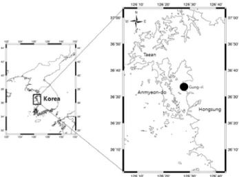
몸체 중앙의 주름부위 (groove) 에서 전편모의 기저부분이 관찰되었고, 후편모는 전편모의 바로 뒤쪽 주름부위에 기저부 분이 위치하고 있었다 (Fig. 4A). 이와 같은 주름은 P.
qugwadi에서도 관찰된 바 있다 (Blackbourn et al., 1998).
본 연구를 통하여 확인된 장편모는 16.34 ± 1.52 μm, 단편 모는 8.25 ± 1.39 μm로 측정되었으며 (Table 1), 전편모의 경우 기존 연구와 비교하여 길며 단편모는 짧은 것으로 나타났 다. 전편모의 경우 P. atlanticus는 12.7 ± 2.4 μm, P.
qugwadi는 9.67 ± 2.07 μm로써 본 조사 결과보다 짧았으 며, 특히 P. chesapeaki의 경우 5.51±1.19 μm로 매우 짧은
Species Host Country
Body length
(μm)
Body width (μm)
Anterior flagellum (μm)
Posterior flagellum
(μm) Method Sources Mean ± SD
Range N
Mean ± SD Range
N
Mean ± SD Range
N
Mean ± SD Range
N P. olseni Ruditapes
philippinarum Korea
3.37 ± 0.33 2.7-4.2
50
1.72 ± 0.22 1.2-2.3
50
16.34 ± 1.52 13.2-21.1
50
8.25 ± 1.39 6-12.4
50
SEM Present study Labyrintho
myxa sp. Macoma
balthica USA
- 3-5
-
- 2-3
-
- 13-17
-
- 7-9
-
SEM Perkins (1968)
P. marinus Crassostrea
virginica USA
- 4-6
-
- 2-3
-
- 10-18
-
- 5-10
-
TEM
Perkins and Menzel
(1996) P.
atlanticus (= olseni)
R. decussatus Portugal 4.5 ± 0.6- 25
2.9 ± 0.4 - 25
12.7 ± 2.4 - 20
10.7 ± 3.2 - 17
SEM/T EM
Azevedo (1989)
P. qugwadi Patinopecten
yessoensis Canada
3.86 ± 0.31 - 7
2.47 ± 0.29 - 7
9.01 ± 1.2 - 5
7.95 ± 1.2 - 2
TEM
Blackboun et al. (1998) P.
chesapeaki Mya arenaria USA
3.73 ± 0.48 2.2-4.7
-
2.41 ± 0.36 1.4-2.5
-
5.51 ± 1.19 - -
3.0 ± 0.65 - -
TEM
Malaughlin et al. (2000) P.
andrewsii Macoma
balthica USA
4.4 ± 0.6 - 10
2.0 ± 0.5 - 10
- - -
- - -
TEM
Coss et al. (2001) P.
mediterran
eus Ostrea edulis Spain 4.4 ± 0.18 - 50
- - -
- - -
- - -
TEM Casas et al. (2004) P.
honshuensi s
Venerupis (R.)
philippinarum Japan
- 5-7
-
- 2-3
-
- - -
- - -
LM
Dungan and Reece
(2006) P. olseni Austrovenus
stutchburyi ZealandNew
- 4.5-6
100
- 2.5-4
100
- - -
- - -
LM Dungan et al. (2007)
P.
beihaiensis C. ariakensis China
- 3-5
-
- - -
- - -
- - -
LM Moss et al. (2008) Table 1. Dimensions of zoospores of P. olseni in the present study and other Perkinsus spp. reported from previous studies.
TEM: transmission electron microscope, LM: light microscope
것이 특징이었다. P. marinus의 경우 본 연구과 유사한 10-18 μm였다. 후편모 (단편모) 의 경우 P. atlanticus는 10.7 ± 3.2 μm로써 본 연구 결과보다 길었으나 P. marinus (6-10 μm) 와 유사하였다. P. chesapeaki의 경우 장편모와 마찬가지로 타 종에 비해 현저히 짧은 후편모 (3.0 ± 0.65 μm) 를 보유하였다.
전편모와 후편모는 각각 basal portion과 apical portion 으로 구성되어 있었다 (Fig. 3). 전편모의 두께는 기저부분이 0.2 μm였으며 basal portion에서는 이 같은 두께가 일정하 게 유지되다 말단으로부터 0.4 μm 앞 지점에서 시작된 apical portion에서 편모의 두께가 급격히 감소한 후 종결되
었다 (Fig. 3). 후편모의 경우 몸체와 붙은 기저부를 포함하는 basal portion은 전편모와 동일한 두께를 나타냈으나, 후편모 의 경우 apical portion은 약 3 μm로 전편모에 비해 다소 길었으며 완만히 감소하다가 종결되었다. 이는 Perkins (1968) 가 보고한 바와 매우 유사한 결과로써 P. marinus의 경우에도 전편모의 apical portion이 후편모의 그것보다 급격 히 종결되는 것으로 보고되었다. Azevedo (1989) 역시 P.
atlanticus의 유주자도 본 연구 결과와 매우 유사한 형태를 갖고 있음을 보고하였으나 후편모의 apical portion이 약 6.7 μm로 전체 후편모 길이의 약 67%를 차지하였으나 본 연구 에서 확인된 P. olseni의 경우 약 40% 이내로 그 비율이 낮았
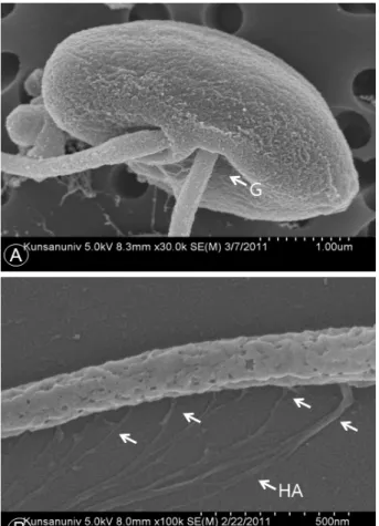
Fig. 4. SEM micrographs of the body (A) showing the insertion point of the flagella, and anterior flagellum with mastigonemes consisting of spur-like structure (arrows) and hair-like appendages (HA). G, groove
Body width Anterior flagellum Posterior flagellum
Body length * ** **
Body width
Anterior flagellum *
*: p<0.05, **: p<0.01 Table 2. Correlations among zoospore body size, anterior and posterior flagella.
다. 유주자 몸체의 장축과 단축, 장편모와 단편모의 길이에 대 한 상관관계를 분석한 결과 체장은 체폭, 장편모 및 단편모와 양의 상관관계를 나타냈으며, 장편모는 단편모와 양의 상관관 계를 나타내었다 (Table 2).
전편모에서 확인되는 편모털은 편모상에 분포하는 미세한 털로 편모의 표면적을 높여 충체의 추진력을 향상시키는 역할 을 하는 것으로 알려져 있다 (Ringo, 1967; Bouck, 1972).
본 연구에서 관찰된 P. olseni 전편모의 경우 다수의 편모털이
분포하고 있었으며 편모털은 편모의 한쪽 면에서만 관찰되었 다 (Fig. 4B). 편모털의 평균 길이는 1.375 ± 0.103 μm 였 으며, spur와 같은 형태의 기저부분이 0.21 ± 0.11 μm 간 격으로 배열되어 있었고 여기에 다수의 미세한 편모털이 부착 된 형태를 띠고 있었다. 현재까지 P. olseni (Ordas and Figueras, 1998), P. qugwadi (Blackbourn et al., 1998) 및 Perkinsus sp. (Perkins, 1968; Coss et al., 2001)에서 편모털이 확인된 바 있으며, 이들 중 P. qugwadi와 Macoma balthica에 기생하는 Perkinsus sp.의 경우 본 조사에서 관 찰된 바와 같이 편모털에 spur 및 미세한 편모털이 부착됨을 보고하였다.
본 연구를 통하여 우리나라 서해안 및 남해안 일대의 바지 락에 기생하는 P. olseni 유주자의 미세구조를 관찰하였다. 특 히 주사전자현미경을 이용한 유주자 외부구조에 관한 관찰은 아시아 권역에 분포하는 P. olseni 경우 최초의 보고로써 그 의의가 있다. 연구결과 우리나라에서 확인된 P. olseni 유주자 의 외형적 특징은 미국과 유럽에서 검출되는 타종 (他種) 과 비교하여 소형이며 동일종인 유럽과 뉴질랜드의 P. olseni와 비교하여도 몸체가 작고 전편모의 길이가 현저히 긴 것으로 나 타났다. 또한 전편모의 basal portion : apical portion의 비 율 역시 유럽에서 확인된 P. olseni와 차이가 있음이 확인되었 다. 그러나 편모털의 존재와 편모털의 구조 및 편모 기저부의 형태는 타 연구 결과와 상당히 유사한 것으로 조사되었다. 이 러한 형태적 특성은 지리적 원근성 및 숙주의 차이에 의한 기 생충-숙주간 상호작용의 변화에 의한 결과로 판단된다.
요 약
Perkinsus spp.는 국내를 비롯한 전 세계에 걸쳐 수산업적 으로 중요한 이매패류에 감염되어 대량폐사를 유발하는 대표 적 기생충이다. 본 연구에서는 바지락에서 검출되는 P. olseni 의 유주자를 광학현미경과 주사전자현미경을 이용해 미세구조 를 관찰한 결과 P. olseni의 유주자는 타원형의 몸체에 1개의 장편모와 1개의 단편모로 구성되어 있었으며, 장편모에는 섬 모털을 보유하고 있었으며, 섬모털은 편모의 한쪽 면에만 분포 하고 있었다. 유주자의 평균 체장은 3.37 ± 0.33 μm, 체폭 은 1.72 ± 0.22 μm 이었으며, 장편모는 16.34 ± 1.52 μ
m, 단편모는 8.25 ± 1.39 μm으로 측정되었다. 이는 유럽과 미국에서 보고된 Perkinsus spp.와 비교 하였을 때 몸체의 크기는 작지만 편모의 길이는 긴 것으로 확인되었다.
사 사
본 연구는 한국연구재단의 일반연구 (2010-0013304) 에 대한 연구비 지원으로 이루어졌습니다.
References
Andrews, J.D. (1996) Histology of Perkinsus marinus, a pathogen of oysters in Chesapeake Bay 1950-1984.
Journal of Shellfish Research, 15(1): 13-16.
Auzoux-Bordenave, S., Vigario, A.M., Ruano, F., Domart-Coulon, I. and Doumenc, D. (1995) In vitro sporulation of the clam pathogen Perkinsus atlanticus (Apicomplexa, Perkinsea) under various environmental conditions. Journal of Shellfish Research, 14: 469-475.
Azevedo, C. (1989) Fine structure of Perkinsus atlanticus n. sp. (Apicomplexa, Perkinsea) parasite of the clam Ruditapes decussatus from Portugal.
Journal of Parasitology, 75(4): 627-635.
Blackbourn, J., Bower, S.M. and Meyer, G.R. (1998) Perkinsus qugwadi sp. nov. (incertae sedis), a pathogenic protozoan parasite of Japanese scallops, Patinopecten yessoensis, cultured in British Columbia, Canada. Canadian Journal of Zoology, 76: 942-953.
Bouck, G.B. (1972) Architecture and assembly of mastigonemes and related structures. In; Advances in Cell and Molecular Biology. (ed. by Dupraw E.
J.). Vol 2: pp. 237-271. Academic Press Incorporated, New York.
Burreson, E.M. and Ragone Calvo, L.M. (1996) Epizootiology of Perkinsus marinus disease of oyster in Chesapeake Bay, with emphasis on data since 1985. Journal of Shellfish Research, 15: 17-34.
Casas, S.M., Grau, A., Reece, K.S., Apakupakul, K., Azevedo, C. and Villalba, A. (2004) Perkinsus mediterraneus n. sp. a protistan parasite of the European flat oyster Ostrea edulis from the Balearic Islands, Mediterranean Sea. Diseases of Aquatic Organisms, 58: 231-244.
Choi, K.-S. and Park, K.-I. (1997) Report on occurrence of Perkinsus sp. in the Manila clam, Ruditapes philippinarum, in Korea. Journal of Aquaculture, 10: 227-237.
Choi, K.-S., Park, K.-I., Cho, M. and Soudant, P. (2005) Diagnosis, pathology, and taxonomy of Perkinsus sp.
isolated from the Manila clam Ruditapes philippinarum in Korea. Journal of Aquaculture, 18:
207-214.
Coss, C.A., Robledo, J.A., Ruiz, G.M. and Vasta, G.R.
(2001) Description of Perkinsus andrewsi n. sp.
isolated from the Baltic clam (Macoma balthica) by
characterization of the ribosomal RNA locus, and development of species-specific PCR-based diagnostic assay. Journal of Eukaryotic Microbiology, 48: 52-61.
Dungan, C.F. and Reece, K.S. (2006) In vitro propagation of two Perkinsus spp. Parasites from Japanese Manila clam Venerupis philippinarum and description of Perkinsus honshuensis n. sp. Journal of Eukaryotic Microbiology, 53: 316-326.
Hamaguchi, M., Suzuki, N. and Usuki, H. (1998) Perkinsus protozoan infection in short-necked clam Tapes (= Ruditapes) philippinarum in Japan. Fish pathology, 33: 473-480.
Lester, R.J.G. and Davis, G.H.G. (1981) A new Perkinsus species (Apicomplexa, Perkinsea) from the abalone, Haliotis ruber. Journal of Invertebrate Pathology, 37: 181-187.
Liang, Y.B., Zhang, X.C., Wang, L.J., Yang, B., Zhang, Y.
and Cai, C.L. (2001) Prevalence of Perkinsus sp. in the Manila clam, Ruditapes philippinarum, along the Northern coast of the Yellow Sea in China.
Chinese Journal of Oceanologia et Limunologia Sinica, 32: 502-511.
Mackin, J.G., Owen, H.M. and Collier, A. (1950) Preliminary note on occurrence of a new protistan parasite, Dermocystidium marinum n. sp., in Crassostrea virginica (Gmelin). Science, 111:328-329.
McLaughlin, S.M., Tall, B.D., Shaheen, A., Elsayed, E.
and Faisal, M. (2000) Zoosporulation of a new Perkinsus species isolated from the gills of the softshell clam Mya arenaria. Parasite, 7: 115-122.
Moss, J.A., Xiao, C.F., Dungan, C.F. and Reece, K.S.
(2008) Description of Perkinsus beihaiensis n. sp. a new Perkinsus sp. parasite in oysters of southern China. Journal of Eukaryotic Microbiology, 55:
117-130.
Odars, M.C. and Figueras, A. (1998) In vitro culture of Perkinsus atlanticus, a parasite of the carpet shell clam Ruditapes decussatus. Diseases of Aquatic Organisms, 33: 129-136.
Park, K.-I. and Choi, K.-S. (2001) Spatial distribution of the protozoan parasite Perkinsus sp. found in the Manila clams, Ruditapes philippinarum, in Korea.
Aquaculture, 203: 9-22.
Park, K.-I., Figueras, A. and Choi, K.-S. (2006) Application of enzyme-linked immunosorbent assay (ELISA) for the study of reproduction in the Manila clam, Ruditapes philippinarum: (Mollusca: Bivalvia):
II. Impacts of Perkinsus olseni on clam reproduction. Aquaculture, 251: 182-191.
Park, K.-I., Park, J.-K., Lee, J. and Choi, K.-S. (2005) Use of molecular markers for species identification of Korean Perkinsus sp. isolated from Manila clam, Ruditapes philippinarum. Diseases of Aquatic Organisms, 66: 255-263.
Perkins, F.O. (1968) Fine structure of zoospores from Labyrinthomyxa sp. parasitizing the clam Macoma balthica. Chesapeake Science, 9: 198-208.
Perkins, F.O. (1996) The structure of Perkinsus marinus
(Mackin, Owen and Collier, 1950) Levine, 1978 with comments on taxonomy and phylogeny of Perkinsus sp. Journal of Shellfish Research, 15: 67-87.
Ray, S.M. and Mackin, J.G. (1954) Studies on transmission and pathogenicity of Dermocystidium
marinum. Texas A&M Research Foundation Project 23, Technical Report. 1-11.
Ringo, D.L. (1967) Flagellar motion and fine structure of the flagella apparatus in Chlamydomonas.
Journal of Cell Biology, 33: 543-571.