Received: November 6, 2012 ; Accepted: November 28, 2012
Corresponding author: Min Woo Park
Tel: +82 (61) 690-8984 e-mail: [email protected] 1225-3480/24453
나프탈렌에 노출된 바지락,
Ruditapes philippinarum
의 생존 및 생식지표 변화진영국, 김태익, 명정인, 황형규, 박민우 국립수산과학원 남서해수산연구소
Change of Survival and Reproductive Indicator of the Manila Clam, Ruditapes philippinarum Following Chronic Exposure to
Naphthalene
Young Guk Jin, Tae Ik Kim, Jeong In Myeong, Hyung Kyu Hwang and Min Woo Park Southwest Sea Fisheries Research Institute, NFRDI, Yeosu 556-823, Korea
ABSTRACT
Manila clam, Ruditapes philippinarum (SL; 32.74 ± 2.18 mm, TW; 8.29 ± 1.41 g, N; 450) were exposed to various concentrations of naphthalene for 20 weeks. Exposure concentrations of naphthalene established control, solvent control (ethanol), 30, 60, 90 and 120 μg Nap L-1 following results of 96h acute exposure. After exposure during 20 weeks, survival rate of the manila clam was lowed in 60, 90 and 120 μg Nap L-1 exposure group compared control group. Also, sex ratio of male was higher in 60 μg Nap L-1 exposure group (χ2 = 5.492, P < 0.05) but lowed 90 μg Nap L-1 exposure group (χ2 = 4.214, P < 0.05) and 120 μg Nap L-1 exposure group (χ2 = 30.118, P < 0.05).
Gonad development was delayed in female (> 60 μg Nap L-1) and male (> 30 μg Nap L-1). Intersex was 16.28% in female, 1.68% in male. In this result, naphthalene caused survival decrease, imbalance of sex ratio, delay of gonad development of the manila clam. Also, chronic exposure to naphthalene suggested abnormal effects in reproduction of the manila clam.
Key words: Manila clam, naphthalene, survival, reproductive indicator
서 론
다환방향족탄화수소 (Polycyclic Aromatic Hydrocarbons:
PAHs) 는 Naphthalene, Fluorene, Pyrene 등과 같은 물질 을 포함하고 있으며, 해수, 저질, 해양생물 등 다양한 해양환경 구성원들에게서 쉽게 발견되지만, 그 독성으로 인해 환경오염 의 중요한 요인으로 인식되고 있다 (Zitko, 1971; Yunker et al., 2002). 그 중 2007년 발생한 허베이스피리트호 원유유출 사고와 같이 PAHs를 포함하고 있는 물질들의 인위적인 환경 오염은 다양한 생물들에게 치명적인 영향을 미친다 (Bingham and Falk, 1969).
원유 유출지역에 영향을 미치는 PAHs의 구성성분 및 농도 는 원유의 생산지, 유출형태, 유출 양 및 기간, 온도 및 기상상 태, 지리학적 위치, 노출된 생물상의 종류 등 다양한 요소들에 따라 매우 상이하게 나타난다 (Clark and Finley, 1977;
Sanborn, 1977). 하지만 여러 가지 노출조건에도 불구하고 PAHs는 다양한 생물들에게 사망률 증가, 성장감소, 유전학적 손상 등의 악영향을 미치는 것으로 보고되고 있다 (Brown et al., 1996; Carls et al., 1999).
특히 원유유출시 회피능력을 가진 유영성 어류들과 달리 이 매패류를 비롯해 연안에 서식지를 가진 해양무척추동물들은 조류의 영향, 서식지, 회피운동 등에 있어 최악의 상황에 노출 되어 있으며, 생존율과 성장 감소 뿐 만 아니라 형태학적 기형 증가 (Wu and Zhou, 1992; Stekoll et al., 1996), 유생발 달 저해 (Hamdoun et al., 2002) 및 생식소발달을 지연시키 는 원인이 된다 (Cajaraville et al., 1992).
여러 가지 여건에 따라 다르지만 일반적으로 PAHs의 구성 성분 중 naphthalene이 가장 많이 포함되어 있다 (Pollino
et al., 2009). 나프탈렌은 광에 의한 산화가 빠르고 휘발성이 강해 잔류성은 낮지만, 유류의 수용성 성분 (Water Soluble Fraction: WSF) 중 독성이 매우 강해 생물들에게 치명적인 영향을 미치는 것으로 보고되고 있다 (Vijayavel and Balasubramanian, 2006).
하지만 나프탈렌이 해양생물에게 미치는 영향에 대한 연구 는 원유유출의 위험성을 항상 가지고 있는 국내의 경우 일부 어종을 제외하고 (Lee and Ryu, 2011) 매우 빈약하며, 특히 연안정착성 이매패류에 대해서는 그 보고를 찾아보기가 힘든 실정이다. 그 중에서도 생식에 대한 연구는 생태계 보전 및 소 득 증가를 위해서도 꼭 필요하다.
따라서 본 연구는 PAHs 중에서도 높은 독성을 가진 나프탈 렌이 바지락, Ruditapes philippinarum의 생존, 성비 및 생 식소발달에 어떠한 영향을 미치는지 조사하여 갈수록 감소하 는 자원량을 회복하고 해양생태계를 관리하는데 기초자료를 제공하고자 하였다.
재료 및 방법
1. 재 료
본 연구에 사용된 바지락, Ruditapes philippinarum은 각장 32.74 ± 2.18 mm, 전중 8.29 ± 1.41 g 1500 개체를 전라남도 고흥군에서 2010년 9월에 채집하여 주사기를 이용 한 Germ Cell Aspiration (GCA) 방법 (Lee et al., 2010) 으로 성 (암: 798개체, 수: 702개체) 을 확인하였다. 그 후 암 컷과 수컷을 분리수용한 후 생식소의 발달단계가 비활성기가 될 때까지 실내 수조에서 사육하였다.
2. 실험방법
1) 실험기간 및 개체수
실험기간은 2011년 2월 15일부터 7월 5일 까지 20주 였 다. 실험 개시 전 분리수용 후 사육중인 바지락 10개체를 무작 위로 선별하여 조직표본을 제작한 다음 비활성기의 생식소 발 달단계를 확인하였다. 실험 개체수는 암컷과 수컷 각각 450개 체로 실험구 당 암·수 25개체씩 1:1의 비율로 설정하였다.
2) 수온 및 먹이공급
실험기간 동안 수온은 자연수온에 의존하였다. 수온은 실험 개시일에 평균 6℃였으며, 실험기간 동안 계속 상승하여 실험 종료일인 7월 5일에는 평균 21℃를 기록하였다. 먹이는 Isochrysis galbana, Chactoceros calcitrans, Tetraselmis tetrathele 3종의 미세조류를 혼합하여 5,000-6,000 cells/mL을 매일 오전 11시에 공급하였다.
3) 시험용액 및 실험구
본 실험에 사용된 시험용액은 나프탈렌 (99% pure, Sigma Chemicals Co.) 을 사용하였으며, 생물에 대한 영향을 최소 화하기 위해 에탄올에 녹인 후 (DiMichele and Taylor, 1978) 증류수로 1 g Nap L-1 표준용액을 만든 다음, 실험농 도별로 희석하여 조제하였다.
노출구의 농도는 96h 급성노출 실험 (LC50: 423.97 μg Nap L-1) 후 장기간 노출임을 감안해 control, solvent control (ethanol), 30, 60, 90, 120 μg Nap L-1를 실험구 로 하였다.
실험은 30 L의 지수형 순환여과식 사각 유리수조를 이용하 여 3반복으로 진행하였으며, 매일 오전 11시에 5 L의 사육수 를 먹이와 섞어 교환하였다. 또한 사육수의 증발을 최소화하기 위해 유리수조 윗면을 랩으로 밀봉하였다.
4) 생존율
생존율은 매일 사망개체를 판별하여 전체 개체수에서 누적 사망개체수를 뺀 값을 생존율로 환산하였다.
5) 성비
성비는 실험종료일에 생존개체를 모두 채집하여 조직학적 방법에 의해 제작된 조직표본을 image measurement system (FOCUS technology, 2005) 이 부착된 광학현미경 (Olympus, CX31) 으로 관찰하여 확정하였다. 또한 암컷과 수컷의 개수를 χ2 (Chi squared) test를 하여 개체군 성비 의 유의성 여부를 판정하였다.
6) 비만도
비만도 (Condition Index: CI) 는 확정된 성을 기준으로 암·수로 나누어 (육중량 (g) × 1000) / 각장3 (mm) 의 식으 로 계산하였다.
7) 생식소 발달단계
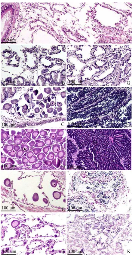
생식소 발달단계는 제작된 조직표본을 이용하여 생식소의 조직상을 관찰한 후 생식세포의 발달정도를 토대로 암·수 모두 비활성기 (Inactive stage: In), 초기활성기 (Early active stage: Ea), 후기활성기 (Late active stage: La), 성숙기 (Ripe stage: R), 산란기 (Spawning stage: S) 및 퇴화흡수 기 (Degeneration: D) 로 나누었으며 (Fig. 1), 그 결과를 백 분율 (%) 로 표시하였다.
8) 생식소지수
생식소지수는 구분된 생식소 발달단계를 토대로 Eversole (1997) 의 방법을 참고하여 암·수로 나눈 후 각 발달단계별 상 수 (In= 0, Ea= 1, La= 2, R= 4, S= 3.5, D= 3) 를 곱
Fig. 1. Gonad development of the manila clam, Ruditapes philippinarum. A:
Inactive stage, B: Early active stage of female, C: Late active stage of female, D: Ripe stage of female, E: Spawing stage of female, F:
Degenerative stage of female, G: Early active stage of male, H: Late active stage of male, I: Ripe stage of male, J: Spent stage of male, K:
Degenerative stage of male. Oc: oocyte.
40 60 80 100
1W 4W 8W 12W 16W 20W
Exposure time (Weeks)
Survival (%)
Control Solvent control
30 ㎍ Nap L¯¹ 60 ㎍ Nap L¯¹ 90 ㎍ Nap L¯¹ 120 ㎍ Nap L¯¹
Fig. 2. Survival rate of the manila clam, Ruditapes philippinarum following exposure to naphthalene during 20 weeks.
0 0.4 0.8 1.2 1.6
Control Solvent control
30 ㎍ Nap L¯¹
60 ㎍ Nap L¯¹
90 ㎍ Nap L¯¹
120 ㎍ Nap L¯¹ Exposure group
Sex ratio
Female Male
Fig. 3. Sex ratio of the manila clam, Ruditapes philippinarum following exposure to naphthalene during 20 weeks.
한 전체의 합을 개체수로 나누었다.
9) Intersex
제작된 조직표본을 광학현미경으로 관찰하여 암컷과 수컷의 생식세포가 동시에 존재하는 조직상을 Intersex로 판별하였으 며, 그 출현율을 백분율 (%) 로 표시하였다.
10) 조직학적 방법
채집된 개체들은 각장, 각고, 각폭, 전중 및 육중을 측정하 였고, 내장낭 부분을 절취하여 Drury and Wallington (1980) 의 방법에 따라 Bouin's solution에 24시간 고정한 후 48시간 이상 수세하였다. 그 후 파라핀 포매법을 거친 다음 microtome으로 4-6 ㎛로 연속절편 하여 조직표본을 제작하 였다. 제작된 조직표본은 Mayer's hematoxylin-eosin (HE) 비교염색을 실시하여 광학현미경으로 관찰하였다.
11) 통계분석
대조구와 노출구간의 차이는 SPSS 10.0 (SPSS, Inc., Chicago, IL) 통계프로그램을 이용하여 일원배치 분산분석 후 사후검정을 하여 그 유의성 여부를 검증하였다.
결 과
1. 생존율
20주 동안 나프탈렌의 다양한 농도에 노출된 바지락의 생존 율은 Fig. 2와 같이 대조구, solvent 대조구 및 30 μg Nap L-1의 경우 노출 종료일에 각각 86.7%, 87.3%, 83.3%로 대 조구와 비교해 별다른 차이가 나타나지 않았다. 하지만, 60 μ g Nap L-1에 노출된 바지락은 노출 12주에 90.7%에서 노출 16주에 79.5%로 급격히 감소하였으며, 노출 종료일인 20주에
68.0%를 나타냈다. 90 μg Nap L-1와 120 μg Nap L-1에 노출된 바지락은 노출 16주에 각각 82.6%와 74.5%를 나타냈 으며, 노출 20주에는 각각 74.0%와 64.7%를 보였다.
2. 성 비
노출 종료 후 성비를 관찰한 결과 Fig. 3과 같이 대조구, solvent 대조구 및 30 μg Nap L-1에 노출된 개체들은 암컷 과 수컷의 비율이 1:0.85 이상으로 암컷이 약간 우세하게 나 타났지만 유의적인 차이는 없었다 (P > 0.05). 하지만, 60 μ g Nap L-1에 노출된 개체들은 암컷과 수컷의 비율이 1:1.37 로 수컷의 비율이 우세하게 나타났다 (χ2 = 5.492, P <
0.05). 90 μg Nap L-1에 노출된 개체들은 암컷의 비율이 높 게 나타났으며 (χ2 = 4.214, P < 0.05), 가장 높은 농도인 120 μg Nap L-1에 노출된 개체들 역시 암컷의 비율이 높은 가운데 가장 많은 차이를 보였다 (χ2 = 30.118, P < 0.05).
3. 비만도
20주 동안 나프탈렌의 다양한 농도에 노출된 바지락의 비만 도는 대조구와 비교해 암컷의 경우 유의적인 차이는 없었다 (P > 0.05). 수컷의 경우 대조구 (0.026) 와 비교해 solvent 대조구, 30, 60 및 120 μg Nap L-1에 노출된 개체들은 별다 른 차이를 보이지 않았지만 (P > 0.05), 60 μg Nap L-1에 노출된 개체들은 0.033으로 높게 나타났다 (P < 0.05) (Fig.
4).
4. 생식소 발달단계
바지락 암컷의 생식소 발달단계는 Fig. 5와 같이 대조구의 경우 휴지기, 산란기 및 퇴화흡수기의 조직상을 보이고 있었으 며, 산란기 및 퇴화흡수기의 비율이 87.5%를 차지하고 있었
**
0 0.01 0.02 0.03 0.04 0.05
Control Solvent control
30 ㎍ Nap L¯¹
60 ㎍ Nap L¯¹
90 ㎍ Nap L¯¹
120 ㎍ Nap L¯¹ Exposure group
CI
Female Male
Fig. 4. Condition index of the manila clam, Ruditapes philippinarum following exposure to naphthalene during 20 weeks. Vertical bar: SD. **: significantly different from male control (P < 0.05).
0 20 40 60 80 100
Control Solvent control
30 ㎍ Nap L¯¹
60 ㎍ Nap L¯¹
90 ㎍ Nap L¯¹
120 ㎍ Nap L¯¹ Exposure group
Frequency (%)
In Ea La R S D
Fig. 5. Gonad development of the female manila clam, Ruditapes philippinarum following exposure to naphthalene during 20 weeks. In: inactive stage, Ea:
early active stage, La: late active stage, R: ripe stage, S:
spawning stage, D: degeneration stage.
0 20 40 60 80 100
Control Solvent control
30 ㎍ Nap L¯¹
60 ㎍ Nap L¯¹
90 ㎍ Nap L¯¹
120 ㎍ Nap L¯¹ Exposure group
Frequency (%)
In Ea La R S D
Fig. 6. Gonad development of the male manila clam, Ruditapes philippinarum following exposure to naphthalene during 20 weeks. In: inactive stage, Ea:
early active stage, La: late active stage, R: ripe stage, S:
spent stage, D: degeneration stage.
* ** *
0 1 2 3 4 5
Control Solvent control
30 ㎍ Nap L¯¹
60 ㎍ Nap L¯¹
90 ㎍ Nap L¯¹
120 ㎍ Nap L¯¹
Exposure group
GI
Female Male
Fig. 7. Gonad index (GI) of the manila clam, Ruditapes philippinarum following exposure to naphthalene during 20 weeks. Vertical bar: SD. *, **: significantly different from control (*: female, **: male, P < 0.05).
다. solvent 대조구와 30 μg Nap L-1에 노출된 개체들은 대 조구와 비교해 별다른 차이를 보이지 않았다. 60 μg Nap L-1 에 노출된 개체들은 대조구와 비교해 비활성기의 비율이 다소 높았으며, 산란기의 조직상을 가진 개체들이 높게 나타났다.
90과 120 μg Nap L-1에 노출된 개체들은 휴지기의 조직상 을 가진 개체들은 관찰되지 않았으며, 대조구와 비교해 산란기 와 성숙기의 조직상을 가진 개체들이 농도 의존적으로 높게 나 타났다.
수컷의 생식소 발달단계는 Fig. 6과 같이 암컷과 마찬가지 로 대조구의 경우 휴지기, 산란기 및 퇴화흡수기의 조직상이 관찰되었다. solvent 대조구는 대조구에 비해 별다른 차이를
보이지 않았다. 30 μg Nap L-1에 노출된 개체들은 대조구에 비해 휴지기의 조직상을 가진 개체들의 비율이 높게 나타났으 며, 성숙기에서 퇴화흡수기에 이르는 다양한 발달상을 보이고 있었다. 60 μg Nap L-1에 노출된 개체들은 휴지기, 성숙기 및 산란기의 발달상을 보이는 가운데 주로 산란기의 조직상을 가진 개체들이 주로 관찰되었다. 90과 120 μg Nap L-1에 노 출된 개체들은 성숙기와 산란기의 조직상을 보이고 있었다.
5. 생식소지수
바지락의 생식소지수는 Fig. 7과 같이 암컷의 경우 대조구 (2.75) 와 비교해 solvent 대조구, 30 및 60 μg Nap L-1에
Fig. 9. Photomicrographs showing gonad sections of the manila clam, Ruditapes philippinarum following exposure to naphthalene during 20 weeks. A: Intersex photomicrographs in male manila clam. B: Intersex photomicrographs in female manila clam. Oc: oocyte, Sz: spermatozoa.
0 2 4 6 8 10
Control Solvent control
30 ㎍ Nap L¯¹
60 ㎍ Nap L¯¹
90 ㎍ Nap L¯¹
120 ㎍ Nap L¯¹ Exposure group
Intersex (%)
Fig. 8. Intersex of the manila clam, Ruditapes philippinarum following exposure to naphthalene during 20 weeks.
노출된 개체들은 각각 2.79, 2.81 및 2.6으로 별다른 차이를 보이지 않았지만 (P > 0.05), 90과 120 μg Nap L-1에 노출 된 개체들은 각각 3.45와 3.68로 높게 나타났다 (P < 0.05).
수컷의 경우 대조구와 비교해 solvent 대조구 및 30 μg Nap L-1에 노출된 개체들은 다소 감소하였고, 60 μg Nap L-1 이 상의 농도에 노출된 개체들은 증가하는 경향을 보였다. 그 중 90 μg Nap L-1에 노출된 개체들은 대조구와 유의적인 차이 를 나타냈다 (P < 0.05).
6. Intersex
노출 종료 후 바지락의 생식소에서 관찰된 intersex는 Fig.
8과 같이 60 μg Nap L-1에 노출된 개체들 중 최종 생존한 102 개체들에서 7.84%의 비율로 나타났다. 그 중 암컷 43개 체에서 7개체로 16.28%, 수컷 59개체에서 1개체로 1.68%를
나타내 암컷에서의 intersex 발현율이 높게 관찰되었다. 관찰 된 intersex 조직상은 암컷의 경우 대부분 방란기 또는 퇴화 흡수기의 조직상을 보이고 있었으며, 수컷의 경우 성숙기에서 퇴화흡수기의 조직상까지 다양한 단계가 관찰되었다 (Fig. 9).
고 찰
나프탈렌이 해양생물의 생존에 미치는 영향농도는 종, 생애 주기, 노출기간 등 여러 가지 여건에 따라 매우 다양하게 나타 난다. 해양생물들에 대한 나프탈렌의 96h LC50은 pink salmon, Oncorhynchus gorbuscha의 경우 1200 μg Nap L-1 (Moles and Rice, 1983), marine shrimp, Pandalus goniurus의 경우 12℃에서 971 μg Nap L-1, 4℃에서 2160 μg Nap L-1 (Korn et al., 1979), Dungeness crab, Cancer magister의 경우 2000 μg Nap L-1 (Caldwell et al., 1997), Pacific oyster, Crassostrea gigas의 경우 199 mg Nap L-1 이다 (LeGore, 1974). 장기간 노출 시 pink salmon, Oncorhynchus gorbuscha은 40일 동안 최소 120 μg Nap L-1부터 생존에 영향을 받으며 (Moles and Rice, 1983), manila clam, Tapes semidecussatus의 경우 40 μ g Nap L-1 가 축적된 저질에서 21일 동안 노출 후 43%의 낮 은 생존율을 나타냈다 (Byrne and O'Halloran, 2004).
본 연구에서 96h LC50은 423.97 μg Nap L-1 였으며, 20 주의 장기간 노출에서는 60 μg Nap L-1 이상의 농도에서 생 존율에 영향을 받는 것으로 나타났다. 또한, 60 μg Nap L-1 이상의 농도에서 수온이 상승하는 시기에 급감하는 생존율의 감소는 수온 상승에 따른 영향으로 보인다 (Clark and Finley, 1977; Korn et al., 1979).
해양환경에 노출된 다양한 오염물질들은 영향 물질 및 농도 에 따라 이매패류의 성비, 생식소발달, 생식소지수 등 생식에
영향을 준다 (Gagne et al., 2005; Matozzo and Marin, 2005; Ortiz-Zarragoitia and Cajaraville, 2010). TBT 오 염지역인 덴마크의 Odense 만에 서식하는 Mya arenaria의 경우 암컷과 수컷의 성비가 1:1.8로 수컷의 비율이 높게 나타 났다 (Gagne et al., 2005). 또한 crude oil은 0.5 mg L-1의 농도에서 blue mussels, Mytilus edulis 암컷의 생식소지수 를 감소시키며, 에스트로겐 유사기능 및 생식소 조직 내에 다 양한 영향을 미친다 (Cooley, 1977; Aarab et al., 2004;
Ortiz-Zarragoitia and Cajaraville, 2010 ).
본 연구에서 60 μg Nap L-1에 노출된 바지락의 성비가 수컷이 높은 반면, 90과 120 μg Nap L-1에 노출된 바지락의 경우 암컷이 높은 결과는 나프탈렌이 이매패류에서 비활성기 후 성의 표현에 영향을 미치는 것으로 보인다. 연구된 결과에 서 암컷은 60 μg Nap L-1이상, 수컷은 30 μg Nap L-1이상 의 농도에 노출된 개체들에서 생식소발달이 지연되는 결과를 보였다. 암컷과 수컷의 생식소발달에 영향을 미치는 나프탈렌 의 영향농도는 그 보고를 찾아보기가 쉽지 않아 직접적인 결론 을 내리기는 어렵지만, crude oil에 노출된 이매패류에서 나타 난 결과들을 볼 때 (Ortiz-Zarragoitia and Cajaraville, 2010), 나프탈렌이 생식소발달에 영향을 미치는 것은 확실해 보인다.
일반적으로 내분비계장애물질은 해양생물에게 intersex를 유발하는 것으로 알려져 있으며, 특히 PAHs 등 원유 오염을 포함해 생태계 환경이 악화되면 그 비율이 증가한다 (Morcillo and Porte, 2000; Chesman and Langston, 2006; Ortiz-Zarragoitia and Cajaraville, 2010). 실제로 원유에 오염된 지역에 서식하는 mussels, Mytilus galloprovincialis의 경우 26%의 높은 intersex 출현율이 보 고되었으며, 암컷의 생식소에서 수컷의 생식세포가 발현된 개 체들의 비율이 높게 나타났다 (Ortiz-Zarragoitia and Cajaraville, 2010).
본 결과에서 나타난 intersex 역시 암컷에서 수컷의 생식세 포가 발현된 개체들의 비율이 높았지만, 일부 수컷에서 암컷의 생식세포를 가진 개체가 나타나는 것을 볼 때 나프탈렌은 바지 락의 생식에 비정상적인 영향을 주는 것으로 보인다.
요 약
나프탈렌이 바지락 (각장 32.74 ± 2.18 mm, 전중 8.29 ± 1.41 g) 의 생존, 성비 및 생식소발달에 미치는 영향에 대해 연구하였다. 노출농도는 대조구, solvent 대조구 (에탄올), 30, 60, 90, 120 μg Nap L-1 였으며, 노출기간은 20주였다. 노 출 종료 후 바지락의 생존율은 60 μg Nap L-1 이상의 농도 에서 감소하였다. 성비는 대조구와 비교해 solvent 대조구 (에 탄올)와 30 μg Nap L-1 실험구는 별다른 차이를 보이지 않
았지만, 60 μg Nap L-1 실험구의 경우 수컷의 비율이 높았 다. 그 후 농도가 높아질수록 수컷의 비율이 감소하였다. 생식 소 발달단계는 암컷의 경우 60 μg Nap L-1 이상의 농도에서, 수컷의 경우 30 μg Nap L-1 이상의 농도에서 발달이 지연되 는 결과를 보였다. 생식소에서 관찰된 intersex는 암컷에서 16.28%, 수컷에서 1.68%를 나타냈다. 이러한 결과들을 볼 때 나프탈렌은 바지락의 생존 뿐 만 아니라 성비와 생식에 영향을 주는 것으로 보인다.
REFERENCES
Aarab, N., Minier, C., Lemaire, S., Unruh, E., Hansen, P.D., Larsen, B.K., Andersen, O.K. and Narbonne, J.F. (2004) Biochemical and histological responses in mussel, Mytilus edulis exposed to North Sea oil and to a mixture of North Sea oil and alkylphenols.
Marine Environmental Research, 58: 437-441.
Bingham, E. and Falk, H.L. (1969) The modifying effect of carcinogens on the threshold response. Archives of Environmental Health, 19: 779-783.
Brown, E.D., Baker, T.T., Hose, J.E., Kocan, R.M., Marty, G.D., McGurk, M.D., Norcross, B.L. and Short, J.W. (1996) Injury to the early fife history stages of Pacific herring in Prince William Sound after the Exxon Valdez oil spill. American Fisheries Society Symposium, 18: 448-462.
Byrne, P.A. and O'Halloran, J. (2004) The impact of ballast water effluent on the manila clam, Tapes semidecussatus. Ecotoxicology, 13: 311-322.
Caldwell, R.S., Caldarone, E.M. and Mallon, M.H.
(1997) Effects of a seawater-soluble fraction of Cook Inlet crude oil and its major aromatic components on larval stages of the Dungeness crab, Cancer magister Dana. In: Wolfe, D.A. (ed.) Fate and effects of petroleum hydrocarvons in marine ecosystems and organisms. Pergamon Press, NY. pp. 210-220.
Carls, M., Rice, S. and Hose, J. (1999) Sensitivity of fish embryos to weathered crude oil: I. Low-level exposure during incubation causes malformations, genetic demage, and mortality in larval Pacific herring, Clupea pallasi. Environmental Toxicology and Chemistry, 18: 481-493.
Chesman, B.S. and Langston, W.J. (2006) Intersex in the clam, Scrobicularia plana: a sign of endocrine disruption in estuaries? Biology Letters, 2: 420-422.
Clark, R.C. and Finley, J.S. (1977) Effects of oil spills in Arctic and Subarctic environments. Chapter 9 In:
Malins, D.C. (ed.) Effects of Petroleum on Arctic and Subarctic Marine Organism. Vol II. biological Effects. New York: Academic Press, Inc., pp.
411-476.
Cooley, J.F. (1977) Oil inhibits reproduction in tests on mussels. Maritimes, 21(4): 12-14.
Drury, R.A.B. and Wallington, E.A. (1980) Carleton's Histological Technique. 520 pp. Oxford University Press, Oxford.
Gagne, F., Blaise, C., Pellerin, J., Pelletier, E. and Strane, J. (2005) Health status of Mya arenaria bivalves collected from contaminated sites in Canada and Denmark during their reproductive period.
Ecotoxicology Environmental Safety, 64: 348-361.
Hamdoun, A.M., Griffin, F.J. and Cherr, G.N. (2002) Tolerance to biodegraded crude oil in marine invertebrate embryos and larvae is associated with expression of a multixenobiotic resistance transporter.
Aquatic Toxicology, 61: 127-140.
Korn, S., Moles, D.A. and Rice, S.D. (1979) Effects of temperature on the median tolerance limit of pink salmon and shrimp exposed to tolunce, naphthalene and Cook Inlet crude oil. Bulletin of Environmental Contamination and Toxicology, 21: 521-525.
Lee, J.S., Ju, S.M., Park, J.S., Jin, Y.G., Shin, Y.K. and Park, J.J. (2010) Germ cell aspiration (GCA) method as a non-fatal technique for sex identification in two bivalves. Devlopment and Reproduction, 1: 7-11.
Lee, K.S. and Ryu, H.M. (2011) The acute toxicity of naphthalene on hematologic properties in juvenile flounder, Paralichthys olivaceus. Journal of the Korean Society of Marine Environment and Safety, 17: 191-196.
LeGore, R.S. (1974) The effect of Alaskan crude oil and selected hydrocarbon compounds on embryonic development of the Pacific oyster, Crassostrea gigas.
Ph. D. Thesis, Universe of Washington.
Matozzo, V. and Marine, M.G. (2005) Can 4-nonylphenol induce vitellogenin-like proteins in the clam, Tapes philipinarum?. Environmental Research, 97: 43-49.
Moles, A. and Rice, S.D. (1983) Effects of crude oil and naphthalene on growth, caloric content and fat content of pink salmon juveniles in seawater.
Transactions of the American Fisheries Society, 112:
205-211.
Morcillo, Y. and Porte, C. (2000) Evidence of endocrine disruption in clams, Ruditapes decussata transplanted to a tributylein polluted environment.
Environmental Pollution, 107: 47-52.
Ortiz-Zarragoitia, M. and Cajaraville, M.P. (2010) Intersex and oocyte atresia in a mussel population from the Biosphere's Reserve of Urdaibai (Bay of Biscay). Ecotoxicology and Environmental Safety, 73:
693-701.
Polino, C.A., Georgiades, E. and Hldway, D.A. (2009) Physiological changes in reproductively active rainbow fish, Melanotaenia fluviatilis following exposure to naphthalene. Ecotoxicology and Environmental Safety, 72: 1265-1270.
Sanborn, H.R. (1977) Effects petroleum on ecosystems.
Chapter 6 In: Malins, D.C. (ed.) Effects of Petroleum on Arctic and Subarctic Marine Organisms. Vol II. biological Effects. New York:
Academic Press, Inc. pp. 337-352.
Stekoll, M.S., Deysher, L., Highsmith, R.C., Saupe, S.M., Guo, Z., Erickson, W.P., McDonald, L. and Strickland, D. (1996) Coastal habitat injury assessment: intertidal communities and the Exxon Valdez oil spill. American Fisheries Society Symposium, 18: 177-192.
Vijayavel, K. and Balasubramanian, M.P. (2006) Changes in oxygen consumption and respiratory enzymes as stress indicators in an estuarine edible crab, Scylla serrrata exposed to naphthalene. Chemosphere, 63:
1523-1531.
Wu, J. and Zhou, X.A. (1992) Study of the effects of petroleum on the early development of mussel, Mytilus edulis. Transactions of Oceanology and Lmnology, 2: 46-50.
Yunker, M.B., Macdonald, R.W., Vingarzan, R., Mitchell, R.H., Goyette, D. and Sylvestre, D. (2002) PAHs in the fraser river Basin: a critical appraisal of PAH ratios as indicators of PAH source and composition.
Organic Geochemistry, 33: 489-515.
Zitko, V. (1971) Determination of residual fuel oil contamination of aquatic animals. Bulletin of Environmental Contamination and Toxicology, 5:
559-564.