Received: March 10, 2013; Accepted: March 20, 2013 Corresponding author : Kyung-Il Park
Tel: +82 (63) 469-1882 e-mail: [email protected] 1225-3480/24465
This is an Open Access article distributed under the terms of the Creative Commons Attribution Non-Commercial License with permits unrestricted non-commercial use, distribution, and reproducibility in any medium, provided the original work is properly cited.
바지락 ( Ruditapes philippinarum ) 혈구의 일산화질소 (nitric oxide) 정량
남기웅1, 양현성2, 박경일1
1군산대학교 해양과학대학 수산생명의학과, 2한국해양과학기술원 동해연구소
Quantification of nitric oxide concentration in the hemocytes of Manila clam Ruditapes philippinarum by using
4,5-diaminofluorescein diacetate (DAF-2) detection method
Ki-Woong Nam1, Hyun-Sung Yang2 and Kyung-Il Park1
1Department of Aquatic Life Medicine, Kunsan National University, Gunsan, Jeonbuk 573-701, Republic of Korea
2East Sea Research Institute, Korea Institute of Ocean Science and Technology (KIOST), Uljin, Kyungbuk 767-813, Republic of Korea
ABSTRACT
Nitric oxide (NO) is an important intra-intercellular signaling molecule that regulates many physiological processes and participates in the development some pathological conditions in animals. In this study, we compared different methods for determining NO concentration in the hemocytes of Manila clam Ruditapes philippinarum. For measuring the intracellular NO levels, we used the specific fluorescent probe 4,5-diaminofluorescein diacetate (DAF-2 DA), and the quantification methods that were compared were based on image analysis, spectrophotometry, and flow cytometry. NO concentration could be determined using all the 3 methods, and the concentration varied significantly depending upon the presence of NO regulators in the hemocytes; NO concentration increased in the presence of L-arginine, while it decreased in the presence of N-nitro-L-arginine methyl ester. In particular, it is found that estimation of NO using a flowcytometry is more economical, reliable and accurate compared to image analysis and spectrophotometry. Accordingly we believe that determining NO concentration by using flowcytometry will be useful in evaluating physiological and pathological conditions in marine bivalves.
Key words: nitric oxide, hemocyte, diaminofluorescein-2-diacetate, flow cytometer, Ruditapes philippinarum
서 론
혈구는 해산 연체동물의 면역 반응을 일으키는 중요한 세포 로 자극 시 oxygen radical과 nitrogen radical을 분비하여 생체방어에 이용한다. 척추동물의 경우 nitrogen radical의 일종인 일산화질소 (nitric oxide, NO) 는 주로 대식세포의
효소인 nitric oxide synthase (NOS) 에 의해 L-arginine 에서 합성되어 생체 내에서 cytokine, bacterial lipopolysaccharide (LPS) 또는 기생충 등과 반응하여 항균 작용, 면역조절, 염증유도 등의 역할을 한다 (Palmer et al., 1987; Bogdan, 2001; Aktan, 2004; Menaka et al., 2009). NOS는 isoform인 endothelial NOS (eNOS), inducible NOS (iNOS) 및 neuronal NOS (nNOS) 로 구 성되며, eNOS와 nNOS는 세포내에서 극소량으로 존재하나 (Clancy et al., 1998; see review of Aktan, 2004), iNOS 에 의해 발생하는 NO는 염증유발 물질에 의해 대량으로 발생 한다 (Cattell and Jansen, 1995; Cirino et al., 2006;
Menaka et al., 2009). 무척추동물의 경우 현재까지 iNOS 만 확인된 상태이다 (Imamura et al., 2002).
이매패류의 NO 측정에 관한 연구는 바지락에 LPS를 주사
Quantification of NO concentration
하거나 염증 유발 박테리아인 Vibrio tapetis에 감염시켰을 때, 기생충 감염 시 또는 수질 악화에 따라 혈림프에서 NO 농 도의 증가가 나타남으로써 병원성 물질의 침입이나 환경악화 에 따라 NO 생성의 변화가 측정되고 있다 (Taffala et al., 2003; Gagne et al., 2005 and 2008; Villamil et al., 2007; Jeffroy and Paillard, 2011). 이러한 사실은 NO 생 성이 생물의 생리학적·병리학적 특성에 따라 민감하게 반응하 며 (Aktan, 2004), 따라서 NO량의 변화는 생물의 생리적 특 성을 진단하는데 중요한 인자로 판단된다.
NO는 반감기가 수초에 불과한 기체로 주로 세포내에 존재 하나 일부는 세포외로 유리되어 이산화질소로 산화되거나 아 질산염과 질산염으로 분해된다 (Bryan and Grisham, 2007). 이러한 특성을 이용한 NO 측정은 주로 DAF assay와 Griess assay에 의해 수행되고 있다 (Bevers, 2006). DAF assay는 세포내 존재하는 NO량을 실시간으로 측정하는 경우 에 이용되며, 세포외로 유리된 NO의 누적량을 측정하는 경우 Griess assay가 주로 이용된다 (Lewis et al., 1996:
Nakatsubo et al., 1998). DAF assay는 4,5-Diamino- fluorescein diacetate (DAF-2DA) 가 esterase에 의해 DAF-2 로 변형된 후 세포가 생성하는 NO와 선택적으로 반응하여 Triazolo-fluorescein (DAF-2T) 라는 형광물질이 되는 원리를 이용한 것으로써 세포내에 NO에 의해 생성되는 형광량을 화 상분석 (image analysis) 이나 형광분광기 (fluorescent spectrophotometer), 또는 유세포 분석기(flow cytometer) 를 이용하여 정량할 수 있다 (Kojima et al., 1998, Navarro-Antolin et al., 2001). 한편 Griess assay는 체액 내 NO가 질산염 (NO3-) 으로 산화된 것을 아질산염 (NO2-) 으로 환원 시킨 후 이를 아조화합물로 치환시켜 측정한다.
현재까지 해산 이매패류 혈액의 NO 농도 정량은 대부분 Griess assay를 이용하여 측정되었다. 따라서 본 연구는 DAF assay에 이용되는 화상분석, 형광분석기 및 유세포 분석 기 등 3가지의 측정 방법을 수행하여 비교함으로써 DAF assay를 이용한 해산 이매패류의 NO 농도 측정이 가능한지 확인하고 이를 위한 최적화된 기술을 습득하기 위하여 수행되 었다.
재료 및 방법 1. 시료 및 NO 생성 유발
실험에 사용된 바지락은 경상북도 울진군에서 채집 후 4℃
에서 보관하여 실험실로 운반하였다. 바지락 (평균 각장 약 40 mm, 평균 각고 30 mm) 은 30 liter 수조에서 수온 20℃와 염분 30 psu에서 사육되었으며, 먹이로는 매일 Isochrysis sp. 와 Chlorella sp. 를 1:1로 혼합하여 약 2.14 x 10⁸ cell/l 를 공급하였다.
바지락 후폐각근에서 혈림프액를 추출한 뒤, 3개의 그룹으 로 나누어 1.5 ml tube에 0.4 ml씩 분주하고 Group A (대 조구) 에는 0.4 ml의 marine saline (MS, 12 mM CaCl2· 2H2O, 11 mM KCl, 26 mM MgCl2·6H2O, 45 mM Tris-HCl, 6.45 mM NaCl, pH 7.4) 을 주입하였고, Group B (NOS 저해) 의 경우 혈림프액에 Nω-Nitro-L-arginine methyl ester hydrochloride (L-NAME) 를 주입하여 최종 농도가 10 mM이 되도록 하였으며, Group C의 경우 (NO 생성 유도) L-arginine을 첨가하여 최종 농도를 10 mM로 제 조하였다. 이후 각 tube에 DAF-2DA (Sigma, D2813) 을 투 여하여 최종농도가 5 μM이 되도록 한 다음 20℃에서 30분 간 반응시켰다.
2. 화상분석
상기 1에서 준비된 각 그룹의 혈구 중 20 μl를 슬라이드 글 라스에 놓은 다음 형광현미경 (Olympus BX43) 하에서 이미 지분석프로그램 (Image J, NIH) 을 이용하여 fluorescence brightness value (BV) 값을 측정하였다 (Ferreira and Rasband, 2012). 이때 사진 촬영은 노출값 1,500으로 고정하 였으며, 바지락 1개체 당 50개 혈구 세포의 평균 BV를 바지락 한 개체의 NO 농도로 결정하였다.
3. Fluorescent spectrophotometer
1에서 준비된 각 그룹의 혈구 중 200 μl를 96well 형광 plate에 3회 반복하여 분주하였고 1시간동안 반응시킨 후 fluorescence spectrophotometer (SepectraMAX Gemini XS, Molecular device, USA) 를 이용하여 excitation 490 nm와 emission 520 nm에서 흡광도를 측정하였다.
4. 유세포 분석
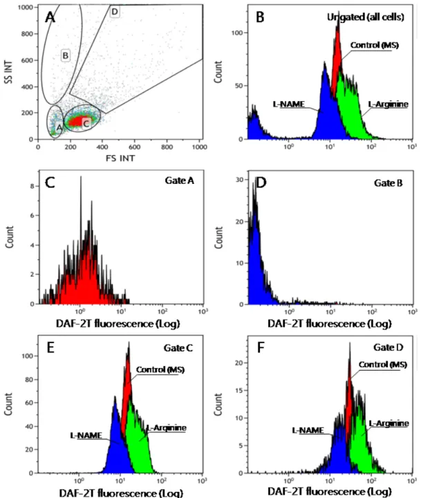
1에서 준비된 각 그룹의 혈구를 선택한 후 혈구 내 NO 생 성량을 유세포 분석기 (Beckman coulter, GiliosTM) 를 이용 하여 측정하였다. 유세포 분석기를 이용한 측정은 100 μl 혈 림프액을 FSC-HLin (gain 2, vlots 106) / SSC-HLin (gain 20, volts 303, Discr. 43) 을 이용하여 density plot 을 작성하였으며, 이때 나타나는 cell sub-population을 세포 크기와 granularity에 따라 4개로 구분하고 각 population의 형광량을 FL1 (gain 2, volts 433) histogram으로 나타냄으 로써 혈구세포에서 나타나는 DAF-2T의 형광값 (relative fluorescence unit, RFU) 을 측정하였다. Data 분석은 Kaluza 1.2 (Beckman coulter) 프로그램을 이용하였다.
5. 통계
각 그룹별 DAF-2T에 의한 NO 농도는 평균 ± SD (표준
Fig. 1. Microscopic observation of diaminotriazolofluorescein (DAF-2T) fluorescence (indicating NO generation) in the hemocytes of Manila clam exposed to NO inducer or inhibitor. Unlike that observed in the control (A, A-1), the fluorescence increased (activated) when the hemocytes were exposed to L-arginine (B, B-1), while it decreased when the hemocytes were treated with L-NAME (C, C-1). The images obtained using a light microscope are in the left lane, and the ones obtained using a fluorescent microscope are in the right lane. Scale bars, 10 μm.
편차)로 나타내었으며, NO 농도는 SPSS12.0 통계분석프로 그램의 one-way ANOVA를 실시 후 Duncan’s multiple range test로 평균 간의 유의성을 95% 신뢰수준에서 확인하 였다.
결과 및 고찰
본 연구는 일산화질소 측정을 위한 DAF-2T 정량에 사용되 는 화상분석, 형광분광기법 및 유세포 분석법 등 3 가지 방법 을 비교함으로써 해산 이매패류의 생리적 특성을 진단하기 위
한 기술 개발을 위하여 수행되었다. 이상의 3가지 분석법은 척 추동물의 NO 정량에 널리 이용되고 있으나 (Yamasaki and Sakihama, 2000; Viani et al., 2001; Raghavan et al., 2003; Kolluru et al., 2006), 해산 이매패류의 경우 대부분 Griess assay가 이용되고 있어 본 연구는 바지락을 대상으로 DAF-2T를 이용한 NO 정량을 최초로 시도하고 최적화 하였 다는데 의의가 있다.
Fig. 1은 DAF-2T에 의해 형광 발색된 혈구를 보여주고 있 다. 대조구 (Group A) 로 사용된 혈구 (A-1) 에 비해 L-arginine에 의해 NO가 활성화된 경우 (Group C) 가장 밝
Quantification of NO concentration
Fig. 2. Determination of NO concentration by using a flow cytometer. A, sub-populations of hemocytes of the Manila clam used for NO quantification; B, Fluorescence histogram of all the hemocytes; C, Fluorescence histogram of SP A; D, Fluorescence histogram of SP B;
E, Fluorescence histogram of SP C; and F, Fluorescence histogram of SP D. (SP, subpopulation)
SP A mean ± SD
SP B mean ± SD
SP C mean ± SD
SP D mean ± SD
All mean ± SD Control 0.262 ± 0.18 0.416 ± 0.223 13.256 ± 2.152 21.380 ± 8.541 6.028 ± 5.313 L-arginine 0.970 ± 0.306 1.466 ± 0.286 24.396 ± 3.483 44.482 ± 9.291 19.028 ± 6.025
L-NAME 0.246 ± 0.138 0.308 ± 0.168 9.838 ± 0.529 19.556 ± 3.900 3.056 ± 2.389 Table 1. Mean relative fluorescence units (RFU) of NO in each subpopulation (SP) of hemocytes exposed to NO regulators. SD,
standard deviation
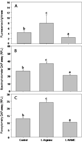
은 형광체를 보여주고 있고 (B-1), L-NAME에 의해 NO 생 성이 저해된 경우 (Group C) 가장 낮은 형광 밝기를 나타내 었다 (C-1). 각 그룹별 바지락 혈구의 형광량을 화상분석을 이 용해 정량한 결과 대조구는 4.273 ± 0.877 BV (Mean ± SD) 이었으나, L-arginine에 의해 활성화 된 경우 7.911 ± 3.20 BV로 증가하였고, 혈구 내 NO 생성이 L-NAME에 의
해 저해된 경우 2.390 ± 0.779 BV로 감소하였다 (Fig. 2A.
p < 0.05). 따라서 NO량은 무처리구 (대조구), NO 활성제 및 NO 저해제의 투여 유무에 따라 다르게 나타남이 화상분석 을 통하여 확인되었다.
형광분광계를 이용하여 각 그룹의 형광량을 측정한 결과 대 조구 (Group A) 의 경우 형광량은 26.551 ± 6.580 RFU 였으며, L-arginine에 의해 활성화된 경우 (Group B) 는 36.797 ± 6.673 RFU로 증가하였으나, 반대로 L-NAME에 의해 NO 생성이 저해된 실험구 (Group C) 는 21.041 ± 4.040 RFU로 감소함으로써 그룹 간 평균 NO 농도의 차이가 발생함이 관찰되었다 (Fig. 2B, p< 0.05).
유세포 분석기의 forward scatter/side scatter를 이용해 혈구의 density plot을 작성하였을 때 Fig. 3A와 같이 4개의 각기 다른 집단이 구별되었다. 혈구 소집단 (sub-population, SP) A의 경우 세포의 크기와 밀도가 작은 blast cell로 추정 되었으며, SP B는 크기는 작으나 밀도가 높은 세포 또는 debris 집단, SP C는 과립구와 무과립구의 혼합체, SP D는 2개 이상의 혈구가 응집된 그룹으로 추정되었다. 각 SP에 속 한 혈구의 수는 Table 1에 나타난 바와 같이 SP A와 B에는 상대적으로 소수의 세포가 분포하였으며, 대부분의 세포는 혈 구세포 SP C에 분포하였다. 모든 세포에 대한 histogram을 작성하고 이에 대한 각 실험조건에 따른 평균 NO량을 측정한 결과 형광량은 3개 그룹으로 구분되었다 (Fig. 3B). 즉 대조 군에 비하여 L-arginine이 투여되어 NO 생성이 촉진된 경우 NO량이 상승하였고, 반대로 L-NAME로 NO 생성이 저해된 경우 대조군에 비하여 NO량이 감소하였다. 이러한 경향은 SP A와 B를 제외한 SP C와 D에서 명확히 관찰되었다 (Fig.
3C, D). SP A와 B 경우 L-arginine이 투여된 경우에도 대부 분 1 RFU 이하의 매우 낮은 형광량을 나타냈다 (Fig. 3A, B, Table 1). 형광량이 높았던 SP C의 경우 대조구에서는 평균 13.256 ± 2.152 RFU를 나타냈으나 NO 유도제가 첨가된 경우 24.396 ± 3.483 RFU로 상승하였고, 반대로 NO 저해 제가 들어간 경우엔 9.838 ± 0.529 RFU로 대조구에 비해 감소하였다 (Fig. 4). SP D의 경우에도 대조구에서는 평균 21.380 ± 8.541 RFU를 나타냈으나 NO 유도제가 첨가된 경우 44.482 ± 9.291 RFU로 상승하였고, 반대로 NO 저해 제가 들어간 경우엔 19.556 ± 3.900 RFU로 대조구에 비해
Fig. 3. Comparison of NO concentration measured by image analysis (A), fluorescent spectrophotometry (B) and flow cytometry (C). Note all results indicate that NO concentration varied according to the NO regulators.
감소하였다.
본 연구결과 바지락 혈구 내 DAF-2T 정량은 화상분석, 형 광분광기 및 유세포 분석기를 이용한 방법 등 본 연구에 사용 된 모든 분석법에서 유효한 결과를 얻었다. 그러나 화상분석의 경우 패류 1개체 당 50 개의 혈구세포만을 측정하는 반면 유 세포 분석기나 형광분광계를 이용한 형광량 측정은 수천-수만
혈구세포의 형광량을 측정하는 만큼 화상분석보다는 형광분광 계나 유세포 분석기를 이용한 측정이 높은 신뢰성을 갖는 것으 로 판단된다. 또한 형광분광계나 유세포 분석기를 이용한 측정 이 화상분석에 비하여 측정소요 시간이 단축된다는 장점을 갖 고 있다.
한편, 유세포 분석기를 이용하여 바지락 혈구세포를 크기와
Quantification of NO concentration
SP A mean ± SD
SP B mean ± SD
SP C mean ± SD
SP D mean ± SD Control 647.0 ± 854.2 (8) 411.8 ± 493.5 (5) 5773.6 ± 1339.9 (70) 1407.4 ± 481.6 (17) L-arginine 200.8 ± 127.1 (2) 279.0 ± 435.6 (3) 6692.0 ± 825.6 (77) 1569.6 ± 624.5 (18) L-NAME 247.6 ± 176.6 (3) 1875.2 ± 1502.7 (23) 4694.4 ± 1297.7 (57) 1358.6 ± 514.2 (17) Table 2. Mean value of the number of hemocytes in the subpopulation of Manila clam hemocytes exposed to NO regulators.
The percentage (%) of hemocytes in each subpopulation has been stated in parentheses. SD, standard deviation
밀도에 따라 구분한 결과 4개의 소집단으로 구분되었고, 이들 의 NO 농도는 집단에 따라 매우 상이함이 확인되었다. 특히 총 세포수의 2-23%를 차지하는 SP A와 B에 속하는 세포는 형광량이 매우 미미하여 이들은 NO 생성에 직접적으로 관여 하지 않는 것으로 나타났다. 반면 SP D의 경우 혈구응집에 의해 혈구의 크기와 밀도가 상승한 것으로 판단되며, 이때 대 조구 (무처리구) 의 NO 농도가 SP C의 그것 보다 높다는 사 실은 혈구응집은 항상성 (homeostasis) 이나 면역관련 혈구 의 비정상적인 상태를 나타내므로 (Auffret and Oubella, 1997) 본 연구에서 유도한 NO 생성량 측정에는 적합하지 않 은 것으로 추정된다. 따라서 혈구 소집단 4개 중 SP A, B, D 는 바지락 혈구의 NO 농도 측정 시 배재하는 것이 바람직한 것으로 판단되었다. 화상분석법 역시 현미경하에서 특정 세포 만을 구분할 수 있기 때문에 유세포 분석기와 마찬가지로 정상 적인 혈구 세포의 형광량을 측정할 수 있다는 장점을 갖고 있 다. 그러나 형광분광계를 이용한 NO 측정은 혈림프 내의 모 든 입자를 사용하기 때문에 해산 이매패류의 경우 혈림프액에 다양한 이물질이 존재하는 경우 (Allam et al., 2002; Araya et al., 2009) 를 감안하면 정확성 높은 NO량을 측정하기 위 한 방법으로는 화상분석법이나 유세포 분석법이 형광분광계를 이용한 분석법에 비해 유리한 것으로 판단된다.
따라서 본 연구를 통하여 DAF-2T의 형광량 측정을 통한 해산 이매패류 혈구의 NO 생성량을 정량한 결과 유세포 분석 기를 이용한 방법이 화상분석법과 형광분광계를 이용한 방법 에 비하여 신속하며, 특정세포만 선택하여 NO량을 측정할 수 있다는 정확성과 다수의 세포로부터 data를 얻을 수 있다는 신뢰성을 담보할 수 있어 가장 우수한 것으로 판단되었다.
요 약
일산화질소 (NO) 는 면역계에서 세포내·외의 신호전달에 관여하는 물질로 생물의 생리적, 병리학적 기작을 조절한다.
본 연구는 바지락 혈구의 NO 농도 측정을 위해 4,5-diaminofluorescein diacetate (DAF-2 DA) 를 이용한 DAF assay의 적용이 가능한지 확인하고자 화상분석법, 형광 흡광도 측정법 및 유세포분석 기법 등을 이용하였다. 연구결과 인위적인 바지락 혈구의 NO 생성을 위해 L-arginine을 첨가
한 경우 대조구에 비하여 NO 생성이 유의적으로 증가하였고, 반대로 NO 저해제인 L-NAME를 첨가한 경우 NO 생성은 급격히 감소하였다. 이러한 결과는 본 조사에 이용된 화상분석 법, 형광흡광도 측정법 및 유세포분석 기법 등 모든 조사 방법 에서 동일하게 확인되었다. 특히 3가지 측정 방법 중 유세포 분석법은 측정의 신속성, 신뢰성 및 정확성을 담보할 수 있는 유용한 방법으로 판단된다. 따라서 유세포 분석기를 이용한 NO 측정은 향후 바지락의 생리적·병리적 특성을 확인하는데 유용한 마커로써 이용될 수 있을 것으로 기대된다.
사 사
본 연구는 한국연구재단의 일반연구자지원사업에 의한 지원 으로 수행되었습니다 (NRF 2010-0013304).
References
Aktan, F. (2004) iNOS-mediated nitric oxide production and its regulation. Life Sciences, 75: 639-653.
Allam, B., Paillard, C. and Ford, S.E. (2002) Pathogenicity of Vibrio tapetis, the etiological agent of brown ring disease in clams. Diseases of Aquatic Organisms, 48: 221-231.
Araya, M.T., Siah, A., Mateo, D.R., Markham, F., McKenna, P., Johnson, G.R. and Berthe, F.C.J.
(2009) Morphological and molecular effects of Vibrio splendidus on hemocytes of softshell clams. Mya arenaria. Journal of Shellfish Research, 28: 751-758.
Auffret, M. and Oubella, R. (1997) Hemocyte aggregation in the oyster Crassostrea gigas: In vitro measurement and experimental modulation by xenobiotics. Comparative Biochemistry and Physiology Part A: Physiology, 118: 705–712.
Bevers, L. (2006) Activity of endothelial nitric oxide synthesis: substrate, modulators and products. Ph.D.
thesis, pp. 31-35, Universiteit Utrecht, The Netherlands.
Bogdan, C. (2001) Nitric oxide and the immune response. Nature Immunology, 2: 907-916.
Bayne, C.J. (1983) Molluscan immunobiology. In; The Mollusca, Vol. 5, Physiology. (ed by Saleuddin, A.S.M. and Wilbur K.M.). pp. 407–486. Academic Press, New York
Bryan, N.S. and Grisham, M.B. (2007) Methods to
detect nitric oxide and its metabolites in biological samples. Free Radical Biology and Medicine, 43:
645–657.
Cattell, V. and Jansen, A. (1995) Inducible nitric oxide synthase in inflammation. The Histochemical Journal, 27: 777-784.
Cirino, G., Distrutti, E. and Wallace, J. (2006) Nitric oxide and inflammation. Inflammation and Allergy Drug Targets, 5: 115-119.
Clancy, M.C., Amin, A.R. and Abramson, S.B. (1998) The role of nitric oxide in inflammation and immunity. Arthritis and Rheumatism, 41: 1141-1151.
Ferreira, T. and Rasb, W.S. (2012) Image J User Guide - IJ 1.46r, imagej.nih.gov/ij/docs/guide/
Gagné, F., André, C., Cejka, P., Hausler, R., Fournier, M.
and Blaise, C. (2008) Immunotoxic effects on freshwater mussels of a primary-treated wastewater before and after ozonation: a pilot plant study.
Ecotoxicology and Environmental Safety, 69:
366-373.
Gagne, F., Berube, E., Fournier, M. and Blaise, C.
(2005) Inflammatory properties of municipal effluents to Elliptio complanata mussels - lack of effects from anti-inflammatory drugs. Comparative Biochemistry and Physiology, 141(C): 332 – 337.
Imamura, M., Yang, J. and Yamakawa, M. (2002) cDNA cloning, characterization and gene expression of nitric oxide synthase from the silkworm, Bombyx mori. Insect Molecular Biology, 11: 257-265.
Jeffroy, F. and Paillard, C. (2011) Involvement of nitric oxide in the in vitro interaction between Manila clam, Ruditapes philippinarum, hemocytes and the bacteriumVibrio tapetis. Fish and Shellfish Immunology, 31: 1137-1141.
Kolluru, G.K., Tamilarasan, K.P., Geetha Priya, S., Durgha, N.P. and Chatterjee, S. (2006) Cadmium induced endothelial dysfunction: Consequence of defective migratory pattern of endothelial cells in association with poor nitric oxide availability under cadmium challenge. Cell Biology International, 30:
427-438.
Lewis, S.E.M., Donnelly, E.T., Sterling, E.S.L., Kennedy, M.S., Thompson, W. and Chakravarthy, U. (1996) Nitric oxide synthase and nitrite production in human spermatozoa: evidence that endogenous nitric oxide is beneficial to sperm motility.
Molecular Human Reproduction, 2: 873-878.
Menaka, K.B., Ramesh, A., Thomas, B. and Kumari, N.S. (2009) Estimation of nitric oxide as an inflammatory maker in periodontitis. Journal of Indian Society of Periodontology, 13: 75-78.
Nakatsubo, N., Kojima, H., Kikuchi, K., Nagoshi, H., Hirata, Y., Maeda, D., Imai, Y., Irimura, T. and Nagano T., (1998) Direct evidence of nitric oxide production from bovine aortic endothelial cells using new fluorescence indicators: diaminofluoresceins.
FEBS Letters, 427: 263-266.
Palmer, R.M. J., Ferrige, A.G. and Moncadab, S. (1987) Nitric oxide release accounts for the biological activity of endothelium derived relaxing factor.
Nature, 327: 524-526.
Raghavan, S.A.V., Sharma, P. and Dikshit, M. (2003) Role of ascorbic acid in the modulation of inhibition of platelet aggregation by polymorphonuclear leukocytes. Thrombosis Research, 110: 117–126.
Taffala, C., Gomez-Leon, J., Novoa, B. and Figueras, A.
(2003). Nitrite oxide production by carpet shell calm (Ruditapes decussatus) hemocytes. Developmental and Comparative Immunology, 27: 197-205.
Viani, P., Giussani, P., Ferraretto, A., Signorile, A., Riboni, L. and Tettamanti, G. (2001) Nitric oxide production in living neurons is modulated by sphingosine: a fluorescence microscopy study. FEBS Letters, 506: 185-190.
Yamasaki, H. and Sakihama, Y. (2000) Simultaneous production of nitric oxide and peroxynitrite by plant nitrate reductase: in vitro evidence for the NR-dependent formation of active nitrogen species.
FEBS Letters, 468: 89-92.