Korean Journal of Microbiology (2014) Vol. 50, No. 4, pp. 381-383 DOI http://dx.doi.org/10.7845/kjm.2014.4081
Copyright ⓒ 2014, The Microbiological Society of Korea
| 단 보|
Bacillus sp. SKU31-1가 생산하는 α-Glucosidase 저해제 분리 및 특성 조사
김신덕
서경대학교 화학생명공학과
Isolation and Characterization of α-Glucosidase Inhibitor Produced by Bacillus sp. SKU31-1 Strain
Shin-Duk Kim
Department of Chemical and Biological Engineering, Seokyeong University, Seoul 136-704, Republic of Korea (Received December 1, 2014 / Accepted December 18, 2014)
In the course of screening for α-glucosidase inhibitor produced by microorganism, the active compound was isolated from the culture filtrate of Bacillus sp. SKU31-1 using a series of chromatography procedures. The structure of the active compound was elucidated as 5-amino-1-hydroxymethyl-1, 2, 3, 4-cyclohexanetetrol on the basis of spectroscopic evidence obtained and comparison with data from the literature. The active compound showed potent inhibitory activity against α-glucosidase with an IC50 value of 1.9 μM for maltose and 4.9 mM for sucrose. A Lineweaver-Burk plot indicated that its inhibition of α-glucosidase was competitive, with a Ki value of 0.15 mM.
Keywords: Bacillus sp., competitive inhibitor, α-glucosidase inhibitor, valiolamine
*For correspondence. E-mail: [email protected]; Tel.: +82-2-940-7171;
Fax: +82-2-940-7019
당뇨병은 비만과 밀접한 관계가 있으며 나이가 들면서 발생 빈도가 높고 심각한 급만성 합병증을 유발하는 만성 질환으로 전 세계적으로 발병률이 지속적으로 상승하는 추세이다. 국내에서도 서구화된 식습관과 인구 노령화에 따라 당뇨병 발생 빈도가 급 속하게 증가함에 따라 당뇨병 치료를 위한 약제와 더불어 치료 보조제로서 기능성 식품 개발의 필요성이 크게 대두되고 있다.
소장에 존재하는 α-glucosidase는 식이 중에 함유된 탄수화물 을 단당류로 전환시켜 흡수를 가능하게 하는 탄수화물 소화에 필수적인 효소이다. α-Glucosidase 저해제는 섭취한 탄수화물의 흡수를 막고 바로 배출시켜 식후 혈당 저하를 유도하므로 type II 인슐린 비의존성 당뇨, 비만과 고지혈증 등의 대사 이상증 치료 제로서의 개발 가능성(Heacock et al., 2005) 뿐만 아니라 항바 이러스와 항전이 활성도 보고되어 다중 치료제로서 관심을 받고 있는 물질이다(Courageot et al., 2000). α-Glucosidase 저해제인 acarbose, miglitol과 voglibose 등이 식후 혈당 저하 물질로 개발 되어 상업적으로 시판되고 있으나 복부 팽만감, 구토 등 부작용 의 문제가 있기 때문에(Playford et al., 2013), 새로운 α -glucosidase 저해제 개발 연구가 지난 몇 년간 활발하게 시도되 었다. α-Glucosidase 저해제를 개발하기 위한 방법으로 화학적
합성, 천연물에서 분리와 미생물에 의한 생산 등을 들 수 있다.
화학합성에 의해 Sulfonamide (Yuasa et al., 2001) xanthone 유 도체 등의 물질이 보고되었으나 간질환과 소화계 이상 등 부정 적인 증상을 나타내고(Fowler, 2007), 천연물 기원의 물질은 대 량생산의 어려움이 있기 때문에 미생물에 의한 저해제 생산이 가장 효과적인 전략이라 판단된다. 본 연구에서는 국내 토양에 서 분리한 균주 3,000주를 대상으로 α-glucosidase 저해제 생산 여부를 탐색한 결과 강력한 저해 활성을 보인 SKU31-1 균주를 선발하여 균주 동정 및 배양조건을 확립하였고, SKU31-1 균주 가 생산하는 α-glucosidase 저해 물질(이하 compound K)를 분 리하여 특성 조사를 실시하였다.
토양 시료 10 g을 2–3시간 정도 풍건하여 멸균수로 10-2, 10-3, 10-4로 희석한 후, nutrient agar 배지에 각각 200 µl씩을 도말하 고 28℃에서 2일간 배양 후 나타난 single colony를 G 배지 (soluble starch 1%, glucose 2%, soybean meal 2.5%, yeast extract 0.5%, NaCl 0.2%, CaCO3 0.2%, pH 7.3)을 사용하여 28℃, 230 rpm으로 5일간 배양한 후 5,000 rpm에서 10분간 원 심 분리한 상등액의 효소 저해 활성을 검정한 결과 가장 강력한 저해 활성을 보인 균주 31-1을 선발하고 형태적, 배양적 특성 및 생화학적 특성 조사와 16S rDNA 염기서열 분석에 의한 균주 동 정을 실시하였다. SKU31-1 균주는 그람 양성의 간균으로, 생화 학적 특성에 의해 Bacillus subtilis/amyloliquefaciens group으로
382 Shin-Duk Kim
Fig. 1. Profiles of a-glucosidase inhibitory activitities during culture of SKU 31-1 in three different media. The data are the means of three replications. Vertical bars indicate standard deviations.
Source of
enzyme Enzyme IC50 (M)
Compound K Acarbose Rat intestinal
Yeast
Maltase Sucrose a-Glucosidase
1.9 × 10⁻⁶ 4.9 × 10⁻³ 1.7 × 10⁻⁴
3.4 × 10⁻⁶ 1.2 × 10⁻⁵ 5.5 × 10⁻⁴ Table 1. Inhibitory effects of compound K and acarbose against a-glucosidases
Fig. 2. Lineweaver-Burk plot analysis of inhibition kinetics of yeast a-glucosidase by compound K. 4-Nitrophenol-α-D-glucopyranoside was used as a substrate. The concentration of compound K was 0 mM (◆) or 0.1 mM (■). The values are expressed as means of triplicate reactions.
분류되었으며 또한 16S rDNA의 염기서열 분석 결과 SKU31-1 균주는 Bacillus subtilis 168와 Bacillus amyloliquefaciens FZB42 와 동일한 98% 수준의 유사성을 보여 Bacillus sp. SKU31-1로 명명하였다(자료 미제시).
SKU31-1 균주의 활성 물질 생산을 위한 최적 배지조건을 확 립하기 위해 G 배지, Tryptic Soy Broth (BactoTM), nutrient broth 배지에 각각 접종하여 230 rpm, 28℃에서 5일간 배양하 며, 매 4시간 마다 pH 변화, 배지 종류와 배양 시간에 따른 균체 밀도와 활성을 측정하였다. 배지 종류에 따른 균체 성장의 차이 는 나타나지 않고 사용한 모든 배지 조건에서 8시간 후 급격하게 증가하기 시작하여 배양 30시간 후 균체 밀도가 최대값(109 CFU/ml) 에 도달한 후 약간 감소 후 유지되었으나, α-glucosidase 저해 활 성은 배지에 따라 큰 차이를 나타내었다. NB 배지에서는 균체 생육은 잘 이루어졌으나 활성물질 생산은 전혀 이루어지지 않았 고, TSB 배지에서는 미미한 정도의 활성을 나타낸 반면에 G배 지에서는 3일 후부터 강한 저해활성을 나타냈다(Fig. 1). 이로서 SKU31-1 균주의 활성 물질 생산에 탄소원으로는 soluble starch, 질소원으로는 soybean meal이 효과적임을 알 수 있었다.
활성물질 생산에 가장 효과적인 G 배지에서 SKU31-1 균주 를 3일간 배양한 배양액(3 L)으로부터 여러 chromatography 방 법들을 사용하여 활성물질을 분리하였다. 배양액을 5,000 rpm 에서 30분간 원심분리 한 후 그 상등액을 Amberlite IRC-50 (H+, 3.5 × 45 cm) column에 흡착시킨 후, 증류수로 세척하고 0.5 N NH4OH로 활성물질을 용출시켜 감압 농축한 다음 butanol:
methanol: CHCl3: conc NH4OH (4:5:2:1)을 용매로 하여 silica gel column chromatography (silica gel 60, Merck, 3.5 × 45 cm) 를 실시하여 활성이 있는 분획(F16–F21)을 모아 감압 건조 시킨 후 50% methanol을 이용하여 Sephadex LH-20 column chromatography를 행하여 활성 peak을 얻었으며 최종 분리 과 정으로 HPLC (Waters μbondapak C18 column, acetonitrile : 1 mM NH4OH = 1 : 9 isocratic solvent system, 206 nm UV detection)을 실시하여 compound K (12.8 mg)을 분리하였다. 분 리된 Compound K는 백색의 무정형 가루로 TLC plate 상에
single band (silica gel F254 Merck; CH3Cl : MeOH : conc NH4OH = 1 : 3 : 2, rf = 0.4; BuOH : MeOH : conc NH4OH = 4 : 2 : 2, rf=0.45)로 나타났고, HPLC에서도 단일 peak (retention time; 26 min)로 나타나 순도를 확인할 수 있었다.
Compound K의 α-glucosidase (Yeast, Sigma) 저해 활성은 인공 기질인 p-nitrophenyl α-D-glucopyranoside (Sigma)를 이 용하여 분광법으로 검정하였다(Kim, 2013). Phosphate buffer (25 mM, pH 6.8) 225 μl, 기질 (20 mM) 20 μl, 시료 25 μl, 효소 (0.75 unit/ml) 15 μl를 혼합하여 37℃에서 20분간 반응시킨 뒤 NH4OH 500 μl를 첨가하여 반응을 종료시킨 후 micro plate reader를 이용하여 405 nm에서 흡광도를 측정하여 α-glucosidase 활성 억제 정도를 검정하였다. Rat small intestinal α-glucosidase 의 저해활성은 maltose (2 mM)와 sucrose (15 mM) 500 μl를 각 각 기질로 하여 37℃에서 20분간 반응시킨 다음 100℃에서 5분 간 처리하여 반응을 정지시킨 후 생성된 glucose 양을 glucose kit를 사용하여 측정하였다. Compound K의 농도에 따른 yeast α-glucosidase, rat small intestinal maltase와 sucrase의 저해율 을 실험한 결과 Table 1과 같이 50%의 저해활성을 나타내는 농 도 IC50이 각각 1.7 × 10⁻⁴, 1.9 × 10⁻⁶, 4.9 × 10⁻³ M (acarbose의 경우는 IC50이 각각 5.5 × 10-4,3.4 × 10-6, 1.2 × 10⁻⁵ M)으로 강 한 활성을 나타내었다. Compound K는 시판 중인 α-glucosidase 저해제인 acarbose 보다 yeast α-glucosidase와 rat small intestinal maltase 활성은 약간 강하였으나 sucrase 활성은 현저 히 낮게 나타났다. 효소와 pre-incubation 에 의해 compound K의 저해활성이 증가하여 효소와 천천히 반응하는 것을 알 수 있었
α-Glucosidase 저해제 분리 및 특성 조사 383
Fig. 3. Structure of compound K (5-amino-1-hydroxymethyl-1,2,3,4- cyclohexanetetrol).
Position δC δH Multiplicity, J 1
2 3 4 5 6 7
78.7 76.4 73.8 76.3 52.9 35.0 68.2
3.25 3.67 3.56 3.23 1.36 1.89 3.34 3.53
( 1H, d) J=9.5 (1H, dd) J=9.5, 9.8 (1H, dd) J=9.8, 4.2 (1H, ddd) J=4.2, 2.9, 3.9 (1H, dd) J=3.9, 15.1 (1H, dd) J=15.1, 2.9
(1H, d) J=11.2
(1H, d) J=11.2
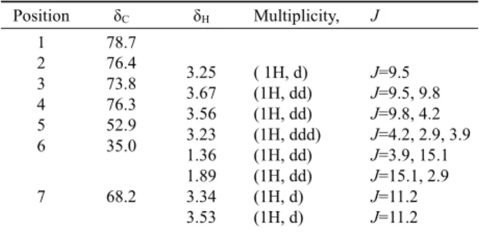
Table 2. 1H and 13C NMR data for compound K in D2O (δ in ppm and J in Hz)
고, 효소활성을 90% 저해하는 compound K 양을 효소와 혼합한 후 투석을 4시간씩 2회(USY-1 ultrafilter, Advantec MFS Inc) 실시하여 반응하지 않은 compound K을 제거하였을 때 효소활 성이 복구되는 것으로 보아 compound K는 가역적 저해제임을 알 수 있었다. Lineweaver-Burk plot에 의해 compound K가 competitive inhibitor임이 밝혀졌으며, Ki 값이 0.15 mM로 강력 한 저해제 임을 확인할 수 있었다(Fig. 2).
Compound K의 구조는 EI-MS, 1H NMR, 13C NMR, 2D COSY 등을 근거로 하여 결정하였다. 분자식은 ESI-MS와 NMR data에 의해 C7H15NO5으로 밝혀졌으며, (M+H)+ peak는 m/z 194에 나타났다. 13C NMR spectrum에서 7개의 signal (2 x methylene, 4 x methine 과 1 x quaternary carbon)을 확인할 수 있었으며, chemical shift는 1H-1H 2D NMR spectra에 의해 결정 하였다(Table 2). 1H NMR spectrum에 의해 six-membered ring 구조와 side chain methylene 존재가 확인되었고, H-2의 doublet splitting으로 C-1 carbon이 proton을 가지고 있지 않음을 알 수 있었다. 이상을 종합하고 문헌조사를 통해 활성 물질 K의 구조 는 5-amino-1-hydroxymethyl-1,2,3,4-cyclohexanetetrol (Fig. 3) 로 Streptomyces hygroscopicus에서 분리 보고된 바 있는 valiolamine (Kameda et al., 1984; Wang et al., 2010)과 동일한 물질임이 밝혀졌다. 그러나 Bacillus에서 violamine의 분리는 아 직 보고된 바 없다. 본 연구에서 선발된 균주 Bacillus sp.
SKU31-1에 의한 violamine의 생산은 Streptomces hygroscopicus 에 비해 배양 시간이 짧아 유리하다고 판단된다. SKU31-1 균주
를 이용한 α-glucosidase 저해제 violamine의 대량 생산 체계를 구축하기 위하여 저렴한 산업용 배지 개발과 대량배양 조건 확 립을 위한 후속 연구가 필요하다고 사료된다.
적 요
미생물 기원의 α-glucosidase 저해제를 탐색하는 과정에서 토 양에서 분리한 균주인 Bacillus sp. SKU31-1 배양액에서 강력한 저해제 compound K을 일련의 크로마토그래피 방법에 의해 분 리 정제하였고 1H NMR, 13C NMR, 1H-1H COSY spectra 분석과 문헌조사를 통해 5-amino-1-hydroxymethyl-1,2,3,4-cyclohexanetetrol 로 동정되었다. Compound K의 α-glucosidase 저해 활성은 IC50
값이 maltose 기질에서는 1.9 μM이고, sucrose 기질 사용시 4.9 mM 이었다. Lineweaver Burk plot에 의해 Ki값이 0.15 mM 로 강력한 경쟁적 저해제 임이 밝혀졌다.
References
Courageot, M.P., Frenkiel, M.P., Dos Santos, C.D., Deubel, V., and Despres, P. 2000. Alpha-glucosidase inhibitors reduce dengue virus production by affecting the initial steps of virion morphogenesis in the endoplasmic reticulum. J. Virol. 74, 564–572.
Fowler, M.J. 2007. Diabetes Treatment, Part 2: Oral agents for glycemic management. Clin. Diabetes 25, 131–134.
Heacock, P.M., Hertzler, S.R., Williams, J.A., and Wolf, B.W. 2005.
Effects of a medical food containing an herbal a-glucosidase inhibitor on postprandial glycemia and insulinemia in healthy adults. J. Am. Diet. Assooc. 105, 65–71.
Kameda, Y., Asano, N., Yoshikawa, M., Takeuchi, M., Yamaguchi, T., and Matsui, K. 1984. Valiolamine, a new a-glucosidase inhibiting aminocyclitol produced by Streptomyces hygroscopicus. J.
Antibiotics 37, 1301–1307.
Kim, S.D. 2013. a-Glucosidase inhibitor from Buthus martensi Karsch.
Food Chem. 136, 297–300.
Playford, R.J., Pither, C., Gao, R., and Middleton, S.J. 2013. Use of the alpha glucosidase inhibitor acarbose in patients with 'Middleton syndrome': normal gastric anatomy but with accelerated gastric emptying causing postprandial reactive hypoglycemia and diarrhea.
Can. J. Gastroenterol. 27, 403–404.
Wang, C., Sun, Y., Wen, Q., Wang, G., Wang, Y., Qu, Q., Yang, G., and Hu, X. 2010. Novel reagents for quantitative analysis of valiolamine in biological samples by high-performance liquid chromatography with pre-column UV derivatization. Talanta 81, 1613–1618.
Yuasa, H., Takada, J., and Hashimoto, H. 2001. Glycosidase inhibition by cyclic sulfonium compounds. Bioorg. Med. Chem. Lett. 11, 1137–1139.